How to Treat Plantar Fasciitis with Physiotherapy? Physiotherapy in Toronto | Rehab Mechanics
How to Treat Plantar Fasciitis with Physiotherapy?
Physiotherapy treats plantar fasciitis by reducing fascial tension, correcting lower extremity biomechanics, and stimulating tissue repair through a combination of targeted clinical modalities. Comprehensive rehabilitation addresses the root cause of the mechanical overload rather than just masking the localized pain.
Key Takeaways:
Primary Symptoms: Sharp, localized heel pain that is most severe during the first steps in the morning or upon weight-bearing after prolonged periods of rest. Pain often presents as a stabbing sensation at the bottom of the heel.
Core Modalities: Evidence-based treatment typically involves a combination of custom orthotics, extracorporeal shockwave therapy, targeted soft tissue release, therapeutic taping, and progressive corrective exercises.
General Timelines: Recovery timelines vary significantly based on individual physiological responses, the chronicity of the injury, and adherence to rehabilitation protocols. While immediate symptom relief can occur early in treatment, true structural remodeling of the fascial tissue often takes several weeks to months of consistent care.
(Visual Asset Placeholder: Clinical diagram of the foot's fascial band) Alt Text: Anatomical diagram of the human foot highlighting the plantar fascia band connecting the heel bone to the toes, illustrating the Windlass mechanism and the common site of inflammation at the medial calcaneal tubercle in plantar fasciitis.
Understanding the Anatomy of Plantar Fasciitis
The plantar fascia is a thick, web-like band of fibrous connective tissue (aponeurosis) that runs across the bottom of your foot. It originates at the medial tubercle of the heel bone (calcaneus) and fans out to connect to the base of your toes. Functionally, it acts as a dynamic shock absorber and is the primary support structure for the longitudinal arch of your foot, bearing up to 14% of the total load of the foot during the walking cycle.
Plantar fasciitis occurs when excessive mechanical stress or repetitive strain leads to microscopic tearing and subsequent inflammation of this tissue, most commonly right at its attachment point on the heel bone. It is one of the most frequent repetitive strain injuries evaluated at our Queen West clinic.
Fasciitis vs. Fasciosis: The Chronic Degeneration Cycle
While the suffix "-itis" implies acute inflammation, current medical research indicates that chronic cases (lasting longer than a few weeks) are more accurately described as plantar fasciosis. Fasciosis involves non-inflammatory structural degeneration. In these persistent cases, the collagen fibers that make up the fascia become disorganized, fragmented, and lose their vascularity (blood supply). Understanding this distinction is crucial because treating chronic cellular degeneration requires active mechanical loading and biological stimulation, rather than simple rest and anti-inflammatory medication.
Risk Factors and the Kinetic Chain
Plantar fasciitis rarely develops in isolation. It is typically the result of compounding biomechanical flaws or environmental factors, including:
Foot Mechanics: Both flat feet (pes planus) and rigid, high arches (pes cavus) alter how weight is distributed, increasing tension on the fascia.
Occupational Hazards: Service industry workers, nurses, or tradespeople who stand for prolonged hours on unyielding surfaces (like concrete) face continuous micro-trauma.
Activity Spikes: Distance runners or athletes who rapidly increase their training volume or change their running surface frequently overload the tissue before it can adapt.
Upstream Restrictions: Deficiencies in ankle mobility or weakness in the gluteal muscles can force the foot to compensate, placing an unnatural burden on the medial arch.
The Biomechanics of "First Step" Morning Pain
A hallmark characteristic of plantar fasciitis is severe pain during the first few steps in the morning. This phenomenon is biomechanically driven. During sleep, the foot naturally rests in a slightly pointed (plantarflexed) position. This allows the inflamed or damaged plantar fascia to artificially shorten and attempt to heal in a contracted state overnight.
When a patient takes their first step out of bed, the sudden upward bending (dorsiflexion) of the foot and toes forcefully stretches this newly healed, contracted tissue. This rapid stretch re-tears the micro-fibers, triggering an acute pain response. As the patient walks and the tissue warms up, it becomes more pliable, and the sharp pain often dulls to a deep ache. However, as tissue fatigue sets in toward the end of the day, a dull, throbbing pain frequently returns.
Clinical Assessment: Moving Beyond a Basic Diagnosis
Effective rehabilitation begins with diagnostic accuracy. Heel pain is not always plantar fasciitis. At our 68 Abell Street facility, care begins with an exhaustive in-person physical assessment. This includes a comprehensive gait analysis, joint mobility testing, and load-testing to rule out competing diagnoses.
Common conditions that mimic plantar fasciitis include:
Fat Pad Atrophy: The degradation of the natural shock-absorbing fat cushion under the heel bone, often presenting as a deep bruise sensation.
Calcaneal Stress Fractures: Micro-fractures in the heel bone caused by repetitive impact.
Tarsal Tunnel Syndrome: The compression of the posterior tibial nerve, which can cause shooting pain, numbness, or tingling down into the heel and arch.
Only after a precise mechanical diagnosis is confirmed, and informed patient consent is obtained, is an individualized treatment plan initiated.
Treatment Modalities at Rehab Mechanics
Rehabilitation for plantar fasciitis is a phased, multimodal process. Passive treatments are utilized to manage acute pain, while active loading is introduced to rebuild long-term tissue resilience.
Soft Tissue Therapy and The Kinetic Chain
Manual soft tissue therapy focuses on releasing tension not just in the plantar fascia itself, but throughout the entire posterior kinetic chain. The human body operates on a system of interconnected tension. The calf muscles (gastrocnemius and soleus) merge into the Achilles tendon, which attaches to the back of the heel bone. If the calf muscles are chronically tight, they exert an upward pulling force on the heel bone, which in turn stretches and strains the plantar fascia attached to the bottom of the same bone.
By utilizing manual release techniques, deep tissue mobilization, or Instrument-Assisted Soft Tissue Mobilization (IASTM) on the calf complex, physiotherapists can reduce this upstream tension. This down-regulates the local nervous system and directly decreases the mechanical pulling force exerted on the injured heel.
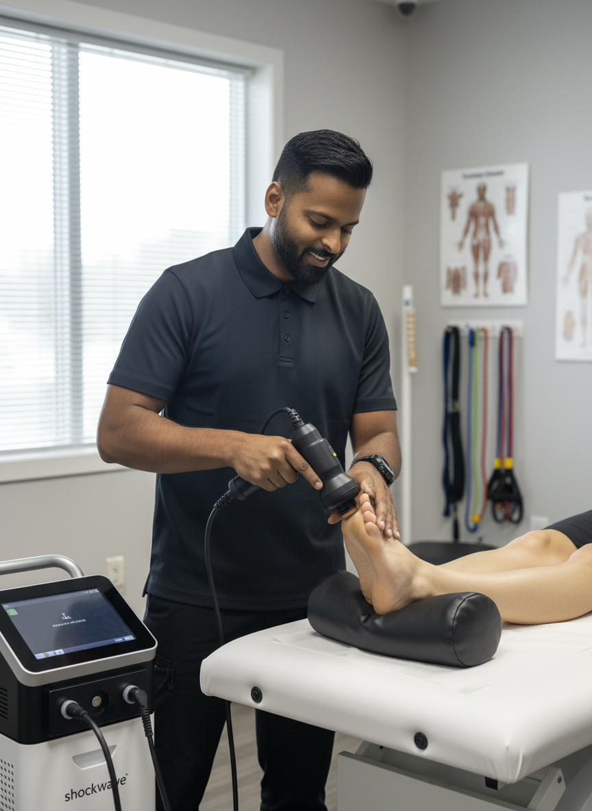
Extracorporeal Shockwave Therapy
Shockwave therapy is a highly effective, evidence-based modality frequently utilized for chronic tendinopathies and stubborn fascial issues (fasciosis) that have failed to respond to conservative stretching.
The treatment involves delivering high-energy acoustic sound waves directly into the injured tissue via a specialized clinical device. These waves create controlled micro-trauma within the fascia. This mechanical stimulus disrupts chronic, stalled healing cycles and triggers a renewed biological repair cascade. Specifically, it stimulates localized blood flow and the formation of new micro-blood vessels (angiogenesis) in a tissue that has poor natural blood supply. Furthermore, the acoustic pulses help to deplete Substance P—a neurotransmitter responsible for relaying pain signals—offering patients functional relief as the tissue rebuilds.
Custom Orthotics and Biomechanical Offloading
For patients whose plantar fasciitis is fundamentally driven by structural biomechanical issues—such as excessive pronation (inward rolling of the foot) or unusually high, rigid arches—custom orthotics provide necessary mechanical support.
Unlike generic, over-the-counter gel inserts which merely compress under body weight, custom orthotics are prescribed medical devices cast to the exact contours of the patient's foot while held in a neutral, biomechanically optimal position. By purposefully redistributing the daily load across the entire surface area of the foot and correcting inward collapse during the walking cycle, orthotics successfully reduce the daily mechanical strain on the healing fascia, allowing it the physiological space it requires to heal.
Taping and Home Management
Between clinical visits, managing daily loads is critical. Our physiotherapists frequently utilize specialized taping techniques, such as low-dye taping, to artificially support the longitudinal arch and compress the heel pad. This temporary strapping acts as an external ligament, reducing the stretch placed on the fascia during essential daily walking.
Patients are also educated on home-management strategies, including rolling the arch of the foot over a frozen water bottle to manage acute inflammatory flare-ups, and the strict avoidance of walking barefoot on hard surfaces, particularly first thing in the morning.
Corrective Exercise Protocols: High-Load Strength Training
Passive treatments alone are rarely sufficient for long-term resolution. The plantar fascia must be physically loaded to build tensile strength and align new collagen fibers correctly.
A cornerstone of modern evidence-based physiotherapy for this condition involves high-load strength training. By placing a rolled towel under the toes to engage the Windlass Mechanism (a natural tightening of the fascia when the toes are bent upward) and performing slow, heavily loaded calf raises, the tissue is subjected to high-load eccentric strengthening.
A typical protocol involves moving very slowly: 3 seconds raising up, a 2-second hold at the top, and 3 seconds lowering down. This slow, controlled tension under heavy load signals the body to lay down stronger, more resilient connective tissue capable of handling the high-impact demands of running, jumping, and daily occupational life.
Comparing Interventions for Plantar Fasciitis
Treatment Modality
Primary Mechanism of Action
Clinical Application
Soft Tissue Therapy
Reduces muscular and fascial tension in the interconnected posterior chain (calves/Achilles).
Immediate symptom management, improving ankle mobility, and reducing "pull" on the heel.
Shockwave Therapy
Stimulates cellular metabolism, breaks down calcifications, and induces local angiogenesis (blood flow).
Chronic, stubborn fascial pain (lasting > 3 months) that resists conservative stretching.
Custom Orthotics
Mechanically corrects foot posture, prevents excessive pronation, and redistributes weight-bearing load.
Long-term biomechanical management and off-loading of the medial arch.
Clinical Taping
Provides temporary structural support to the longitudinal arch.
Acute pain management during essential daily weight-bearing activities.
Corrective Exercises
Increases load tolerance and guides the alignment of new collagen fibers through eccentric loading.
Long-term functional rehabilitation, rebuilding tensile strength, and injury prevention.
Author BiographyWritten by Mr. Sanjay Attwala (BSC, MSC, RPT), Registered Physiotherapist. Sanjay Attwala manages patient care at Rehab Mechanics (S. Attwala Physiotherapy Professional Corporation) located at 68 Abell Street, Toronto. He is in good standing with the College of Physiotherapists of Ontario (CPO). Learn more about our clinical team here.
Medical Disclaimer:The content provided in this article is for general educational and informational purposes only and does not constitute formal medical advice. Individual physiological responses to physiotherapy vary, and Rehab Mechanics does not guarantee specific treatment outcomes. An in-person assessment is legally and clinically required to develop an individualized treatment plan and obtain informed consent before commencing care.