
Can Poor Ergonomics Permanently Damage My Spine and Posture?
Poor ergonomics forces the body into a continuous state of flexion, overloading the posterior spinal musculature and restricting joint mobility. Physiotherapy assists in managing this condition through a targeted Exercise Prescription that restores Extension, strengthens the core, and rebuilds postural endurance. The biomechanical root cause is the chronic yielding of extensor muscles against gravity, leading to rigid fascial adaptations and severe joint compression.
The Physical Toll of the Toronto Desk Job
In the bustling corporate towers of the Financial District and the creative studios of Queen West and Liberty Village, long hours at a desk are a mandatory part of the job. Over time, professionals begin to notice a deep, burning ache between their shoulder blades, a rigid lower back, and a noticeable forward slump in their posture that they cannot seem to correct, no matter how hard they try to "sit up straight."

What many Torontonians do not realize is that this is not just muscle fatigue; it is a structural failure driven by poor Ergonomics. The human body is highly adaptable. When you force it into a rounded, hunched position for eight to ten hours a day, the connective tissues literally remodel themselves to lock you into that shape.
Even teenagers gaming or studying for hours are at risk, as poor sitting mechanics place unnatural sheer stress on developing bones, including the vulnerable Epiphyseal Plate in growing youths. At Rehab Mechanics, we view poor posture as a treatable biomechanical dysfunction. Through highly specific physical therapy and ergonomic restructuring, we can reverse the tissue adaptations and restore a tall, powerful, and pain-free spine.
Structural / Biomechanical Analysis
To understand why standing up straight hurts, we must perform a detailed biomechanical analysis of what prolonged sitting does to your skeletal foundation.

The Loss of Spinal Extension
The human spine requires a balance of forward bending (flexion) and backward bending (Extension).
The Flexion Trap: Typing at a keyboard forces the thoracic (mid) spine into a permanent state of deep flexion.
The Structural Block: Over time, the facet joints in the back of the spine become glued shut with fibrotic scar tissue. You physically lose the mechanical ability to arch backward (Extension).
The Exhaustion of the Posterior Chain
Your body fights gravity every second of the day.
The Erector Spinae: The Erector Spinae is a massive, thick column of muscle running parallel to your spine. Its primary job is to hold your trunk upright. When you lean forward to look at a monitor, this muscle is violently overstretched and forced into a continuous, exhausting isometric contraction.
The Tipping Point: The Erector Spinae eventually fails. It loses its blood supply, goes into a state of chemical hypoxia, and develops excruciating, rock-hard trigger points.
Core and Shoulder Collapse
The front of the body is equally impacted by poor Ergonomics.
The External Obliques: The abdominal wall, specifically the External Obliques, becomes neurologically dormant due to the slouched posture. Without this lateral core stability, the lower back takes all the compressive load of sitting.
Shoulder Internal Rotation: The chest muscles shorten, violently pulling the shoulders forward. This destroys the shoulder's ability to achieve External Rotation, making it painful or impossible to reach overhead or open your chest fully.
Clinical Red Flags
We must look for specific signs that poor ergonomics has caused a deeper structural injury rather than just a simple muscle ache:
The "Hump" Deformity: A visible, hard accumulation of fibro-fatty tissue at the base of the neck (C7/T1 junction).
Radiating Numbness: Tingling or "pins and needles" shooting down the arm, indicating that the slouched posture has compressed a cervical nerve.
Extension Intolerance: A sharp, stabbing pain in the mid-back when taking a deep breath or attempting to arch backward.
Breathing Restriction: An inability to expand the ribcage fully, leading to shallow, panicked neck-breathing.
Loss of Grip Strength: The forward shoulders compress the brachial plexus, leading to noticeable weakness in the hands.
Primary Source Proof
Clinical research definitively proves that combining targeted ergonomic interventions with motor control exercises is vastly superior to passive rest for resolving work-related musculoskeletal disorders.
The Rehab Mechanics Corrective Protocol
At our clinic, we provide a customized Exercise Prescription designed to mechanically reverse the specific tissue adaptations caused by your workstation.
Phase 1 — Load Modification
Ergonomic Overhaul: We assess your workstation height, monitor placement, and chair mechanics to ensure your spine is visually neutral, instantly stopping the mechanical overload.
Joint Unlocking: Our physiotherapists use high-grade manual manipulations to break the rigid scar tissue in the thoracic facet joints, mechanically restoring the lost Extension.
Phase 2 — Pelvic Fortification
Reactivating the Core: Utilizing targeted breathing drills to wake up the deep transversus abdominis and the External Obliques, providing a solid foundation for the ribcage to rest upon.
Hip Flexor Release: Sitting locks the hips in flexion. We manually release the psoas to stop the pelvis from pulling the lower back into a painful arch.
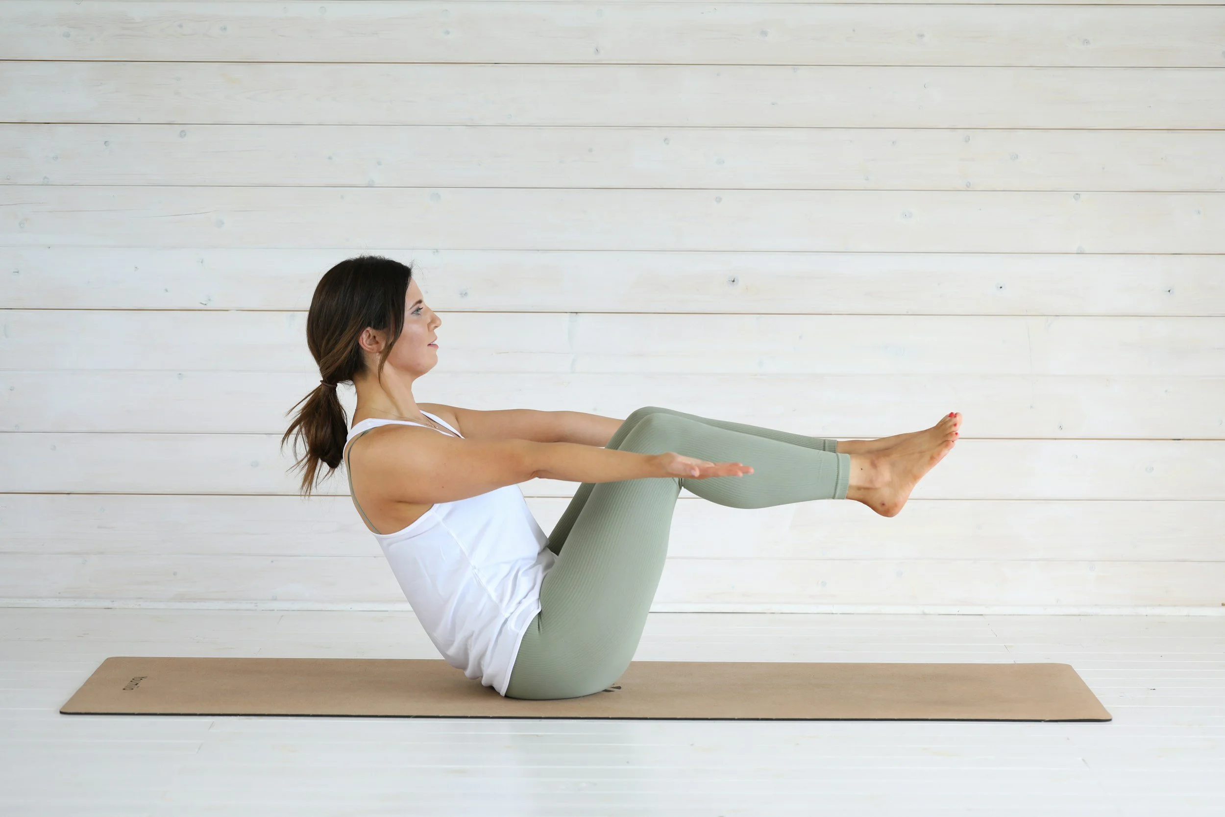
Phase 3 — Gait Retraining / Mechanics Correction
Scapular Retraction: Training the lower trapezius and rhomboids to pull the shoulder blades down and back, actively reversing the slouched posture.
Restoring External Rotation: Using resistance bands to strengthen the infraspinatus and teres minor, pulling the arm bone back into a neutral, centralized position to stop shoulder impingement.
Phase 4 — Return-to-Activity Strategy
Endurance Building: Posture requires biological endurance. We progress to heavy farmer's carries and prone positional holds to build massive stamina in the Erector Spinae.
Long-Term Exercise Prescription: Providing a 5-minute daily mobility protocol that can be executed at the desk to maintain fascial hydration and prevent the joints from freezing again.
Related Conditions We Treat
Thoracic Outlet Syndrome
Cervicogenic Headaches
Scapular Dyskinesis
Lumbar Disc Herniation
Myofascial Pain Syndrome
Rotator Cuff Tendinopathy
Related Blogs
"The 'Tech Neck' Trigger Point: How Posture Creates Unbreakable Knots"
"Does Poor Posture Cause Sharp Pain in the Middle of the Back?"
"Can Physiotherapy Cure Chronic Tension Headaches from Desk Work?"
Services Used in Treatment
Manual Therapy
Soft Tissue Release
Myofascial Release
Shockwave Therapy
Gait Retraining
Neuromuscular Re-Education
Custom Orthotics
Strengthening Programs
FAQ Section
1. Can physiotherapy assist in managing pain from poor ergonomics?
Yes. Physiotherapy helps address contributing factors by mobilizing stiff joints, releasing tight muscles, and strengthening postural stabilizers to support an upright spine.
2. What is an Exercise Prescription?
An Exercise Prescription is a highly specific, customized rehabilitation plan built by your physiotherapist. It details the exact movements, loads, and frequencies required to support recovery for your specific biomechanical faults.
3. Why do my upper back muscles burn when I sit?
Your Erector Spinae and trapezius muscles are fighting gravity to hold your head up. When you slouch, they are overstretched and overworked. We help reduce this mechanical overload by improving your ergonomic setup and strengthening your core.
4. How does core strength support my posture?
Muscles like the External Obliques stabilize your trunk from the front and sides. We focus on neuromuscular re-education to ensure these muscles fire automatically, taking the strain off your lower back.
5. Why can't I lift my arms overhead without pain?
Hunching forward causes your shoulders to roll inward, leading to impingement. We assist in managing this by restoring External Rotation to the shoulder joint, clearing the space for the tendons to move freely.
6. Can bad posture affect young adults and teenagers?
Yes. Heavy backpacks and hours of gaming place massive shear forces on developing spines, potentially irritating the Epiphyseal Plate (growth plate) in younger individuals. We optimize movement early to prevent long-term structural changes.
7. Does stretching fix a slouched posture?
Stretching the chest helps, but it is not enough. You must actively build endurance in the back muscles to hold the correction. We utilize targeted strengthening programs to build lasting resilience.
8. Will a standing desk cure my back pain?
A standing desk is a great ergonomic tool, but standing with poor mechanics can still cause pain. We help you optimize your standing and walking posture to ensure your joints remain safe under load.
How Physiotherapy Helps
Reducing tissue irritation in the overstretched mid-back muscles
Correcting pelvic drop and anterior tilt
Improving cadence and arm swing mechanics
Strengthening stabilizers in the deep core and Erector Spinae
Reducing mechanical overload on the cervical spine
Improving foot mechanics to provide a solid standing foundation
Contact Us Today — All you have to lose is the pain
Book a comprehensive biomechanical assessment with our clinical team today. Located inside the Prime Medical Centre at 68 Abell Street, Queen West.
Email: info@rehabmechanics.com Phone: (416) 533-3900
Hyperlinked to our glossary terms:
a. Epiphyseal Plate: The growth plate; a hyaline cartilage plate at each end of a long bone in growing individuals.
b. Erector Spinae: A large muscle group running vertically along the spine responsible for straightening and rotating the back.
c. Exercise Prescription: A customized fitness and rehabilitation plan designed by a physiotherapist to address a patient's specific injuries and functional goals.
d. Extension: A straightening movement that increases the angle between two bones.
e. External Obliques: Large, superficial lateral abdominal muscles that aid in twisting and compressing the abdomen.
. External Rotation: Turning a limb outward away from the center of the body
About the Author
Mr. Sanjay Attwala (B.Sc., M.Sc., RPT) is a Registered Physiotherapist, clinical director, and the founder of Rehab Mechanics in Toronto. With over 15 years of registered clinical practice and a deep specialization in complex musculoskeletal rehabilitation, Sanjay synthesizes rigorous international academic training with advanced evidence-based therapeutics to guide his clinical practice and patient education initiatives.
Academic Background & Credentials
Master of Science (M.Sc.) in Physiotherapy – University of Keele, United Kingdom (2010).
Bachelor of Science (B.Sc.) – University of Waterloo, Ontario, Canada.
Registered Physiotherapist (RPT) – Regulated health professional in excellent standing with the College of Physiotherapists of Ontario (CPO).
Corporate Entity – Operating officially under the S. Attwala Physiotherapy Professional Corporation with a DBA of Rehab Mechanics.
Clinical Expertise & Philosophy
Sanjay’s clinical approach rejects passive symptom management in favor of identifying underlying biomechanical root causes. His diverse expertise spans advanced manual therapies, personalized corrective exercise prescription, and modern physical modalities. At the Rehab Mechanics Toronto Queen West clinic, he routinely diagnoses and treats complex conditions including:
Spinal & Discogenic Pathology – Cervical, thoracic, and lumbar disc injuries, sciatica, and sacroiliac joint (SIJ) dysfunction.
Upper & Lower Extremity Injuries – Rotator cuff tears, frozen shoulder, tennis/golfer’s elbow, carpal tunnel syndrome, and complex ankle/foot pathologies.
Perinatal & Pelvic Health Rehabilitation – Specialized assessment and rehabilitation protocols tailored specifically for women during pregnancy and the post-partum period, addressing pelvic girdle pain, diastasis recti, and core stabilization.
Specialized Rehabilitation – Pelvic health therapy, TMJ dysfunction, post-surgical rehabilitation (including Total Hip and Total Knee Replacements), and custom orthotics dispensing.
Shockwave Therapy: with advanced cutting edge technological devices to suit your needs.
Interdisciplinary Practice & Patient Care
Sanjay practices an integrated model of healthcare, working closely alongside medical doctors inside the Prime Medical Centre on Abell Street to streamline patient recovery pathways. He maintains a human-centric, communication-first clinical framework, ensuring that care remains fully customized rather than automated.
His clinical caseload encompasses a broad operational spectrum under Ontario's regulatory frameworks, including:
Motor Vehicle Accident (MVA) Claims – Rehabilitation navigating Ontario’s statutory accident benefits schedule.
Workplace Safety and Insurance Board (WSIB) – Occupational injury management and return-to-work screening.
Extended Health Care (EHC) & Private Practice – Multi-tier insurance coordination and long-term athletic development plans.
Commitment to Research & Community
Outside of his clinical caseload at Rehab Mechanics and his additional practice affiliations in Etobicoke, Sanjay is an active health writer and community educator. He translates contemporary peer-reviewed medical research into accessible, actionable guidance on his professional blog. As a dedicated father and husband, he mirrors his professional advice in his personal life, focusing on structural mobility, cross-training, and longevity to help his family and his community thrive. Naturally he takes he a keen interest in rehabilitation for women who are pregnant and post-partum.
Disclaimer:
The information provided on this blog is intended for educational and informational purposes only. It does not constitute medical advice, diagnosis, or a treatment plan. Always seek the direct advice of a Registered Physiotherapist, physician, or other qualified health provider regarding any medical condition or physical rehabilitation routine.
Google MyBusiness for Instant Posts, Photos, Updates, Offers and Communication
Be sure to check out Rehab Mechanics’ Google My Business Profile for daily posts, location and directions in addition to the directions on the website!
We love interacting with prospective and current patients, and love honest feedback and even constructive criticism and areas for improvement!
Can Shockwave Therapy Replace PRP Injections for Chronic Tendinitis?
Yes. Extracorporeal Shockwave Therapy (ESWT) is a highly effective, non-invasive alternative to PRP injections. Physiotherapy utilizes these acoustic waves to biologically regenerate degenerated tendons, stimulating massive neovascularization to structurally support recovery without the pain and risks of invasive needle procedures.
The Needle vs. The Sound Wave
In the highly active and intensely demanding environments of Toronto's Queen West, Liberty Village, and the Financial District, chronic tendon injuries are a daily reality. Whether it is a severe case of tennis elbow from typing 10 hours a day, Achilles tendinopathy from marathon training, or debilitating plantar fasciitis, tendon pain has a notorious reputation for being incredibly stubborn.
When months of passive rest, ice, and generic stretching fail to provide relief, frustrated patients frequently begin researching regenerative sports medicine. They inevitably stumble upon Platelet-Rich Plasma (PRP) injections—a procedure where a doctor extracts your blood, spins it down, and injects the concentrated healing factors directly into your painful tendon with a needle.
While PRP can be effective, it is highly invasive, exceptionally expensive, and causes a significant spike in acute post-injection pain. At Rehab Mechanics, we offer a non-invasive, technologically advanced alternative that achieves the exact same biological goal: Extracorporeal Shockwave Therapy (ESWT). By harnessing the power of high-energy acoustic sound waves, our specialized physiotherapists can physically shatter scar tissue and biologically restart the stalled healing process of your tendons without ever piercing the skin.
Structural / Biomechanical Analysis
To understand how sound waves can regenerate tissue as effectively as a blood injection, we must perform a deep cellular analysis of chronic tendinopathy and the science of mechanotransduction.
The Cellular Decay (Tendinosis)
Cellular Decay
Why do chronic tendon injuries refuse to heal?
The Avascular Nature: Tendons (like the Achilles or the lateral epicondyle in the elbow) have notoriously terrible blood supply.
Failed Healing: When a tendon is repetitively overloaded, it sustains micro-tears. Because there is little blood flow to deliver healing nutrients, the body patches the tears with chaotic, weak, disorganized scar tissue.
The "Dead" Tissue: Chronic tendinitis is rarely inflamed; it is actually in a state of active cellular decay (tendinosis). The tendon physically thickens, loses its elasticity, and becomes choked with dysfunctional nerve endings.
The Biological Goal: Neovascularization
To cure a "dead" tendon, you must force the body to supply it with fresh blood. This is the exact premise of both PRP injections and Shockwave Therapy: create a localized trauma that tricks the body into hitting the "re-start" button on the healing cascade.
The Shockwave Solution (Mechanotransduction)
Extracorporeal Shockwave Therapy (ESWT) achieves this biologically without a needle.
The Acoustic Hammer: The ESWT device generates high-energy, rapid acoustic sound waves that travel safely through the skin and hit the dense, fibrotic scar tissue deep inside the tendon.
Micro-Trauma and Fragmentation: These sound waves act like a microscopic jackhammer. They physically break down the disorganized scar tissue and shatter any calcium deposits (calcific tendinitis) that have formed.
Cellular Regeneration: The mechanical force of the sound waves is converted into chemical signals by your cells (a process called mechanotransduction). The body is triggered to release growth factors (like VEGF) which stimulate the growth of brand-new capillary blood vessels (neovascularization) directly into the starving tendon.
Clinical Red Flags
Shockwave therapy is a powerful clinical tool, but it must be applied to the correct pathology. We look for these clinical signs indicating a tendon is ready for regenerative intervention:
The Chronicity Factor: Pain that has been consistently present and unchanging for greater than 3 months, completely failing standard rest and ice protocols.
The Palpable Nodule: A hard, thickened, "gristly" lump that can be physically felt on the tendon (common in the Achilles or Patellar tendon), representing thick scar tissue.
The "Warm-Up" Illusion: Pain that is severe during the first 10 minutes of activity, slowly fades as blood is forced into the area, but returns with agonizing, throbbing intensity hours after the activity stops.
Calcific Deposits: Confirmed via ultrasound or X-ray that hard calcium has formed inside the rotator cuff or heel.
Absence of Acute Rupture: We must rule out a full-thickness, acute tear of the tendon, as shockwave is designed for degenerated, intact tissue, not fully severed anatomy.
Primary Source Proof
Decades of peer-reviewed orthopedic and sports medicine literature definitively prove that high-energy Extracorporeal Shockwave Therapy is highly effective at stimulating tendon regeneration and providing long-term pain resolution, often demonstrating clinical efficacy equal to or exceeding that of PRP injections for chronic tendinopathies.
The Rehab Mechanics Corrective Protocol
Shockwave therapy is the catalyst, but biomechanical rehabilitation is the cure. We pair acoustic regeneration with an aggressive, phased mechanical loading protocol.
Phase 1 — Acoustic De-Calcification and Pain Modulation (Weeks 1-4)
ESWT Application: Administering weekly sessions of high-energy radial shockwave therapy directly to the degenerated tendon to shatter the scar tissue and trigger the massive influx of blood flow.
Pain Relief: Shockwave overstimulates the local nerve endings, dramatically dropping the concentration of Substance P (a pain neurotransmitter), providing significant, immediate pain relief after the session.
Isometric Loading: Utilizing heavy, static holds (like holding a heavy dumbbell without moving the wrist) to safely engage the muscle-tendon unit without causing painful friction.
Phase 2 — Soft Tissue De-Tethering and Pelvic Fortification
Instrument-Assisted Soft Tissue Mobilization (IASTM): Breaking down the tight, hypertonic muscle bellies located above the injured tendon (e.g., releasing the calf to take tension off the Achilles) to provide mechanical slack to the healing insertion point.
Proximal Stability: If treating the knee or foot, we aggressively strengthen the deep core and glutes. If the pelvis is stable, the lower extremity joints absorb significantly less shear force.
Phase 3 — Heavy Slow Resistance (HSR) Remodeling (Weeks 4-8)
Eccentric and Concentric Loading: This is where the structural thickening occurs. Tendons only lay down new, parallel collagen fibers under slow, heavy tension. We prescribe highly specific resistance training (e.g., 4 seconds lifting, 4 seconds lowering) to rebuild the tendon's tensile capacity.
Phase 4 — Return-to-Activity Strategy (Weeks 8+)
Energy Storage and Plyometrics: Tendons act like biological springs. Before you return to running or tennis, we must train the tendon to absorb and release kinetic energy rapidly through skipping, bounding, and rapid deceleration drills.
Biomechanical Optimization: Correcting the specific faulty movement patterns—whether it is your running gait or your desk ergonomics—that caused the tendon to degenerate in the first place.
Related Conditions We Treat
Plantar Fasciitis
Lateral Epicondylitis (Tennis Elbow)
Medial Epicondylitis (Golfer's Elbow)
Achilles Tendinopathy
Patellar Tendinopathy (Jumper's Knee)
Calcific Tendinitis of the Shoulder
Related Blogs
"Can Physiotherapy Cure Tennis Elbow If I Never Play Tennis?"
"Can Shockwave Therapy Dissolve Calcium Buildup in My Shoulder?"
"Your Achilles Heel Could Be Too Much Rest and Not Enough Physio"
Services Used in Treatment
Shockwave Therapy
Manual Therapy
Soft Tissue Release
Myofascial Release
Gait Retraining
Neuromuscular Re-Education
Custom Orthotics
Strengthening Programs
FAQ Section
1. Can shockwave therapy assist in managing chronic tendon pain?
Yes. Shockwave therapy supports recovery by utilizing acoustic energy to break down rigid, degenerated scar tissue and stimulate the growth of new blood vessels, helping to optimize the biological healing process.
2. Is shockwave therapy better than a cortisone injection?
Cortisone masks pain and is scientifically proven to weaken tendon tissue over time. We assist in managing tendinopathy using shockwave therapy because it aims to structurally regenerate the tendon rather than just suppress inflammation.
3. Does shockwave therapy hurt?
The treatment creates a deep, intense mechanical pulsing sensation that can be uncomfortable, but it is highly tolerable and adjusted to your comfort level. The minor discomfort is necessary to help reduce long-term tissue irritation.
4. How many shockwave sessions do I need?
While pain modulation can occur after the first session, stimulating meaningful cellular regeneration typically requires a protocol of 3 to 6 sessions, depending on the chronicity of the injury.
5. Do I still need to do exercises if I get shockwave therapy?
Absolutely. Shockwave provides the blood flow, but Heavy Slow Resistance (HSR) training is required to teach the new collagen fibers how to align properly. We focus on strengthening programs to build permanent resilience.
6. Can shockwave break down a bony heel spur?
Yes. ESWT acts like an acoustic hammer, making it highly effective at helping to reduce calcific deposits in the shoulder or heel, allowing the body to naturally reabsorb the fragments.
7. How long does a shockwave session take?
The actual application of the shockwave device takes only 5 to 10 minutes. It is integrated seamlessly into your broader, comprehensive physiotherapy session.
8. Are there side effects to shockwave therapy?
Side effects are minimal. You may experience slight redness, mild bruising, or a temporary ache in the treated area for 24 hours, which is the expected, desired biological response that supports recovery.
How Physiotherapy Helps
Reducing tissue irritation through acoustic neovascularization
Correcting pelvic drop to support global kinetic alignment
Improving cadence and force absorption mechanics
Strengthening stabilizers via Heavy Slow Resistance (HSR)
Reducing mechanical overload on degenerated tendon anchors
Improving foot mechanics to restore explosive spring capacity
Contact Us Today — All you have to lose is the pain
Book a comprehensive shockwave and biomechanical assessment with our clinical team today. Located inside the Prime Medical Centre at 68 Abell Street, Queen West.
Email: info@rehabmechanics.com Phone: (416) 533-3900
About the Author
Mr. Sanjay Attwala (B.Sc., M.Sc., RPT) is a Registered Physiotherapist, clinical director, and the founder of Rehab Mechanics in Toronto. With over 15 years of registered clinical practice and a deep specialization in complex musculoskeletal rehabilitation, Sanjay synthesizes rigorous international academic training with advanced evidence-based therapeutics to guide his clinical practice and patient education initiatives.
Academic Background & Credentials
Master of Science (M.Sc.) in Physiotherapy – University of Keele, United Kingdom (2010).
Bachelor of Science (B.Sc.) – University of Waterloo, Ontario, Canada.
Registered Physiotherapist (RPT) – Regulated health professional in excellent standing with the College of Physiotherapists of Ontario (CPO).
Corporate Entity – Operating officially under the S. Attwala Physiotherapy Professional Corporation with a DBA of Rehab Mechanics.
Clinical Expertise & Philosophy
Sanjay’s clinical approach rejects passive symptom management in favor of identifying underlying biomechanical root causes. His diverse expertise spans advanced manual therapies, personalized corrective exercise prescription, and modern physical modalities. At the Rehab Mechanics Toronto Queen West clinic, he routinely diagnoses and treats complex conditions including:
Spinal & Discogenic Pathology – Cervical, thoracic, and lumbar disc injuries, sciatica, and sacroiliac joint (SIJ) dysfunction.
Upper & Lower Extremity Injuries – Rotator cuff tears, frozen shoulder, tennis/golfer’s elbow, carpal tunnel syndrome, and complex ankle/foot pathologies.
Perinatal & Pelvic Health Rehabilitation – Specialized assessment and rehabilitation protocols tailored specifically for women during pregnancy and the post-partum period, addressing pelvic girdle pain, diastasis recti, and core stabilization.
Specialized Rehabilitation – Pelvic health therapy, TMJ dysfunction, post-surgical rehabilitation (including Total Hip and Total Knee Replacements), and custom orthotics dispensing.
Shockwave Therapy: with advanced cutting edge technological devices to suit your needs.
Interdisciplinary Practice & Patient Care
Sanjay practices an integrated model of healthcare, working closely alongside medical doctors inside the Prime Medical Centre on Abell Street to streamline patient recovery pathways. He maintains a human-centric, communication-first clinical framework, ensuring that care remains fully customized rather than automated.
His clinical caseload encompasses a broad operational spectrum under Ontario's regulatory frameworks, including:
Motor Vehicle Accident (MVA) Claims – Rehabilitation navigating Ontario’s statutory accident benefits schedule.
Workplace Safety and Insurance Board (WSIB) – Occupational injury management and return-to-work screening.
Extended Health Care (EHC) & Private Practice – Multi-tier insurance coordination and long-term athletic development plans.
Commitment to Research & Community
Outside of his clinical caseload at Rehab Mechanics and his additional practice affiliations in Etobicoke, Sanjay is an active health writer and community educator. He translates contemporary peer-reviewed medical research into accessible, actionable guidance on his professional blog. As a dedicated father and husband, he mirrors his professional advice in his personal life, focusing on structural mobility, cross-training, and longevity to help his family and his community thrive. Naturally he takes he a keen interest in rehabilitation for women who are pregnant and post-partum.
Disclaimer:
The information provided on this blog is intended for educational and informational purposes only. It does not constitute medical advice, diagnosis, or a treatment plan. Always seek the direct advice of a Registered Physiotherapist, physician, or other qualified health provider regarding any medical condition or physical rehabilitation routine.
Google MyBusiness for Instant Posts, Photos, Updates, Offers and Communication
Be sure to check out Rehab Mechanics’ Google My Business Profile for daily posts, location and directions in addition to the directions on the website!
We love interacting with prospective and current patients, and love honest feedback and even constructive criticism and areas for improvement!














Should I Put My Rolled Ankle in a Walking Boot to Heal?
Prolonged immobilization in a walking boot weakens ligaments and destroys proprioception. Physiotherapy assists in managing ankle sprains by utilizing early, protected weight-bearing and aggressive neuromuscular re-education, helping to support recovery, restore structural stability, and prevent chronic ankle instability.
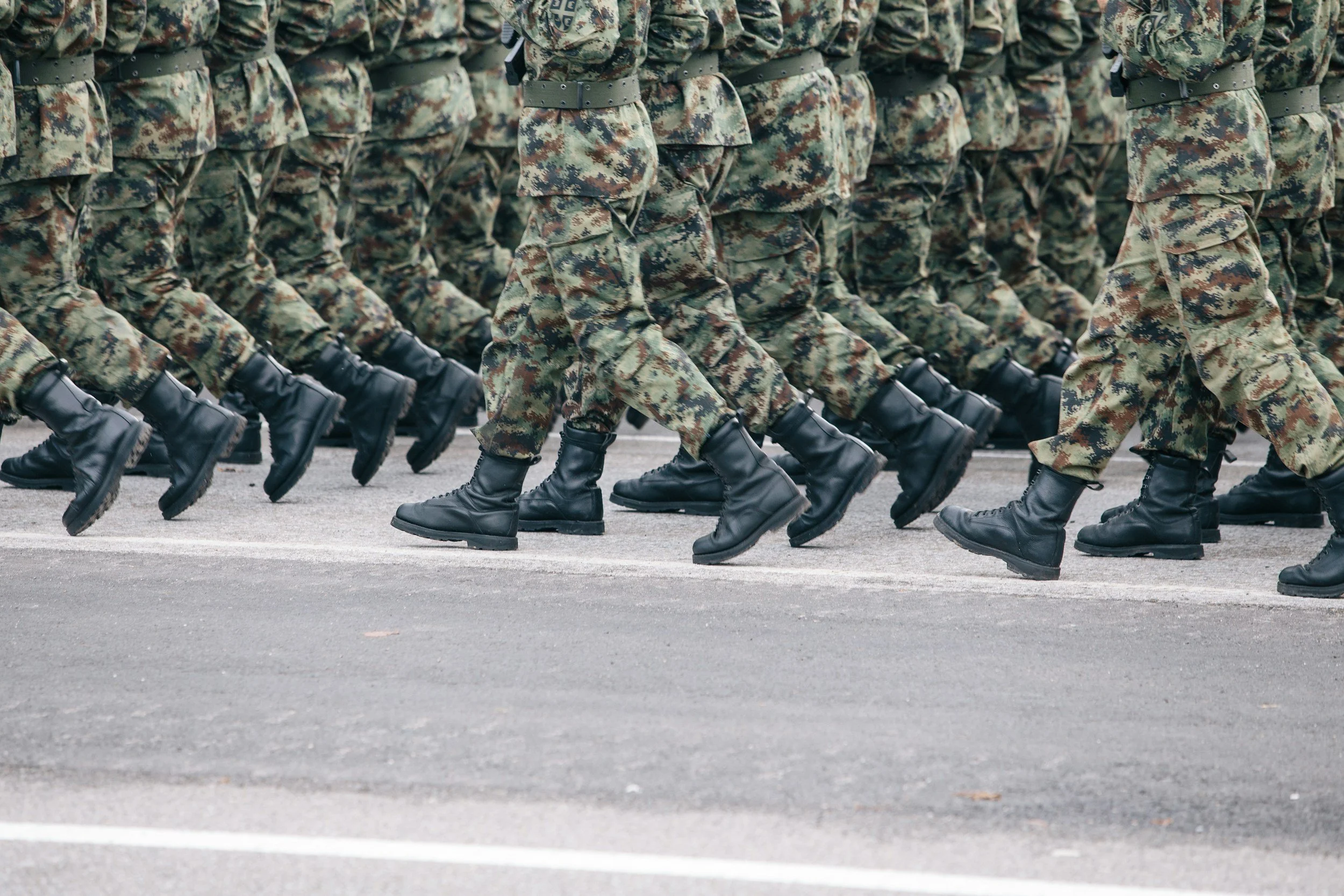
Boots on the ground…
The Immobilization Trap
In a bustling city like Toronto, an ankle sprain is an incredibly common occurrence. Whether you rolled your foot stepping off a streetcar in Liberty Village, hiking a rugged trail in High Park, or playing recreational basketball in Trinity Bellwoods, the initial pain and swelling are intense.
The standard response for many patients is to visit an urgent care clinic, where they are often provided with a rigid CAM walking boot or crutches and told to "stay off it" for four to six weeks. The patient complies, assuming that absolute rest is the safest way to let the torn ligaments heal.
However, when they finally remove the boot weeks later, they discover a new nightmare: their ankle feels profoundly stiff, their calf muscle has visibly shrunken, and their ankle feels terrifyingly wobbly and weak. The moment they step on an uneven sidewalk, they roll it again.
At Rehab Mechanics, we manage high-level sports trauma daily. We know that modern sports medicine explicitly advises against prolonged immobilization for standard ankle sprains. While a boot may be necessary for severe fractures or high syndesmotic tears, putting a standard inversion sprain in a rigid boot causes rapid tissue atrophy and neurological disconnect. By utilizing early, protected physical therapy, we actively rebuild your torn ligaments and save you from a lifetime of chronic instability.
Structural / Biomechanical Analysis
To understand why a walking boot causes long-term harm, we must perform a biomechanical analysis of the ankle's defense systems and how biological tissues actually heal.
The Lateral Ligament Complex
When you "roll" your ankle, the foot typically turns inward (inversion). This violently stretches or tears the ligaments on the outside of your ankle.
The ATFL and CFL: The Anterior Talofibular Ligament and Calcaneofibular Ligament are the primary passive stabilizers.
The Healing Process: Ligaments require mechanical stress to align their collagen fibers properly as they heal. If you lock the ankle in a boot, the new scar tissue heals in a chaotic, weak, and shortened clump, leaving the joint permanently unstable.
The Neurological Disaster: Proprioceptive Loss
The most severe damage from an ankle sprain is neurological, not structural.
Mechanoreceptors: Your ankle ligaments are packed with millions of nerve endings that tell your brain exactly where your foot is in space (proprioception).
The Disconnect: When you tear the ligament, you tear these nerves. If you put the foot in a rigid boot, the brain completely "forgets" how to communicate with the ankle.
The Tipping Point (Chronic Ankle Instability): When you take the boot off, your brain cannot sense uneven ground fast enough to fire your protective muscles. This sensory delay guarantees you will continually re-sprain the ankle, a devastating condition known as Chronic Ankle Instability (CAI).
Muscular Atrophy
The peroneal muscles on the outside of your calf act as dynamic stirrups; they are the active brakes that stop your ankle from rolling. Immobilization causes these muscles to rapidly atrophy. A weak muscle combined with a loose ligament is a recipe for permanent mechanical failure.
Clinical Red Flags
We must perfectly differentiate a standard sprain from a severe fracture or surgical injury before determining the loading protocol. We use strict criteria (like the Ottawa Ankle Rules):
Inability to Bear Weight: If you are physically unable to take four consecutive steps immediately after the injury or in the clinic, a fracture must be ruled out.
Point Tenderness on the Bone: Exquisite, sharp pain when pressing directly on the bony prominences of the medial or lateral malleolus (ankle bones), rather than just the soft tissue below them.
The Squeeze Test Failure: Sharp pain radiating up the shin when the calf is compressed, indicating a High Ankle (Syndesmotic) Sprain, which does require specialized immobilization.
Massive, Immediate Edema: Swelling that is so severe and rapid that it pools deeply into the toes within hours.
Primary Source Proof
Decades of peer-reviewed orthopedic and sports medicine literature definitively prove that early functional mobilization and protected weight-bearing yield vastly superior functional outcomes, faster return to sport, and fewer re-injuries compared to prolonged rigid immobilization for acute lateral ankle sprains.
The Rehab Mechanics Corrective Protocol
We replace passive resting with active, protected mechanical loading. Our goal is to forge strong, aligned ligaments and razor-sharp reflexes.
Phase 1 — Protected Weight Bearing and Edema Control (Weeks 1-2)
Functional Bracing (Not Booting): We transition you out of a rigid boot and into a semi-rigid lace-up brace. This prevents the ankle from rolling side-to-side (protecting the torn ligament) but allows the ankle to bend forward and backward (dorsiflexion/plantarflexion), ensuring early collagen alignment.
Manual Lymphatic Drainage: Our physiotherapists utilize targeted soft tissue massage to push the massive, stagnant pool of swelling out of the foot and up into the lymphatic system of the leg.
Pain-Free Active ROM: Implementing precise "alphabet drawing" exercises with the toes to maintain joint lubrication and prevent the joint capsule from freezing.
Phase 2 — Pelvic and Core Fortification
Proximal Stability: An unstable ankle forces the entire leg to compensate. We aggressively strengthen the gluteus medius and deep core. If the hip is stable, it prevents the leg from wildly swaying, reducing the compensatory sheer force on the healing ankle joint.
Phase 3 — Gait Retraining / Proprioceptive Re-Syncing (Weeks 3-6)
Neuromuscular Re-Education: This is the absolute core of the cure. We utilize balance boards, BOSU balls, and uneven surface training. We must force the damaged nerve endings in the ankle to rebuild their high-speed connection to the brain.
Peroneal Strengthening: Using resistance bands to heavily load the muscles on the outside of the calf, ensuring they are strong enough to actively brake against a sudden twist.
Phase 4 — Return-to-Activity Strategy (Weeks 6+)
Dynamic Perturbations: The physiotherapist applies sudden, unexpected pushes to your body while you balance, training your spinal cord to react in milliseconds.
Plyometric Bounding: Progressing to lateral skater jumps, agility ladders, and deceleration drills. We do not clear you for sports until your injured ankle handles rapid, multi-directional shock identically to your healthy ankle.
Related Conditions We Treat
Chronic Ankle Instability (CAI)
High Ankle Sprains (Syndesmotic Sprains)
Peroneal Tendinopathy
Plantar Fasciitis
Achilles Tendinopathy
Tibial Stress Fractures
Related Blogs
"Is My Chronic Outer Ankle Pain a Lingering Sprain or a Torn Tendon?"
"Do I Always Need Physiotherapy for a Rolled Ankle?"
"Why Is My High Ankle Sprain Taking Months to Heal?"
Services Used in Treatment
Manual Therapy
Soft Tissue Release
Myofascial Release
Gait Retraining
Neuromuscular Re-Education
Custom Orthotics
Strengthening Programs
Shockwave Therapy
FAQ Section
1. Can physiotherapy assist in managing an acute ankle sprain?
Yes. Physiotherapy supports recovery by utilizing early, protected movement to ensure ligaments heal with proper alignment, while restoring the critical balance reflexes required to optimize movement safely.
2. Should I wear a walking boot for a rolled ankle?
For standard inversion sprains, prolonged boot use causes severe muscle wasting and joint stiffness. We assist in managing your recovery by using functional bracing, which protects the ligament while allowing safe, healing movement.
3. Why does my ankle still feel weak months after a sprain?
This is Chronic Ankle Instability, caused by the loss of proprioceptive nerve endings during the initial tear. We focus on neuromuscular re-education to rebuild the brain-to-ankle connection and support structural confidence.
4. How does hip strength prevent ankle rolling?
Your glute muscles control the alignment of your entire leg. We focus on strengthening these stabilizers to ensure your leg remains perfectly straight over your foot, reducing mechanical overload on the ankle ligaments.
5. Is it safe to walk on a swollen ankle?
Yes, if properly supported and within pain limits. Early, controlled weight-bearing acts as a mechanical pump, which actually helps reduce tissue irritation and flush swelling out of the joint faster than absolute rest.
6. What are peroneal muscles and why do they matter?
The peroneal muscles run down the outside of your calf and act as active "brakes" against ankle rolling. We utilize specific strengthening programs to fortify these muscles, providing a biological defense against future sprains.
7. Can custom orthotics help chronic ankle instability?
If severe overpronation (flat feet) is causing your ankle to constantly cave inward, custom orthotics can help optimize movement by supporting your arch and providing a neutral foundation for your kinetic chain.
8. How long does it take to fully recover from an ankle sprain?
While the acute pain may subside in weeks, rebuilding the dense collagen fibers and the high-speed neurological balance reflexes typically requires 6 to 8 weeks of targeted, progressive rehabilitation.
How Physiotherapy Helps
Reducing tissue irritation through active lymphatic drainage
Correcting pelvic drop to stabilize the entire kinetic chain
Improving cadence and confident weight-bearing mechanics
Strengthening stabilizers in the peroneal and calf complex
Reducing mechanical overload on the healing lateral ligaments
Improving foot mechanics to restore high-speed proprioception
Contact Us Today — All you have to lose is the pain
Book a comprehensive biomechanical ankle assessment with our clinical team today. Located inside the Prime Medical Centre at 68 Abell Street, Queen West.
Email: info@rehabmechanics.com Phone: (416) 533-3900
About the Author
Mr. Sanjay Attwala (B.Sc., M.Sc., RPT) is a Registered Physiotherapist, clinical director, and the founder of Rehab Mechanics in Toronto. With over 15 years of registered clinical practice and a deep specialization in complex musculoskeletal rehabilitation, Sanjay synthesizes rigorous international academic training with advanced evidence-based therapeutics to guide his clinical practice and patient education initiatives.
Academic Background & Credentials
Master of Science (M.Sc.) in Physiotherapy – University of Keele, United Kingdom (2010).
Bachelor of Science (B.Sc.) – University of Waterloo, Ontario, Canada.
Registered Physiotherapist (RPT) – Regulated health professional in excellent standing with the College of Physiotherapists of Ontario (CPO).
Corporate Entity – Operating officially under the S. Attwala Physiotherapy Professional Corporation with a DBA of Rehab Mechanics.
Clinical Expertise & Philosophy
Sanjay’s clinical approach rejects passive symptom management in favor of identifying underlying biomechanical root causes. His diverse expertise spans advanced manual therapies, personalized corrective exercise prescription, and modern physical modalities. At the Rehab Mechanics Toronto Queen West clinic, he routinely diagnoses and treats complex conditions including:
Spinal & Discogenic Pathology – Cervical, thoracic, and lumbar disc injuries, sciatica, and sacroiliac joint (SIJ) dysfunction.
Upper & Lower Extremity Injuries – Rotator cuff tears, frozen shoulder, tennis/golfer’s elbow, carpal tunnel syndrome, and complex ankle/foot pathologies.
Perinatal & Pelvic Health Rehabilitation – Specialized assessment and rehabilitation protocols tailored specifically for women during pregnancy and the post-partum period, addressing pelvic girdle pain, diastasis recti, and core stabilization.
Specialized Rehabilitation – Pelvic health therapy, TMJ dysfunction, post-surgical rehabilitation (including Total Hip and Total Knee Replacements), and custom orthotics dispensing.
Shockwave Therapy: with advanced cutting edge technological devices to suit your needs.
Interdisciplinary Practice & Patient Care
Sanjay practices an integrated model of healthcare, working closely alongside medical doctors inside the Prime Medical Centre on Abell Street to streamline patient recovery pathways. He maintains a human-centric, communication-first clinical framework, ensuring that care remains fully customized rather than automated.
His clinical caseload encompasses a broad operational spectrum under Ontario's regulatory frameworks, including:
Motor Vehicle Accident (MVA) Claims – Rehabilitation navigating Ontario’s statutory accident benefits schedule.
Workplace Safety and Insurance Board (WSIB) – Occupational injury management and return-to-work screening.
Extended Health Care (EHC) & Private Practice – Multi-tier insurance coordination and long-term athletic development plans.
Commitment to Research & Community
Outside of his clinical caseload at Rehab Mechanics and his additional practice affiliations in Etobicoke, Sanjay is an active health writer and community educator. He translates contemporary peer-reviewed medical research into accessible, actionable guidance on his professional blog. As a dedicated father and husband, he mirrors his professional advice in his personal life, focusing on structural mobility, cross-training, and longevity to help his family and his community thrive. Naturally he takes he a keen interest in rehabilitation for women who are pregnant and post-partum.
Disclaimer:
The information provided on this blog is intended for educational and informational purposes only. It does not constitute medical advice, diagnosis, or a treatment plan. Always seek the direct advice of a Registered Physiotherapist, physician, or other qualified health provider regarding any medical condition or physical rehabilitation routine.
Google MyBusiness for Instant Posts, Photos, Updates, Offers and Communication
Be sure to check out Rehab Mechanics’ Google My Business Profile for daily posts, location and directions in addition to the directions on the website!
We love interacting with prospective and current patients, and love honest feedback and even constructive criticism and areas for improvement!
Why Does My Kneecap Feel Loose and Click When I Walk Down Stairs?
Patellar instability causes the kneecap to mal-track and click during movement. Physiotherapy assists in managing this condition by correcting pelvic drop, releasing tight lateral fascia, and strengthening the vastus medialis oblique (VMO) to restore optimal joint tracking and reduce mechanical wear on the retropatellar cartilage.
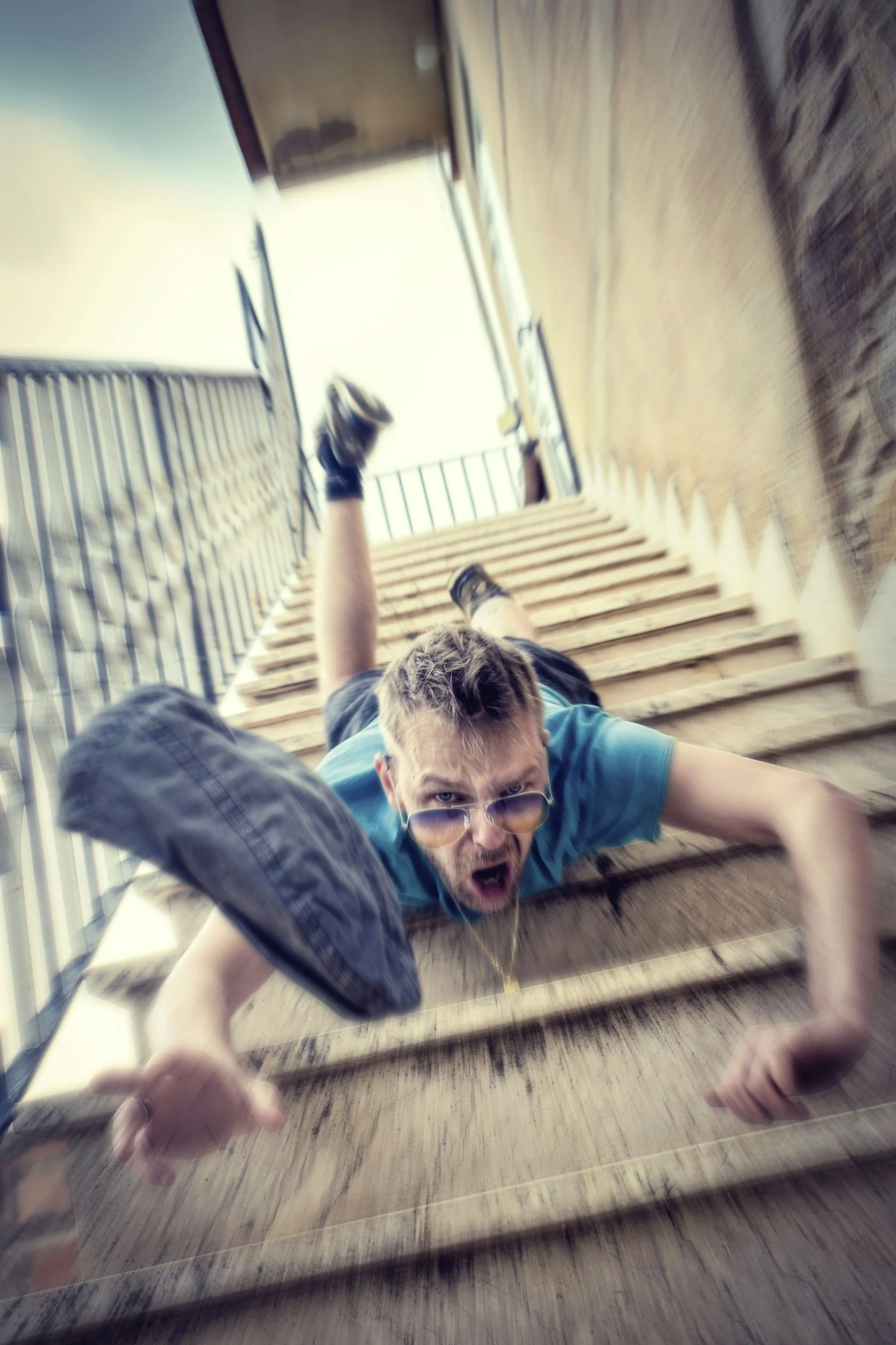
No we said walking down the stairs - not head first - but this will also hurt your knees going ‘down the stairs’
The Downward Staircase Dilemma
For active residents in Toronto, descending stairs is an unavoidable part of daily life. Whether you are navigating the deep steps of a TTC subway station, leaving your Queen West apartment, or hiking the trails in High Park, walking downward places immense sheer force on your lower body.
Many individuals experience a deeply unsettling phenomenon during this specific movement: their kneecap audibly "clicks," "pops," or physically feels as though it is sliding out of place before snapping back. This "giving way" or loose sensation triggers immediate anxiety, leading patients to rigidly lock their legs or avoid stairs altogether.
Walk-in clinics often dismiss this as generic "Runner's Knee" and prescribe a compressive sleeve. However, at Rehab Mechanics, we know that a clicking, shifting kneecap is a specific biomechanical failure known as Patellar Instability or Patellar Mal-tracking. If ignored, this chronic shifting acts like sandpaper, prematurely wearing away the cartilage behind your kneecap. By analyzing and correcting the muscular forces acting on your knee, specialized physical therapy can securely anchor your patella and restore your confidence on the stairs.
Structural / Biomechanical Analysis
To understand why your kneecap is shifting, we must perform a detailed structural analysis of the knee extensor mechanism and the forces that guide it.
The Trochlear Groove
Your kneecap (patella) is a floating bone embedded inside your massive quadriceps tendon.
The Track: It is designed to glide up and down within a specific V-shaped bony trench on the front of your thigh bone (femur) called the trochlear groove.
The Balance of Forces: For the kneecap to stay perfectly centered in this groove, the muscles pulling it from the inside, outside, top, and bottom must possess perfectly balanced tension.
The Pathology of Patellar Mal-Tracking
In patellar instability, this balance is destroyed, and the kneecap is violently pulled off-center (usually toward the outside of the leg).
Vastus Medialis Oblique (VMO) Weakness
The VMO is the teardrop-shaped muscle on the inside of your thigh, just above the knee. Its exclusive job is to pull the kneecap inward during movement.
The Shutdown: If you have experienced knee swelling in the past, or if you sit at a desk all day, the VMO rapidly weakens and becomes neurologically delayed. It fails to fire fast enough to keep the kneecap centered.
Lateral Fascial Tightness
On the outside of your leg, the Iliotibial (IT) band and the lateral retinaculum exert an outward pull.
The Imbalance: If the VMO is weak and the outer structures are tight, the kneecap is physically dragged outward. As you bend your knee to step down a stair, the misaligned kneecap catches on the bony ridge of the groove, then loudly "clicks" or "pops" as it forces its way past the obstruction.
The Pelvic Drop (Dynamic Valgus)
The root cause is almost always higher up the kinetic chain.
If your side-hip muscle (gluteus medius) is weak, your thigh bone rotates inward when you bear weight on one leg (like stepping down a stair). This inward rotation physically knocks the groove out from underneath the kneecap, causing massive instability and cartilage wear.
Clinical Red Flags
We meticulously differentiate between simple patellofemoral pain and true structural instability:
The "J Sign": When extending the leg from a bent position, the kneecap visibly jumps outward in a "J" pattern at the very end of the movement.
Apprehension Test: Extreme anxiety or defensive muscle guarding when a physiotherapist gently attempts to push the kneecap outward.
Catching on Descent: An audible, painful click that reliably occurs when the knee is loaded eccentrically (walking down stairs or hiking downhill), but not when walking up.
Recurrent Subluxations: A history of the knee actually "giving out" or fully buckling during pivoting movements on a sports field.
Primary Source Proof
Clinical biomechanical research consistently demonstrates that targeted strengthening of the VMO and the gluteal complex is the definitive first-line intervention for stabilizing the patella and preventing long-term cartilage degradation.
The Rehab Mechanics Corrective Protocol
We do not just hand you a knee brace. We rebuild the muscular scaffolding required to hold the kneecap perfectly in place.
Phase 1 — Load Modification and Tissue De-Tethering
Lateral Release Techniques: Our physiotherapists use advanced manual therapy and instrument-assisted soft tissue mobilization (IASTM) to release the tight IT band, vastus lateralis, and lateral retinaculum, physically removing the outward pull on the patella.
McConnell Taping: Utilizing rigid kinesiology tape to mechanically pull the kneecap back into the center of the groove, providing immediate relief and stability when navigating stairs during the early rehab phase.
Phase 2 — Pelvic Fortification
Gluteus Medius Isolation: We must stop the thigh bone from rotating inward. We prescribe targeted banded lateral walks and clamshells to build a massive, stable lateral hip.
Core-to-Floor Integration: Ensuring the deep abdominal core fires properly to hold the pelvis level during single-leg weight-bearing.
Phase 3 — Gait Retraining / Mechanics Correction (VMO Resurrection)
VMO Biofeedback: Utilizing tactile cues and localized isometric holds (like Spanish Squats) to force the nervous system to re-engage the vastus medialis oblique, training it to fire before the outer thigh muscles.
Terminal Knee Extension (TKE): Heavy resistance band drills focusing on the final 15 degrees of leg straightening, which is where the VMO is most active in stabilizing the kneecap.
Phase 4 — Return-to-Activity Strategy
Eccentric Deceleration: We must train your body to handle the exact movement that causes the click. We implement slow, heavily controlled step-downs and eccentric split squats, closely monitoring your knee alignment.
Plyometric Stability: Progressing to rapid bounding and jumping mechanics, proving to the nervous system that the kneecap is securely anchored during high-velocity sports.
Related Conditions We Treat
Patellofemoral Pain Syndrome (Runner's Knee)
Chondromalacia Patellae
Patellar Tendinopathy (Jumper's Knee)
Iliotibial (IT) Band Syndrome
Knee Osteoarthritis
Meniscus Tears
Related Blogs
"Does Pain Behind the Kneecap Mean My Cartilage is Gone?"
"Will Foam Rolling Fix My IT Band Syndrome and Outer Knee Pain?"
"Solving Runner's Knee: A Deep Dive into Patellofemoral Mechanics"
Services Used in Treatment
Neuromuscular Re-Education
Gait Retraining
Strengthening Programs
Manual Therapy
Soft Tissue Release
Myofascial Release
Custom Orthotics
Shockwave Therapy
FAQ Section
1. Can physiotherapy assist in managing a clicking kneecap?
Yes. Physiotherapy supports recovery by addressing the muscular imbalances pulling the kneecap off-center, helping to optimize movement and safely restore proper tracking within the joint groove.
2. Is it dangerous if my knee clicks without pain?
Painless clicking is common, but it indicates a mechanical tracking issue. Over time, this friction can contribute to cartilage wear. We assist in managing these mechanics to support long-term joint health.
3. Will wearing a knee brace stop my patella from slipping?
A brace offers temporary support, but it does not fix the underlying muscular weakness. We focus on neuromuscular re-education to build your own "biological brace" for durable stability.
4. Why does my knee only pop when walking down stairs?
Descending stairs requires intense eccentric contraction of your quadriceps, which places the highest pressure on the kneecap. If your tracking is off, the patella catches and pops. We help reduce this mechanical overload.
5. How does hip strength impact my kneecap?
Your glute muscles control the angle of your thigh bone. We focus on strengthening these stabilizers to prevent the inward rotation that violently forces the kneecap out of alignment.
6. Do I need surgery for a loose kneecap?
In many cases, no. Unless you have suffered a catastrophic dislocation tearing the medial ligaments, conservative physical therapy helps address contributing factors and restore stability without surgical intervention.
7. Can tight outer leg muscles cause patellar instability?
Absolutely. A tight IT band and lateral structures physically drag the kneecap outward. We utilize soft tissue release to provide mechanical slack and support optimal joint alignment.
8. How long does it take to stabilize the patella?
Rebuilding neurological motor control takes time. While taping provides immediate relief, building the VMO and gluteal strength required for permanent stability typically involves 6 to 10 weeks of progressive strengthening programs.
How Physiotherapy Helps
Reducing tissue irritation through targeted lateral release
Correcting pelvic drop to prevent dynamic valgus collapse
Improving cadence and eccentric stair-descent mechanics
Strengthening stabilizers in the VMO and gluteus medius
Reducing mechanical overload on the retropatellar cartilage
Improving foot mechanics to anchor a stable kinetic chain
Contact Us Today — All you have to lose is the pain
Book a comprehensive biomechanical knee assessment with our clinical team today. Located inside the Prime Medical Centre at 68 Abell Street, Queen West.
Email: info@rehabmechanics.com Phone: (416) 533-3900
Can I Recover From Golfer's Elbow Without Quitting Weightlifting?
Absolute rest is detrimental to tendon healing. Medial epicondylitis (Golfer's Elbow) requires active, progressive loading to stimulate collagen repair. Physiotherapy assists in managing this condition by utilizing heavy slow resistance, isometric holds, and targeted soft tissue release to structurally rebuild the tendon, allowing you to lift weights safely while the tissue recovers.
Looking to get back to or go to Pebble Beach
The Patient’s Story
In Parkdale, Queen West, and Liberty Village, the local fitness culture is intense. Between heavy barbell deadlifts, advanced kettlebell training, rock climbing, and high-volume computer work, Toronto residents demand massive, sustained pulling power from their upper bodies.
However, this relentless demand on grip strength often results in a sharp, burning, and severely limiting pain on the inside of the elbow—a condition medically known as Medial Epicondylitis, or "Golfer's Elbow." Despite the name, the vast majority of our patients with this condition have never swung a golf club.
When inner elbow pain strikes, the traditional advice from walk-in clinics is universally frustrating: "Stop lifting weights, ice it, wear a heavy brace, and wait three months." But for an active urban professional, abandoning the gym is unacceptable and, clinically, it is the exact wrong approach. Complete rest causes tendons to atrophy and weaken. At Rehab Mechanics, we resolve Medial Epicondylitis not by resting the tendon into oblivion, but by actively remodeling it to handle heavier loads, allowing you to stay in the gym safely while you heal.
Structural / Biomechanical Analysis
To understand why passive rest fails to fix Golfer's Elbow, we must perform a detailed biomechanical analysis of the forearm flexors and the true cellular nature of chronic tendinopathy.
The Flexor-Pronator Mass
Your forearm contains a complex network of muscles that control your wrist and grip. The muscles responsible for bending your wrist forward (flexion) and turning your palm face down (pronation) all merge into a single, thick common tendon.
The Anatomical Anchor: This common flexor tendon anchors directly onto the medial epicondyle—the prominent bony bump on the inside of your elbow.
The Mechanical Overload: Every time you grip a heavy deadlift bar, perform a pull-up, or even type furiously on an un-ergonomic keyboard, massive tension is transferred through these muscles directly into that small bony attachment.
The Pathology of Tendinopathy (Tendinosis)
Golfer's elbow is rarely an acute, inflammatory "tendinitis." It is almost always a chronic, degenerative "tendinosis."
Failed Cellular Healing
When the flexor tendon is overloaded repetitively, it sustains microscopic tears.
The Chaotic Repair: Instead of laying down strong, perfectly parallel Type I collagen fibers to fix the tears, an exhausted body patches the area with weak, disorganized Type III scar tissue.
Angiofibroblastic Degeneration: The tendon physically thickens, loses its elasticity, and becomes infiltrated with highly sensitive, dysfunctional nerve endings and weak, chaotic blood vessels. It is actively decaying, not just inflamed.
The Valgus Stress Factor
In sports and lifting, poor shoulder or wrist mechanics often create "valgus stress"—a lateral, outward-pulling force on the elbow joint.
If your shoulder lacks external rotation during a heavy lift, your elbow is forced to flare out. This violently yanks on the medial epicondyle with every repetition, drastically accelerating the tendon degeneration.
Clinical Red Flags
We differentiate tendinopathy from a simple muscle ache or a ligament sprain by looking for specific, highly localized mechanical failures:
Exquisite Point Tenderness: Breathtaking, sharp pain specifically when pressing a thumb directly into the bony bump on the inside of the elbow.
Pain with Resisted Flexion: Severe pain triggered in the elbow when a therapist asks you to aggressively curl your wrist forward against their resistance.
The Grip Drop: A sudden, inexplicable loss of absolute grip strength, making a previously easy dumbbell feel dangerously heavy or causing you to drop a coffee mug.
Morning Stiffness: The inner elbow joint feels incredibly rigid, achy, and "crunchy" for the first 30 minutes after waking up.
Ulnar Nerve Symptoms: Occasional tingling in the pinky and ring fingers, as the ulnar nerve runs directly behind the inflamed medial epicondyle (Cubital Tunnel overlap).
Primary Source Proof
Orthopedic research explicitly supports progressive, heavy mechanical loading over passive rest or corticosteroid injections for the long-term cure and structural remodeling of chronic tendinopathies.
Review the Clinical Evidence on PubMed: The Efficacy of Heavy Slow Resistance Training in Medial Epicondylitis (National Institutes of Health)
Review the Clinical Evidence on PubMed: Conservative Management and Eccentric Exercise in Epicondylopathy (National Institutes of Health)
Review the Clinical Evidence on PubMed: Extracorporeal Shockwave Therapy for Chronic Tendinopathies (National Institutes of Health)
The Rehab Mechanics Corrective Protocol
To cure Golfer’s Elbow, we must break down the chaotic scar tissue and stimulate the growth of new, aligned collagen fibers. We achieve this through a rigorous, phased clinical pathway while keeping you active.
Phase 1 — Pain Modulation and Neovascularization
Extracorporeal Shockwave Therapy (ESWT): Utilizing high-energy acoustic sound waves to literally shatter the fibrotic scar tissue at the medial epicondyle. This mechanical stimulus forces the body to grow new, healthy blood vessels (neovascularization) into the deadened tendon.
Isometric Loading (The Painkiller): Having you hold a heavy dumbbell in a slightly flexed wrist position without moving for 45-second intervals. Isometrics safely engage the muscle-tendon unit, rapidly signaling the brain to down-regulate pain sensitivity without irritating the joint.
Phase 2 — Pelvic Fortification and Scapular Control
Shoulder Anchoring: You cannot have strong elbows without a strong base. We strengthen the mid-back (lower trapezius and rhomboids) to ensure your shoulder blade remains stable during heavy pulling, preventing the elbow from absorbing compensatory sheer forces.
Core-to-Arm Transfer: Training the deep core to stabilize the torso, ensuring that rotational lifting power comes from the hips rather than the fragile forearm tendons.
Phase 3 — Gait Retraining / Mechanics Correction (Heavy Slow Resistance)
Eccentric Wrist Flexion: Focusing specifically on the "lowering" phase of a wrist curl. Eccentric contractions physically pull the chaotic collagen fibers into perfectly parallel alignment, rebuilding the tendon's tensile strength.
Pronation/Supination Loading: Using offset weights (like a hammer or specialized club) to strengthen the rotational muscles of the forearm, which are critical for stabilizing the elbow during heavy lifting.
Phase 4 — Return-to-Activity Strategy
Grip Modification Coaching: We teach you how to modify your grip (e.g., using lifting straps temporarily, or switching to neutral-grip pull-ups) to continue training your back and legs at 100% capacity without aggravating the medial epicondyle.
Progressive Barbell Re-Integration: Slowly reintroducing heavy deadlifts and cleans under strict biomechanical supervision to ensure the newly remodeled tendon can handle maximum load.
Related Conditions We Treat
Lateral Epicondylitis (Tennis Elbow)
Cubital Tunnel Syndrome
Carpal Tunnel Syndrome
De Quervain's Tenosynovitis
Thoracic Outlet Syndrome
Biceps Tendinopathy
Related Blogs
"Can Physiotherapy Cure Tennis Elbow If I Never Play Tennis?"
"Does Numbness in Your Pinky Finger Mean You Have Cubital Tunnel Syndrome?"
"Can Physiotherapy Cure 'Mommy Thumb' and Texting Wrist Without Cortisone?"
Services Used in Treatment
Manual Therapy
Soft Tissue Release
Myofascial Release
Shockwave Therapy
Gait Retraining
Neuromuscular Re-Education
Custom Orthotics
Strengthening Programs
FAQ Section
1. Can physiotherapy assist in managing Golfer's Elbow?
Yes. Physiotherapy supports recovery by utilizing heavy slow resistance and targeted soft tissue release to structurally rebuild the degenerated tendon, helping to optimize movement and safely restore grip strength.
2. Should I completely stop lifting weights to let my elbow heal?
No. Complete rest weakens the tendon. We assist in managing your load by modifying your grip and utilizing specific isometric exercises, ensuring you can continue training safely while the tissue recovers.
3. Is Golfer's Elbow just inflammation?
Rarely. Chronic inner elbow pain is usually tendinosis—a state of cellular decay and chaotic scar tissue formation. We focus on strengthening programs to help remodel this tissue back into healthy collagen.
4. Why does my elbow hurt more when I turn my palm down?
The muscles that pronate (turn the palm down) anchor directly to the medial epicondyle. We help reduce tissue irritation by specifically strengthening these rotational muscles with offset loading.
5. Can shockwave therapy help chronic elbow pain?
Yes. Extracorporeal Shockwave Therapy (ESWT) can help break down stubborn scar tissue and support localized blood flow, accelerating cellular repair in tendons that have failed to heal naturally.
6. Does a weak shoulder cause elbow pain?
Absolutely. If your shoulder stabilizers are weak, your elbow is forced to overwork and absorb excess valgus stress. We address contributing factors by heavily fortifying the scapular and rotator cuff muscles.
7. Are cortisone shots a good permanent fix for tendon pain?
Cortisone can provide temporary relief but is clinically proven to degrade tendon collagen over time. We support recovery through mechanical loading, which provides a much more durable, long-term biological repair.
8. How long does it take to rebuild a degenerated tendon?
Tendons have poor blood supply and heal slowly. While isometric holds provide rapid pain modulation, building the structural resilience to handle heavy deadlifts typically requires 8 to 12 weeks of consistent strengthening programs.
How Physiotherapy Helps
Reducing tissue irritation through targeted shockwave therapy
Correcting pelvic drop to support global lifting posture
Improving cadence and kinetic chain energy transfer
Strengthening stabilizers in the shoulder blade and forearm
Reducing mechanical overload on the medial epicondyle
Improving foot mechanics to anchor a stable lifting foundation
Contact Us Today — All you have to lose is the pain
Book a comprehensive upper extremity biomechanical assessment with our clinical team today. Located inside the Prime Medical Centre at 68 Abell Street, Queen West.
Email: info@rehabmechanics.com Phone: (416) 533-3900
About the Author
Mr. Sanjay Attwala (B.Sc., M.Sc., RPT) is a Registered Physiotherapist, clinical director, and the founder of Rehab Mechanics in Toronto. With over 15 years of registered clinical practice and a deep specialization in complex musculoskeletal rehabilitation, Sanjay synthesizes rigorous international academic training with advanced evidence-based therapeutics to guide his clinical practice and patient education initiatives.
Academic Background & Credentials
Master of Science (M.Sc.) in Physiotherapy – University of Keele, United Kingdom (2010).
Bachelor of Science (B.Sc.) – University of Waterloo, Ontario, Canada.
Registered Physiotherapist (RPT) – Regulated health professional in excellent standing with the College of Physiotherapists of Ontario (CPO).
Corporate Entity – Operating officially under the S. Attwala Physiotherapy Professional Corporation with a DBA of Rehab Mechanics.
Clinical Expertise & Philosophy
Sanjay’s clinical approach rejects passive symptom management in favor of identifying underlying biomechanical root causes. His diverse expertise spans advanced manual therapies, personalized corrective exercise prescription, and modern physical modalities. At the Rehab Mechanics Toronto Queen West clinic, he routinely diagnoses and treats complex conditions including:
Spinal & Discogenic Pathology – Cervical, thoracic, and lumbar disc injuries, sciatica, and sacroiliac joint (SIJ) dysfunction.
Upper & Lower Extremity Injuries – Rotator cuff tears, frozen shoulder, tennis/golfer’s elbow, carpal tunnel syndrome, and complex ankle/foot pathologies.
Perinatal & Pelvic Health Rehabilitation – Specialized assessment and rehabilitation protocols tailored specifically for women during pregnancy and the post-partum period, addressing pelvic girdle pain, diastasis recti, and core stabilization.
Specialized Rehabilitation – Pelvic health therapy, TMJ dysfunction, post-surgical rehabilitation (including Total Hip and Total Knee Replacements), and custom orthotics dispensing.
Shockwave Therapy: with advanced cutting edge technological devices to suit your needs.
Interdisciplinary Practice & Patient Care
Sanjay practices an integrated model of healthcare, working closely alongside medical doctors inside the Prime Medical Centre on Abell Street to streamline patient recovery pathways. He maintains a human-centric, communication-first clinical framework, ensuring that care remains fully customized rather than automated.
His clinical caseload encompasses a broad operational spectrum under Ontario's regulatory frameworks, including:
Motor Vehicle Accident (MVA) Claims – Rehabilitation navigating Ontario’s statutory accident benefits schedule.
Workplace Safety and Insurance Board (WSIB) – Occupational injury management and return-to-work screening.
Extended Health Care (EHC) & Private Practice – Multi-tier insurance coordination and long-term athletic development plans.
Commitment to Research & Community
Outside of his clinical caseload at Rehab Mechanics and his additional practice affiliations in Etobicoke, Sanjay is an active health writer and community educator. He translates contemporary peer-reviewed medical research into accessible, actionable guidance on his professional blog. As a dedicated father and husband, he mirrors his professional advice in his personal life, focusing on structural mobility, cross-training, and longevity to help his family and his community thrive. Naturally he takes he a keen interest in rehabilitation for women who are pregnant and post-partum.
Disclaimer:
The information provided on this blog is intended for educational and informational purposes only. It does not constitute medical advice, diagnosis, or a treatment plan. Always seek the direct advice of a Registered Physiotherapist, physician, or other qualified health provider regarding any medical condition or physical rehabilitation routine.
Google MyBusiness for Instant Posts, Photos, Updates, Offers and Communication
Be sure to check out Rehab Mechanics’ Google My Business Profile for daily posts, location and directions in addition to the directions on the website!
We love interacting with prospective and current patients, and love honest feedback and even constructive criticism and areas for improvement!
Is My Shoulder Pain a Rotator Cuff Tear or Just Tendinopathy?
A deep ache in the shoulder during lifting often mimics a torn rotator cuff, but it is frequently just tendinopathy or impingement. Rushing to an MRI is often unnecessary. Physiotherapy assists in managing this condition by correcting scapular dyskinesis, decompressing the subacromial space, and utilizing targeted eccentric loading to safely rebuild the irritated tendon without surgical intervention.
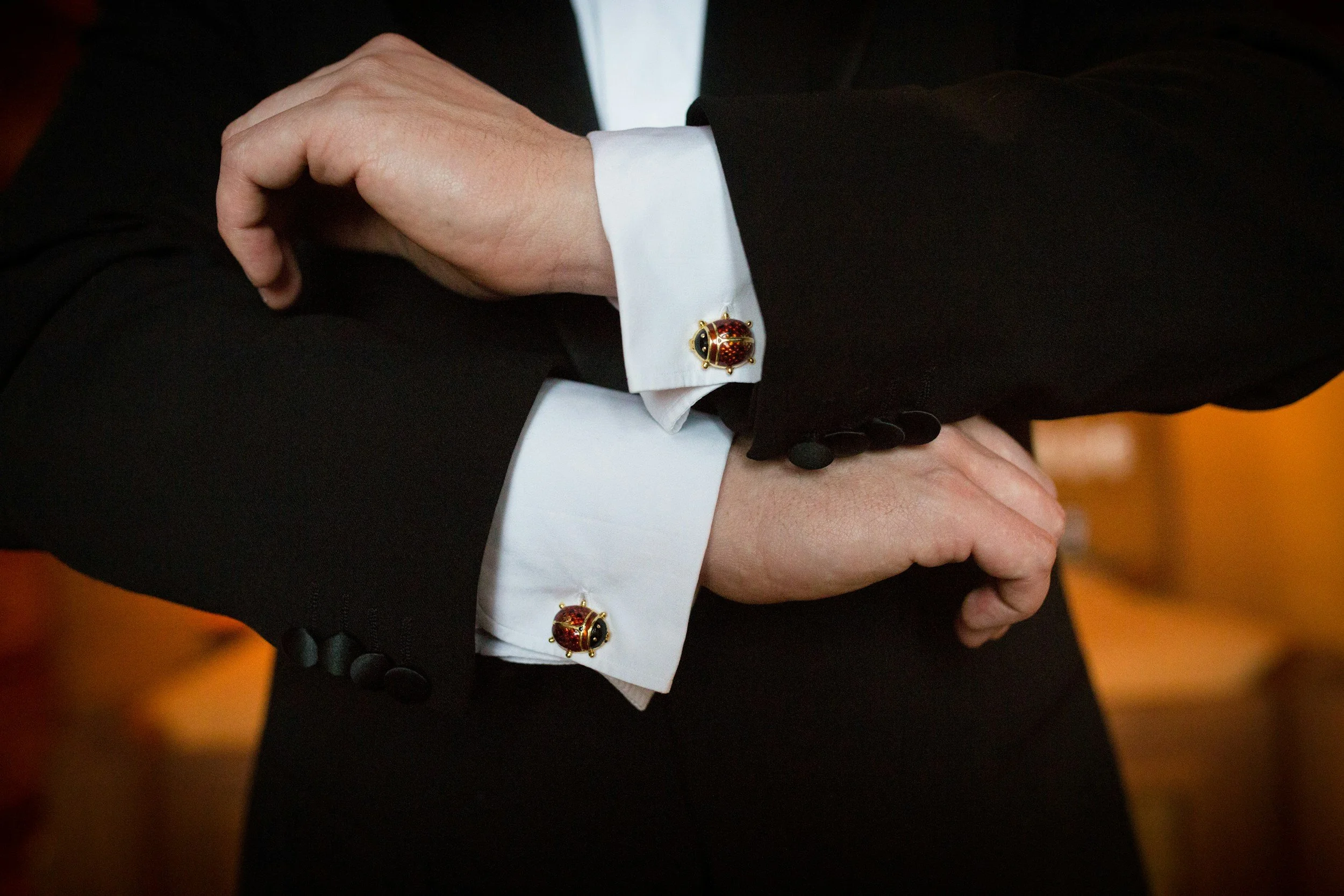
You know it is bad when you cannot put these “cuffs” on…
The Patient’s Story
For the highly driven athletic community in Liberty Village and the professionals in Queen West, shoulder health dictates their ability to stay active. Whether executing heavy overhead presses at a local CrossFit box or carrying a heavy commuter bag on the TTC, the shoulder endures relentless, daily stress.
When a sudden, deep ache develops inside the shoulder—especially one that spikes sharply when lifting a weight or reaching into the back seat of a car—the immediate reaction is usually sheer panic. Patients frantically search for an MRI, terrified that they have suffered a massive, catastrophic tear of their rotator cuff that will require invasive surgery and months of immobilization.
At Rehab Mechanics, we frequently intervene to stop this panic. In sports medicine, we emphasize advanced clinical testing over immediate imaging because a massive percentage of healthy, pain-free adults actually have partial rotator cuff tears on their MRIs. The pain you are experiencing is highly likely to be Rotator Cuff Tendinopathy (cellular decay from overuse) or Subacromial Impingement. By actively rebuilding your shoulder blade mechanics and progressively loading the tendon, expert physiotherapy can silence the pain and restore your overhead power entirely non-operatively.
Structural / Biomechanical Analysis
To understand why your shoulder hurts so intensely without being fully torn, we must perform a detailed biomechanical analysis of the rotator cuff and the narrow tunnel it must navigate.
The Anatomy of the Rotator Cuff
The rotator cuff is a group of four small muscles (Supraspinatus, Infraspinatus, Teres Minor, Subscapularis).
The Dynamic Depressors: Their absolute primary job is not to lift the arm; it is to actively pull the ball of the shoulder bone downward and hold it perfectly centered in the socket while the larger muscles (like the deltoid) do the lifting.
The Fraying Process (Tendinosis vs. Tear)
Most shoulder pain is not a sudden rupture; it is a gradual breakdown of the tissue.
The Impingement Zone: The supraspinatus tendon must travel through a narrow bony tunnel at the top of the shoulder.
The Friction: If the rotator cuff is fatigued, it fails to pull the ball downward. The arm bone slides upward too early, physically crushing the tendon against the bony roof (acromion).
Cellular Decay: This relentless, repetitive crushing causes microscopic tears. The tendon becomes thick, swollen, and chemically inflamed (tendinitis), eventually degenerating into a state of chaotic scar tissue (tendinosis). It feels like a massive tear because the highly sensitive tissue is being constantly pinched.
The Tipping Point: Scapular Dyskinesis
Why did the rotator cuff fail in the first place? It is almost always a failure of the foundation: the shoulder blade (scapula).
The Hunch: Sitting at a desk rounds the upper back and pulls the shoulder blade forward and downward.
The Biomechanical Block: When the shoulder blade is stuck in this position, the bony roof over the rotator cuff physically drops. The subacromial tunnel is instantly narrowed, virtually guaranteeing that the tendon will be crushed the next time you lift your arm overhead.
Clinical Red Flags
We differentiate between an inflamed, irritated tendon (tendinopathy) and a massive structural tear through precise mechanical testing:
The Painful Arc: A sharp, breathtaking pinch specifically occurring midway through lifting the arm out to the side (between 60 and 120 degrees), which signifies impingement of a swollen tendon.
Weakness vs. Pain Inhibition: If you cannot physically lift the arm against gravity (the "Drop Arm" sign), it may indicate a full-thickness tear. If you can lift it, but it just hurts intensely, it is likely tendinopathy.
Night Pain: A deep, throbbing ache that makes it impossible to sleep on the affected shoulder is a hallmark of an inflamed subacromial bursa or highly irritated tendon.
Passive vs. Active Freedom: If a physiotherapist can easily lift your relaxed arm overhead without a hard, bony block, you do not have a frozen shoulder, confirming the issue is muscular/tendinous.
Primary Source Proof
Orthopedic consensus strongly dictates that structured, active physiotherapy is the highly effective, first-line standard of care for rotator cuff tendinopathy and partial tears, successfully preventing unnecessary surgical interventions.
Review the Clinical Evidence on PubMed: Efficacy of Exercise Therapy in the Management of Rotator Cuff Tendinopathy (National Institutes of Health)
Review the Clinical Evidence on PubMed: Non-Operative Treatment of Partial-Thickness Rotator Cuff Tears (National Institutes of Health)
Review the Clinical Evidence on PubMed: The Role of Scapular Kinematics in Subacromial Pain Syndrome (National Institutes of Health)
The Rehab Mechanics Corrective Protocol
Treating tendinopathy requires progressive, active mechanical load. Resting the shoulder in a sling will only cause the tendon to weaken and atrophy further.
Phase 1 — Load Modification and Decompression
Subacromial Space Creation: We must stop the crushing force immediately. Our physiotherapists apply intense ischemic compression to the pectoralis minor (chest) to stop it from dragging the shoulder blade forward, instantly creating more room for the tendon.
Pain-Free Isometrics: We utilize sub-maximal static holds (pushing the arm against a wall without moving). Isometrics are scientifically proven to drop the pain signals in an angry tendon without irritating the joint through friction.
Phase 2 — Pelvic and Scapular Fortification
Serratus Anterior Activation: Implementing targeted "scapular push-ups" and wall slides to re-awaken the muscle that holds the shoulder blade tight to the ribcage.
Thoracic Spine Unlocking: Utilizing high-grade joint manipulations to forcefully restore mid-back extension. If the upper back is upright, the shoulder blade can rotate backward smoothly out of the arm's way.
Phase 3 — Gait Retraining / Mechanics Correction (Rotator Cuff Loading)
Heavy Slow Resistance (HSR): Once the impingement is cleared, we must physically rebuild the frayed tendon. We prescribe slow, heavy eccentric (lowering) movements. This heavy tension forces the chaotic scar tissue to remodel into thick, healthy, parallel collagen fibers.
Rhythmic Stabilization: We apply unexpected, rapid pushes to your arm while you hold it in space. Your nervous system must react instantly, training the rotator cuff to automatically center the ball in the socket.
Phase 4 — Return-to-Activity Strategy
Overhead Kinetic Integration: Progressing to heavy overhead pressing, kettlebell snatches, and dynamic athletic movements.
Energy Transfer Coaching: Ensuring that the power for a heavy lift comes from the hips and core, preventing the fragile shoulder from absorbing isolated, sheer stress.
Related Conditions We Treat
Rotator Cuff Tears (Partial & Full)
Subacromial Impingement Syndrome
Adhesive Capsulitis (Frozen Shoulder)
Scapular Dyskinesis
Biceps Tendinopathy
Thoracic Outlet Syndrome
Related Blogs
"Do I Really Need Another Cortisone Shot for My Shoulder, or Is There a Better Way?"
"Is My Stabbing Shoulder Pain a Torn Rotator Cuff or a Frozen Shoulder?"
"Can Physiotherapy Fix Shoulder Impingement Without Surgery?"
Services Used in Treatment
Manual Therapy
Soft Tissue Release
Myofascial Release
Shockwave Therapy
Gait Retraining
Neuromuscular Re-Education
Custom Orthotics
Strengthening Programs
FAQ Section
1. Can physiotherapy assist in managing rotator cuff tendinopathy?
Yes. Physiotherapy supports recovery by addressing the poor shoulder blade mechanics causing the impingement and utilizing progressive resistance to structurally rebuild the irritated tendon without surgery.
2. Should I get an MRI for my aching shoulder?
In many non-traumatic cases, an immediate MRI is unnecessary and can cause undue anxiety over normal, age-related wear. We utilize precise clinical testing to diagnose and assist in managing the mechanical pain directly.
3. Why does my shoulder hurt more at night?
Inflammatory fluid pools in the joint when you are inactive, and lying on the shoulder physically compresses the irritated bursa and tendon. We help address contributing factors by optimizing sleep posture and reducing daytime friction.
4. Can I still lift weights if I have rotator cuff pain?
Yes, but load modification is critical. We will temporarily guide you toward pain-free, neutral-grip exercises while we aggressively strengthen stabilizers in your mid-back to support a safe return to overhead lifting.
5. How does a tight chest cause back-of-shoulder pain?
Tight chest muscles pull your shoulders forward, overstretching and exhausting the muscles in your upper back. We utilize soft tissue release to open the chest, naturally reducing mechanical overload on the rotator cuff.
6. What are the best exercises for a fraying tendon?
Tendons heal best with Heavy Slow Resistance (HSR), specifically emphasizing the eccentric (lowering) phase. This type of loading supports recovery by forcing the body to lay down new, healthy collagen fibers.
7. Is frozen shoulder the same as a rotator cuff tear?
No. A tear causes profound weakness when trying to lift the arm, whereas a frozen shoulder is a severe thickening of the joint capsule that creates an absolute, physical block to movement. We assist in managing both distinctly.
8. How long does it take for a tendon to heal?
Tendons have poor blood supply and heal slowly. While postural corrections provide rapid pain relief, structurally remodeling the collagen matrix typically requires 8 to 12 weeks of consistent neuromuscular re-education.
How Physiotherapy Helps
Reducing tissue irritation through targeted subacromial decompression
Correcting pelvic and thoracic posture to support the shoulder blade
Improving cadence and total-body kinetic linking
Strengthening stabilizers in the serratus anterior and lower trapezius
Reducing mechanical overload on the vulnerable supraspinatus tendon
Improving foot mechanics to build a strong lifting foundation
Contact Us Today — All you have to lose is the pain
Book a comprehensive biomechanical shoulder assessment with our clinical team today. Located inside the Prime Medical Centre at 68 Abell Street, Queen West.
Email: info@rehabmechanics.com Phone: (416) 533-3900
About the Author
Mr. Sanjay Attwala (B.Sc., M.Sc., RPT) is a Registered Physiotherapist, clinical director, and the founder of Rehab Mechanics in Toronto. With over 15 years of registered clinical practice and a deep specialization in complex musculoskeletal rehabilitation, Sanjay synthesizes rigorous international academic training with advanced evidence-based therapeutics to guide his clinical practice and patient education initiatives.
Academic Background & Credentials
Master of Science (M.Sc.) in Physiotherapy – University of Keele, United Kingdom (2010).
Bachelor of Science (B.Sc.) – University of Waterloo, Ontario, Canada.
Registered Physiotherapist (RPT) – Regulated health professional in excellent standing with the College of Physiotherapists of Ontario (CPO).
Corporate Entity – Operating officially under the S. Attwala Physiotherapy Professional Corporation with a DBA of Rehab Mechanics.
Clinical Expertise & Philosophy
Sanjay’s clinical approach rejects passive symptom management in favor of identifying underlying biomechanical root causes. His diverse expertise spans advanced manual therapies, personalized corrective exercise prescription, and modern physical modalities. At the Rehab Mechanics Toronto Queen West clinic, he routinely diagnoses and treats complex conditions including:
Spinal & Discogenic Pathology – Cervical, thoracic, and lumbar disc injuries, sciatica, and sacroiliac joint (SIJ) dysfunction.
Upper & Lower Extremity Injuries – Rotator cuff tears, frozen shoulder, tennis/golfer’s elbow, carpal tunnel syndrome, and complex ankle/foot pathologies.
Perinatal & Pelvic Health Rehabilitation – Specialized assessment and rehabilitation protocols tailored specifically for women during pregnancy and the post-partum period, addressing pelvic girdle pain, diastasis recti, and core stabilization.
Specialized Rehabilitation – Pelvic health therapy, TMJ dysfunction, post-surgical rehabilitation (including Total Hip and Total Knee Replacements), and custom orthotics dispensing.
Shockwave Therapy: with advanced cutting edge technological devices to suit your needs.
Interdisciplinary Practice & Patient Care
Sanjay practices an integrated model of healthcare, working closely alongside medical doctors inside the Prime Medical Centre on Abell Street to streamline patient recovery pathways. He maintains a human-centric, communication-first clinical framework, ensuring that care remains fully customized rather than automated.
His clinical caseload encompasses a broad operational spectrum under Ontario's regulatory frameworks, including:
Motor Vehicle Accident (MVA) Claims – Rehabilitation navigating Ontario’s statutory accident benefits schedule.
Workplace Safety and Insurance Board (WSIB) – Occupational injury management and return-to-work screening.
Extended Health Care (EHC) & Private Practice – Multi-tier insurance coordination and long-term athletic development plans.
Commitment to Research & Community
Outside of his clinical caseload at Rehab Mechanics and his additional practice affiliations in Etobicoke, Sanjay is an active health writer and community educator. He translates contemporary peer-reviewed medical research into accessible, actionable guidance on his professional blog. As a dedicated father and husband, he mirrors his professional advice in his personal life, focusing on structural mobility, cross-training, and longevity to help his family and his community thrive. Naturally he takes he a keen interest in rehabilitation for women who are pregnant and post-partum.
Disclaimer:
The information provided on this blog is intended for educational and informational purposes only. It does not constitute medical advice, diagnosis, or a treatment plan. Always seek the direct advice of a Registered Physiotherapist, physician, or other qualified health provider regarding any medical condition or physical rehabilitation routine.
Google MyBusiness for Instant Posts, Photos, Updates, Offers and Communication
Be sure to check out Rehab Mechanics’ Google My Business Profile for daily posts, location and directions in addition to the directions on the website!
We love interacting with prospective and current patients, and love honest feedback and even constructive criticism and areas for improvement!
Why Does My Hip Pinch Sharply When I Squat Deep?
A sharp, blocking pain deep in the groin during a squat is the classic hallmark of Femoroacetabular Impingement (FAI). This bone-on-bone friction damages the cartilage and labrum over time. Physiotherapy assists in managing this condition by correcting pelvic tilt, strengthening deep hip rotators, and optimizing movement mechanics to physically decompress the hip joint and safely restore squatting depth.
Muscle working pain is “good” - never pain “not good”.
The Patient’s Story
For the active fitness community in Queen West and Liberty Village, heavy lifting and deep squatting are foundational to their training. However, many athletes suddenly encounter a terrifying limitation: every time they try to squat past 90 degrees, a sharp, stabbing, blocking sensation fires deep in the front crease of their hip or groin.
Patients often assume they have suffered a "groin strain" or a "sports hernia." They take weeks off, stretch their inner thighs relentlessly in Trinity Bellwoods, and try to foam roll away the pain. Yet, the moment they return to the barbell, the exact same sharp pinch occurs.
At Rehab Mechanics, we frequently identify this issue as Femoroacetabular Impingement (FAI). You cannot stretch away a structural bone-on-bone collision. Attempting to force your hips through this mechanical block will actively shred your labrum. By utilizing precise human mechanics, we can alter the resting angle of your pelvis and modify your lifting geometry to permanently clear the joint space, allowing you to squat heavy and pain-free.
Structural / Biomechanical Analysis
To understand the sharp pinch, we must perform a detailed structural analysis of your hip joint and the specific bony changes that cause impingement.
The Ball and Socket Mechanics
Your hip is a massive, deep ball-and-socket joint.
The Fit: The femoral head (the ball) should glide perfectly within the acetabulum (the socket), lubricated by synovial fluid and cushioned by a cartilaginous ring called the labrum.
The Mechanism of Impingement (FAI)
Impingement occurs when extra bone growth alters this perfect spherical fit.
CAM Lesion: Excess bone forms on the neck of the femur (the ball). As you bend your hip up into a deep squat, this non-spherical bump forcefully jams into the rim of the socket.
Pincer Lesion: Excess bone grows out over the rim of the socket, creating a bony "overhang" that prematurely crashes into the femur.
The Joint Compression Pattern
When these bony abnormalities violently crash into each other under the heavy load of a squat, they trap the delicate labrum and articular cartilage between them.
The Tearing Process: This relentless mechanical crushing causes the labrum to fray and tear.
The Anterior Pelvic Tilt Trap: The tipping point for FAI pain is often postural. If you sit at a desk all day, your hip flexors tighten, pulling your pelvis forward (anterior pelvic tilt). This forward tilt physically lowers the "roof" of your hip socket, drastically closing the joint space and guaranteeing that impingement will occur much earlier in the squatting motion.
Clinical Red Flags
We differentiate structural FAI from a simple muscle strain by looking for specific, mechanical signs of joint conflict:
The "C-Sign": The patient naturally grips their hip with their thumb behind the trochanter and fingers deep in the front groin, forming a "C" to show where the deep pain lives.
FADIR Test Positive: Sharp, pinching pain is triggered when the hip is passively forced into Flexion, Adduction, and Internal Rotation.
Loss of Internal Rotation: A profound, physical restriction when trying to rotate the thigh bone inward.
The "Squat Block": A literal inability to achieve a deep squat without the torso severely shifting or the lower back painfully rounding (butt wink) to compensate for the jammed hip.
Mechanical Catching: Sharp clicking, catching, or giving-way sensations deep within the joint during pivoting movements.
Primary Source Proof
Clinical guidelines in orthopedics definitively support conservative, biomechanically driven physical therapy—specifically focusing on pelvic alignment and hip abductor strengthening—as a highly effective first-line intervention for Femoroacetabular Impingement.
Review the Clinical Evidence on PubMed: The Efficacy of Physiotherapist-Led Rehabilitation in Femoroacetabular Impingement Syndrome (National Institutes of Health)
Review the Clinical Evidence on PubMed: Conservative Management of Non-Arthritic Hip Pain and FAI (National Institutes of Health)
Review the Clinical Evidence on PubMed: Biomechanical Factors and Pelvic Tilt in Hip Impingement (National Institutes of Health)
The Rehab Mechanics Corrective Protocol
We cannot shave down the bone without surgery, but we can completely alter how that bone interacts with the socket. We utilize a highly phased, decompressive approach.
Phase 1 — Load Modification and Joint Distraction
Manual Joint Decompression: Our physiotherapists use heavy, hands-on traction bands to gently pull the femur out of the socket. This immediately relieves the pressure on the crushed labrum and stretches the incredibly dense, tight posterior hip capsule.
Hip Flexor Release: Aggressive manual soft tissue mobilization on the psoas and rectus femoris to release the "brakes" that are pulling the pelvis into a dangerous anterior tilt.
Phase 2 — Pelvic Fortification
Posterior Pelvic Tilt Training: Utilizing targeted lower abdominal training (transversus abdominis) to teach the nervous system how to rotate the pelvis backward, instantly lifting the roof of the socket away from the impingement zone.
Deep Rotator Activation: Strengthening the tiny muscles deep under the glutes to act as the rotator cuff of the hip, holding the ball perfectly centered in the socket during movement.
Phase 3 — Gait Retraining / Mechanics Correction
Squat Stance Modification: We meticulously analyze your lifting geometry. Often, slightly widening your stance and externally rotating your toes allows the femoral neck to physically bypass the bony impingement block, restoring pain-free depth.
Gluteus Maximus Dominance: Training you to initiate hip extension entirely with the glutes, preventing the hip flexors from firing excessively and jamming the joint.
Phase 4 — Return-to-Activity Strategy
Progressive Loading: Moving from box squats back to full-depth barbell squats while constantly monitoring for pelvic tilt and "butt wink."
Kinetic Integration: Ensuring your core, pelvis, and ankles are working synergistically to prevent any single joint from absorbing excessive sheer force during heavy athletic performance.
Related Conditions We Treat
Hip Labral Tears
Athletic Pubalgia (Sports Hernia)
Osteitis Pubis
Gluteal Tendinopathy
Snapping Hip Syndrome (Coxa Saltans)
Sacroiliac Joint (SIJ) Dysfunction
Related Blogs
"Does Human Mechanics Physical Therapy Fix Chronic Groin Pain and Sports Hernias?"
"Can Physiotherapy Prevent Surgery for a Hip Labral Tear?"
"Can Fixing Your Pelvic Tilt Cure Chronic Lower Back Pain?"
Services Used in Treatment
Manual Therapy
Soft Tissue Release
Myofascial Release
Shockwave Therapy
Gait Retraining
Neuromuscular Re-Education
Custom Orthotics
Strengthening Programs
FAQ Section
1. Can physiotherapy assist in managing hip impingement?
Yes. Physiotherapy supports recovery by addressing the biomechanical faults—like pelvic tilt and weak glutes—that cause the bone-on-bone friction, helping to optimize movement and reduce joint irritation.
2. Why does my groin pinch only when I squat deep?
Deep squatting forces the thigh bone upward. If you have extra bone growth (FAI) or a forward-tilted pelvis, the bones physically crash into each other at the bottom of the movement, causing a sharp pinch.
3. Will stretching my hip flexors cure the pinching?
While releasing tight hip flexors is crucial to correct pelvic alignment, aggressive stretching can sometimes irritate the joint further. We focus on stabilization and movement modification to support recovery.
4. Do I absolutely need surgery for FAI?
In many cases, no. By optimizing your pelvic mechanics and building robust hip stability, conservative physical therapy helps reduce mechanical overload, frequently rendering surgical intervention unnecessary.
5. How does my core strength affect my hip joint?
Your core muscles anchor the top of your pelvis. If they are weak, the pelvis tilts forward, lowering the roof of the hip socket and causing immediate impingement. We fortify the core to open the joint space.
6. Can a sports hernia feel like hip impingement?
Yes. Both cause deep, chronic groin pain. However, impingement is a joint restriction, while a sports hernia is a tear in the abdominal wall. We use precise testing to differentiate and manage contributing factors for both.
7. Why do I need to change my squat stance?
Adjusting your foot width and toe angle physically changes how the ball fits into the socket during descent. We help you find a modified stance that allows you to squat heavy without bone-on-bone collision.
8. How long does it take to squat without pain?
While manual joint distraction provides immediate relief, building the neuromuscular control to hold the pelvis stable during heavy lifting typically requires several weeks of targeted strengthening programs.
How Physiotherapy Helps
Reducing tissue irritation through targeted joint distraction
Correcting anterior pelvic tilt to open the joint space
Improving cadence and dynamic hip tracking
Strengthening stabilizers in the deep rotators and glutes
Reducing mechanical overload on the labrum and cartilage
Improving foot mechanics to optimize lower-body lifting geometry
Contact Us Today — All you have to lose is the pain
Book a comprehensive biomechanical hip assessment with our clinical team today. Located inside the Prime Medical Centre at 68 Abell Street, Queen West.
Email: info@rehabmechanics.com Phone: (416) 533-3900
About the Author
Mr. Sanjay Attwala (B.Sc., M.Sc., RPT) is a Registered Physiotherapist, clinical director, and the founder of Rehab Mechanics in Toronto. With over 15 years of registered clinical practice and a deep specialization in complex musculoskeletal rehabilitation, Sanjay synthesizes rigorous international academic training with advanced evidence-based therapeutics to guide his clinical practice and patient education initiatives.
Academic Background & Credentials
Master of Science (M.Sc.) in Physiotherapy – University of Keele, United Kingdom (2010).
Bachelor of Science (B.Sc.) – University of Waterloo, Ontario, Canada.
Registered Physiotherapist (RPT) – Regulated health professional in excellent standing with the College of Physiotherapists of Ontario (CPO).
Corporate Entity – Operating officially under the S. Attwala Physiotherapy Professional Corporation with a DBA of Rehab Mechanics.
Clinical Expertise & Philosophy
Sanjay’s clinical approach rejects passive symptom management in favor of identifying underlying biomechanical root causes. His diverse expertise spans advanced manual therapies, personalized corrective exercise prescription, and modern physical modalities. At the Rehab Mechanics Toronto Queen West clinic, he routinely diagnoses and treats complex conditions including:
Spinal & Discogenic Pathology – Cervical, thoracic, and lumbar disc injuries, sciatica, and sacroiliac joint (SIJ) dysfunction.
Upper & Lower Extremity Injuries – Rotator cuff tears, frozen shoulder, tennis/golfer’s elbow, carpal tunnel syndrome, and complex ankle/foot pathologies.
Perinatal & Pelvic Health Rehabilitation – Specialized assessment and rehabilitation protocols tailored specifically for women during pregnancy and the post-partum period, addressing pelvic girdle pain, diastasis recti, and core stabilization.
Specialized Rehabilitation – Pelvic health therapy, TMJ dysfunction, post-surgical rehabilitation (including Total Hip and Total Knee Replacements), and custom orthotics dispensing.
Shockwave Therapy: with advanced cutting edge technological devices to suit your needs.
Interdisciplinary Practice & Patient Care
Sanjay practices an integrated model of healthcare, working closely alongside medical doctors inside the Prime Medical Centre on Abell Street to streamline patient recovery pathways. He maintains a human-centric, communication-first clinical framework, ensuring that care remains fully customized rather than automated.
His clinical caseload encompasses a broad operational spectrum under Ontario's regulatory frameworks, including:
Motor Vehicle Accident (MVA) Claims – Rehabilitation navigating Ontario’s statutory accident benefits schedule.
Workplace Safety and Insurance Board (WSIB) – Occupational injury management and return-to-work screening.
Extended Health Care (EHC) & Private Practice – Multi-tier insurance coordination and long-term athletic development plans.
Commitment to Research & Community
Outside of his clinical caseload at Rehab Mechanics and his additional practice affiliations in Etobicoke, Sanjay is an active health writer and community educator. He translates contemporary peer-reviewed medical research into accessible, actionable guidance on his professional blog. As a dedicated father and husband, he mirrors his professional advice in his personal life, focusing on structural mobility, cross-training, and longevity to help his family and his community thrive. Naturally he takes he a keen interest in rehabilitation for women who are pregnant and post-partum.
Disclaimer:
The information provided on this blog is intended for educational and informational purposes only. It does not constitute medical advice, diagnosis, or a treatment plan. Always seek the direct advice of a Registered Physiotherapist, physician, or other qualified health provider regarding any medical condition or physical rehabilitation routine.
Google MyBusiness for Instant Posts, Photos, Updates, Offers and Communication
Be sure to check out Rehab Mechanics’ Google My Business Profile for daily posts, location and directions in addition to the directions on the website!
We love interacting with prospective and current patients, and love honest feedback and even constructive criticism and areas for improvement!
Can a Torn Meniscus Heal Without Arthroscopic Surgery?
A torn meniscus often heals or becomes entirely asymptomatic without arthroscopic surgery. While traumatic tears in young athletes may require surgical repair, degenerative tears in older adults respond exceptionally well to conservative physiotherapy. This approach assists in managing the condition by reducing joint effusion, restoring terminal extension, and building massive quadriceps strength to dynamically offload the damaged cartilage. The biomechanical root cause is often a failure of the surrounding musculature to absorb impact shock.
Here’s a polished caption option:
Not that kind of “Torn.”
But let’s be honest — we’ve all felt it in one way or another. At Rehab Mechanics, we focus on biomechanical improvement for conditions like a torn meniscus. And while better movement often supports better mental and emotional well-being too, we know our lane — and we stay in it.
The Panic of the MRI Report
For the active residents of Toronto, receiving an MRI report that reads "complex meniscal tear" often feels like a definitive end to an active lifestyle. Whether you felt a sharp pinch while crouching in your Liberty Village condo, or experienced a deep, dull ache after a weekend run through High Park, the immediate assumption is that the torn cartilage must be surgically removed or sewn back together.
For decades, standard medical practice dictated that orthopedic surgeons routinely perform arthroscopic surgeries to "clean up" or trim torn menisci. However, modern medical imaging and rigorous, long-term clinical trials have radically challenged this approach.
When patients are told they need a "knee scope," they often search for second opinions. At Rehab Mechanics in Queen West, we want to deliver a message of profound clinical reassurance: unless your knee is physically, immovably locked in place, rushing into surgery is often highly detrimental. Removing cartilage rapidly accelerates early-onset osteoarthritis. By prioritizing deep biomechanical rehabilitation, we can stabilize the joint, eliminate the clicking pain, and save your natural cartilage from the scalpel.
Structural / Biomechanical Analysis
To understand why conservative physiotherapy is the international standard of care for many meniscus tears, we must perform a detailed biomechanical analysis of the knee joint and its internal shock absorbers.
The Anatomy of the C-Shaped Cushions
The knee is a massive hinge joint where your thigh bone (femur) rests directly on top of your shin bone (tibia). Sandwiched between these two hard bones are two pieces of tough, rubbery fibrocartilage known as the medial (inside) and lateral (outside) menisci.
Mechanical Function: They act as crucial shock absorbers and load distributors. They increase the surface area of the joint, preventing the bones from grinding directly against one another.
The Two Types of Tears (Traumatic vs. Degenerative)
The mechanism of injury dictates the path to recovery.
Acute Traumatic Tears: Common in young athletes, this occurs when the knee is violently twisted while the foot is planted. The cartilage rips, often flipping into the hinge and physically locking the joint. These "bucket-handle" tears often require surgical intervention.
Degenerative Tears: Common in adults over 40. Over decades of use, the cartilage naturally dries out and frays—like a worn-out car tire. A simple movement, like squatting to pick up a box, can cause a frayed edge to tear.
The Blood Supply Dilemma
The primary reason surgery is heavily debated lies in the meniscus's unique blood supply.
The Red Zone: The outer third of the meniscus has a rich blood supply. Tears here can biologically heal and scar back together with proper protection.
The White Zone: The inner two-thirds lack a direct blood supply. Tears here will never physically "knit" back together. However, they do not need to. The pain comes from inflammation and instability, not the tear itself.
The Biomechanical Solution: Muscular Offloading
If the cartilage is torn in the white zone, how can physiotherapy stop the pain? The answer is dynamic stabilization.
If your quadriceps (front thigh muscles) are weak, your knee absorbs 100% of the shock of walking, grinding the torn cartilage. If we build massive strength in the quadriceps and glutes, the muscles absorb the shock before it reaches the joint. The joint is shielded, and the tear becomes completely painless and asymptomatic.
Clinical Red Flags
We must carefully differentiate between a degenerative tear that requires physiotherapy and a severe mechanical block that requires an immediate surgical consult:
True Knee Locking: The knee is physically jammed. You absolutely cannot straighten the leg, no matter how hard you push. This indicates a piece of cartilage has flipped into the hinge.
The "Giving Way" Sensation: A sudden, unpredictable buckling of the knee when twisting or walking down stairs, causing a massive loss of structural confidence.
Joint Line Tenderness: Exquisite, sharp pain when pressing directly into the soft space between the bones on the side of the knee.
Delayed Effusion: The knee slowly puffs up with fluid (looking like a water balloon) 24 to 48 hours after the initial tweak or heavy activity.
Pain with Deep Squatting: A sharp, pinching pain specifically at the very bottom of a deep squat.
Primary Source Proof
Massive, landmark orthopedic trials (including the ESCAPE trial) have definitively proven that for degenerative meniscus tears, structured, supervised physical therapy yields pain relief and functional outcomes that are identical to arthroscopic partial meniscectomy surgery, without subjecting the patient to surgical risks or accelerating arthritis.
The Rehab Mechanics Corrective Protocol
Our clinical approach avoids aggressive bending and focuses strictly on mechanical decompression, reducing joint effusion, and building a muscular exoskeleton.
Phase 1 — Load Modification and Effusion Management
Joint Decompression: We use targeted soft tissue release on the calf, hamstring, and IT band to reduce the resting compressive forces pulling the knee joint tightly together over the tear.
Lymphatic Drainage: Utilizing manual techniques to push the stagnant joint fluid (effusion) out of the knee capsule, instantly reducing the feeling of pressure.
Restoring Terminal Extension: We use precise manual overpressure to ensure you can lock the knee perfectly straight, preventing a chronic, cartilage-grinding limp.
Phase 2 — Pelvic Fortification and Quadriceps Reactivation
Arthrogenic Muscle Inhibition (AMI) Reversal: Swelling physically paralyzes your quadriceps. We use Neuromuscular Electrical Stimulation (NMES) and pain-free isometric holds to force the quad to fire again.
Gluteal Stabilization: Strengthening the side glutes (gluteus medius) prevents the knee from caving inward (valgus collapse), which is the primary movement that crushes the medial meniscus.
Phase 3 — Gait Retraining / Mechanics Correction
Closed-Chain Loading: Introducing controlled mini-squats, step-ups, and leg presses to re-teach the leg how to absorb gravity and body weight safely without twisting the joint.
Cadence Optimization: If walking causes pain, we adjust your step rate to reduce the peak impact forces striking the knee joint.
Phase 4 — Return-to-Activity Strategy
Proprioceptive Retraining: Utilizing balance boards and uneven surfaces to retrain the micro-stabilizers in the knee, ensuring the joint reacts instantly to sudden changes in direction on a soccer pitch.
Heavy Slow Resistance (HSR): Building massive, absolute strength in the quadriceps so they can permanently act as the primary shock absorber for the leg.
Related Conditions We Treat
Knee Osteoarthritis
ACL and MCL Sprains
Patellofemoral Pain Syndrome (Runner's Knee)
Pes Anserine Bursitis
Baker's Cyst
Iliotibial (IT) Band Syndrome
Related Blogs
"Is My Inner Knee Pain a Meniscus Tear or Pes Anserine Bursitis?"
"What Does a 'Boggy End Feel' in My Knee Actually Mean?"
"Can Physiotherapy Prevent Knee Replacement Surgery for Osteoarthritis?"
Services Used in Treatment
Manual Therapy
Soft Tissue Release
Myofascial Release
Shockwave Therapy
Gait Retraining
Neuromuscular Re-Education
Custom Orthotics
Strengthening Programs
FAQ Section
1. Can physiotherapy assist in managing a torn meniscus?
Yes. Physiotherapy supports recovery by utilizing targeted strengthening and fluid management to reduce joint friction, helping to optimize movement and stabilize the knee without surgery.
2. Will my torn meniscus ever fully heal?
Tears in the outer "red zone" can structurally heal. Tears in the inner "white zone" may not heal structurally, but we help address contributing factors by building massive muscular support, rendering the tear completely pain-free.
3. Should I force my knee straight if it feels physically blocked?
No. A physical block indicates a piece of cartilage may be caught in the hinge. Forcing it can cause damage. We utilize careful manual assessment to support the safe return of mobility.
4. Why did my knee swell up two days after I hurt it?
This delayed swelling is a classic sign of meniscal irritation. The joint produces excess fluid to bathe the damaged tissue. We assist in managing this effusion through targeted manual therapy and movement.
5. How does hip strength protect my torn meniscus?
Your hip muscles control your thigh bone. We focus on strengthening these stabilizers to prevent your knee from collapsing inward, a motion that aggressively crushes the menisci.
6. Is it safe to exercise with a torn meniscus?
Yes, but specific load management is required. We guide you through safe, closed-chain exercises that strengthen the leg while strictly avoiding the deep bending and twisting that irritates the tear.
7. Why did my thigh muscle shrink so fast after the injury?
Massive joint swelling triggers a reflex that shuts down your quadriceps. We utilize neuromuscular re-education to optimize movement and safely "wake up" the muscle to prevent further atrophy.
8. Can custom orthotics help my knee pain?
If severe flat feet are causing your knees to naturally cave inward, custom orthotics can help optimize movement by supporting your arches and perfectly aligning your lower kinetic chain.
How Physiotherapy Helps
Reducing tissue irritation through targeted effusion drainage
Correcting pelvic drop to prevent destructive inward knee collapse
Improving cadence and shock absorption during walking
Strengthening stabilizers in the quadriceps and gluteal complex
Reducing mechanical overload on the damaged cartilage
Improving foot mechanics to anchor a stable kinetic foundation
Contact Us Today — All you have to lose is the pain
Book a comprehensive biomechanical knee assessment with our clinical team today. Located inside the Prime Medical Centre at 68 Abell Street, Queen West.
Email: info@rehabmechanics.com Phone: (416) 533-3900
About the Author
Mr. Sanjay Attwala (B.Sc., M.Sc., RPT) is a Registered Physiotherapist, clinical director, and the founder of Rehab Mechanics in Toronto. With over 15 years of registered clinical practice and a deep specialization in complex musculoskeletal rehabilitation, Sanjay synthesizes rigorous international academic training with advanced evidence-based therapeutics to guide his clinical practice and patient education initiatives.
Academic Background & Credentials
Master of Science (M.Sc.) in Physiotherapy – University of Keele, United Kingdom (2010).
Bachelor of Science (B.Sc.) – University of Waterloo, Ontario, Canada.
Registered Physiotherapist (RPT) – Regulated health professional in excellent standing with the College of Physiotherapists of Ontario (CPO).
Corporate Entity – Operating officially under the S. Attwala Physiotherapy Professional Corporation with a DBA of Rehab Mechanics.
Clinical Expertise & Philosophy
Sanjay’s clinical approach rejects passive symptom management in favor of identifying underlying biomechanical root causes. His diverse expertise spans advanced manual therapies, personalized corrective exercise prescription, and modern physical modalities. At the Rehab Mechanics Toronto Queen West clinic, he routinely diagnoses and treats complex conditions including:
Spinal & Discogenic Pathology – Cervical, thoracic, and lumbar disc injuries, sciatica, and sacroiliac joint (SIJ) dysfunction.
Upper & Lower Extremity Injuries – Rotator cuff tears, frozen shoulder, tennis/golfer’s elbow, carpal tunnel syndrome, and complex ankle/foot pathologies.
Perinatal & Pelvic Health Rehabilitation – Specialized assessment and rehabilitation protocols tailored specifically for women during pregnancy and the post-partum period, addressing pelvic girdle pain, diastasis recti, and core stabilization.
Specialized Rehabilitation – Pelvic health therapy, TMJ dysfunction, post-surgical rehabilitation (including Total Hip and Total Knee Replacements), and custom orthotics dispensing.
Shockwave Therapy: with advanced cutting edge technological devices to suit your needs.
Interdisciplinary Practice & Patient Care
Sanjay practices an integrated model of healthcare, working closely alongside medical doctors inside the Prime Medical Centre on Abell Street to streamline patient recovery pathways. He maintains a human-centric, communication-first clinical framework, ensuring that care remains fully customized rather than automated.
His clinical caseload encompasses a broad operational spectrum under Ontario's regulatory frameworks, including:
Motor Vehicle Accident (MVA) Claims – Rehabilitation navigating Ontario’s statutory accident benefits schedule.
Workplace Safety and Insurance Board (WSIB) – Occupational injury management and return-to-work screening.
Extended Health Care (EHC) & Private Practice – Multi-tier insurance coordination and long-term athletic development plans.
Commitment to Research & Community
Outside of his clinical caseload at Rehab Mechanics and his additional practice affiliations in Etobicoke, Sanjay is an active health writer and community educator. He translates contemporary peer-reviewed medical research into accessible, actionable guidance on his professional blog. As a dedicated father and husband, he mirrors his professional advice in his personal life, focusing on structural mobility, cross-training, and longevity to help his family and his community thrive. Naturally he takes he a keen interest in rehabilitation for women who are pregnant and post-partum.
Disclaimer: The information provided on this blog is intended for educational and informational purposes only. It does not constitute medical advice, diagnosis, or a treatment plan. Always seek the direct advice of a Registered Physiotherapist, physician, or other qualified health provider regarding any medical condition or physical rehabilitation routine.
Google MyBusiness for Instant Posts, Photos, Updates, Offers and Communication
Be sure to check out Rehab Mechanics’ Google My Business Profile for daily posts, location and directions in addition to the directions on the website!
We love interacting with prospective and current patients, and love honest feedback and even constructive criticism and areas for improvement!
Why Do Female Athletes Tear Their ACLs More Often Than Men?
August 23, 2026
Female athletes are statistically at a much higher risk for non-contact ACL tears due to specific anatomical and biomechanical vulnerabilities. Physiotherapy assists in managing this risk by correcting landing mechanics, strengthening the gluteus medius to control the wider Q-angle, and utilizing neuromuscular re-education to prevent the knee from collapsing inward during explosive sports. The biomechanical root cause is often a combination of pelvic width and landing strategies that place massive rotational torque on the knee joint.
The Unseen Danger on the Pitch
Do not try this at home…ahem…or even outside no matter how injured you or NOT!
For the highly dedicated female athletes playing in competitive soccer leagues at Lamport Stadium, basketball in Trinity Bellwoods, or intense CrossFit boxes in Liberty Village, the statistics are alarming. A female athlete is between four to eight times more likely to suffer a non-contact Anterior Cruciate Ligament (ACL) tear than a male athlete participating in the exact same sport.
This devastating injury often occurs in a seemingly innocuous moment: a sudden stop, a quick pivot, or landing from a rebound. There is a loud "pop," the knee gives way, and an entire athletic season is lost.
At Rehab Mechanics, we believe that understanding why this happens is the first step in stopping it. The disparity in injury rates is not due to a lack of effort or conditioning; it is driven by deeply ingrained anatomical, hormonal, and biomechanical differences. By actively screening for these vulnerabilities and implementing highly targeted "human mechanics" physical therapy before the season starts, we can drastically reduce the risk of catastrophic knee failure.
Structural / Biomechanical Analysis
To understand why the female knee is more vulnerable, we must perform a detailed structural analysis of the pelvic girdle and the kinetic chain running down to the foot.
The Anatomical Vulnerability: The Q-Angle
The primary structural difference between male and female athletes is the width of the pelvis.
The Wider Pelvis: Women naturally possess a wider pelvis for childbirth.
The Q-Angle (Quadriceps Angle): Because the hips are wider, the femur (thigh bone) must angle inward more sharply to meet the knee. This creates a larger "Q-Angle."
The Mechanical Stress: A larger Q-angle naturally predisposes the knee to collapse inward toward the midline (valgus alignment) under heavy load.
The Biomechanical Failure: Valgus Collapse
The ACL is designed to stop the shin bone from sliding forward. It is highly vulnerable to rotational twisting.
The Landing Strategy: When landing from a jump, male athletes often land with their knees slightly bent and tracking straight over their toes, using their hamstrings to absorb the shock. Female athletes often tend to land with straighter, stiffer knees, relying heavily on their quadriceps.
The "Whip" Effect: When a female athlete with a wider Q-angle lands with stiff knees and weak side-hip muscles (gluteus medius), the femur violently rotates inward. The knee completely caves in (dynamic valgus).
The Rupture: This inward collapse places a massive, instant, twisting shear force directly onto the ACL, snapping it in milliseconds.
The Hormonal Factor
Research indicates that fluctuations in estrogen and relaxin during the menstrual cycle can temporarily increase ligamentous laxity. While we cannot change hormones, this laxity makes flawless muscular control even more critical during certain phases of the month.
Clinical Red Flags
During an injury prevention screening, we look for specific, visible mechanical faults that indicate a high risk for an ACL tear:
Dynamic Valgus on Landing: The knees visibly knock together when landing from a simple box jump.
Quad Dominance: A profound inability to activate the hamstrings or glutes during a squat, relying entirely on the front of the thighs.
Poor Deceleration Mechanics: Landing with a loud, heavy footfall and straight, stiff knees, indicating an inability to absorb kinetic energy properly.
The Trendelenburg Sign: The pelvis noticeably drops on one side when standing on a single leg, indicating severe gluteus medius weakness.
Core Instability: The torso leans heavily to the side or twists uncontrollably when changing direction.
Primary Source Proof
Sports medicine literature overwhelmingly supports the implementation of targeted neuromuscular and proprioceptive training programs to significantly reduce the incidence of non-contact ACL injuries in female athletes.
The Rehab Mechanics Corrective Protocol
We do not just treat injuries; we prevent them. Our prophylactic ACL protocol is designed to overwrite dangerous landing software and build bulletproof hip mechanics.
Phase 1 — Load Modification and Neuromuscular Screening
High-Speed Video Analysis: We record you performing drop jumps and cutting maneuvers to measure your exact Q-angle tracking and identify moments of dynamic valgus.
Isolating Weakness: Utilizing manual muscle testing to find exactly where the kinetic chain is breaking down (usually the gluteus medius or the deep core).
Phase 2 — Pelvic Fortification (The Top-Down Fix)
Gluteus Medius Hypertrophy: The side hip muscle is the only thing stopping the knee from caving in. We utilize heavy, banded lateral walks and single-leg deadlifts to build massive lateral stability.
Posterior Chain Dominance: Shifting reliance away from the quadriceps by aggressively strengthening the hamstrings and gluteus maximus (the primary hip extenders) so they absorb the shock of landing.
Phase 3 — Gait Retraining / Mechanics Correction
Deceleration Mastery: Teaching the nervous system how to land softly. We practice landing mechanics with strict instructions to absorb force by hinging at the hips and keeping the knees tracking perfectly over the second toe.
Anti-Rotational Core Control: If the upper body twists wildly during a cut, it forces the knee to twist. We use heavy Pallof presses and dynamic medicine ball throws to lock the torso down.
Phase 4 — Return-to-Activity Strategy
Reactive Perturbation: We apply sudden, unexpected pushes to the athlete while they balance or land, forcing their nervous system to fire the stabilizing muscles instantly without thinking.
Sport-Specific Agility: Integrating the new, safe landing and cutting mechanics directly into soccer drills or basketball maneuvers to ensure the healthy software holds up under fatigue.
Related Conditions We Treat
ACL and PCL Sprains
Patellofemoral Pain Syndrome (Runner's Knee)
Iliotibial (IT) Band Syndrome
Meniscus Tears
Chronic Ankle Instability
Gluteal Tendinopathy
Related Blogs
"Can I Heal a Torn ACL and Return to Sports Without Surgery?"
"Does Human Mechanics Physical Therapy Fix Chronic Groin Pain and Sports Hernias?"
"Does Pain Behind the Kneecap Mean My Cartilage is Gone?"
Services Used in Treatment
Neuromuscular Re-Education
Gait Retraining
Strengthening Programs
Manual Therapy
Soft Tissue Release
Myofascial Release
Custom Orthotics
Shockwave Therapy
FAQ Section
1. Can physiotherapy assist in managing the risk of an ACL tear?
Yes. Physiotherapy supports injury prevention by utilizing targeted neuromuscular training to correct poor landing mechanics and strengthen the hip stabilizers, helping to reduce the sheer force on the knee.
2. What is the Q-angle and why does it matter?
The Q-angle is the angle from the pelvis to the knee. Because women typically have wider hips, this angle is larger, which can mechanically force the knee inward during athletic movements. We help address contributing factors through targeted strengthening.
3. Why is glute strength so important for my knees?
Your glute muscles control the alignment of your thigh bone. We focus on strengthening these stabilizers to prevent the inward knee collapse (valgus) that violently twists and strains the ACL.
4. Can custom orthotics help prevent knee injuries?
If severe flat feet (overpronation) are causing your knee to cave inward, custom orthotics can help optimize movement by supporting your arch and aligning your lower kinetic chain.
5. How do hormones affect my ACL risk?
Fluctuations in hormones like estrogen can temporarily increase ligament laxity. We assist in managing this risk by building a highly responsive, strong muscular brace that does not rely solely on the passive ligaments.
6. Does landing with stiff knees cause injuries?
Yes. Landing stiffly forces the joints to absorb the shock. We utilize neuromuscular re-education to teach athletes how to bend their hips and knees deeply to let the muscles safely absorb the kinetic energy.
7. Why do I need to strengthen my hamstrings?
The hamstrings physically pull the shin bone backward, acting as the primary muscular backup to the ACL. We support your joint stability by aggressively fortifying this posterior chain.
8. How long does an injury prevention program take?
While mechanics can be assessed immediately, building the reflexive strength to hold a safe knee position during explosive sports typically requires 6 to 8 weeks of specialized, progressive programming.
How Physiotherapy Helps
Reducing tissue irritation by correcting dynamic valgus collapse
Correcting pelvic drop to optimize total leg alignment
Improving cadence and soft-landing mechanics
Strengthening stabilizers in the gluteus medius and hamstrings
Reducing mechanical overload on the passive ACL ligament
Improving foot mechanics to anchor a stable landing base
Contact Us Today — All you have to lose is the pain
Book a comprehensive biomechanical assessment with our clinical team today. Located inside the Prime Medical Centre at 68 Abell Street, Queen West.
Email: info@rehabmechanics.com Phone: (416) 533-3900
About the Author
Mr. Sanjay Attwala (B.Sc., M.Sc., RPT) is a Registered Physiotherapist, clinical director, and the founder of Rehab Mechanics in Toronto. With over 15 years of registered clinical practice and a deep specialization in complex musculoskeletal rehabilitation, Sanjay synthesizes rigorous international academic training with advanced evidence-based therapeutics to guide his clinical practice and patient education initiatives.
Academic Background & Credentials
Master of Science (M.Sc.) in Physiotherapy – University of Keele, United Kingdom (2010).
Bachelor of Science (B.Sc.) – University of Waterloo, Ontario, Canada.
Registered Physiotherapist (RPT) – Regulated health professional in excellent standing with the College of Physiotherapists of Ontario (CPO).
Corporate Entity – Operating officially under the S. Attwala Physiotherapy Professional Corporation with a DBA of Rehab Mechanics.
Clinical Expertise & Philosophy
Sanjay’s clinical approach rejects passive symptom management in favor of identifying underlying biomechanical root causes. His diverse expertise spans advanced manual therapies, personalized corrective exercise prescription, and modern physical modalities. At the Rehab Mechanics Toronto Queen West clinic, he routinely diagnoses and treats complex conditions including:
Spinal & Discogenic Pathology – Cervical, thoracic, and lumbar disc injuries, sciatica, and sacroiliac joint (SIJ) dysfunction.
Upper & Lower Extremity Injuries – Rotator cuff tears, frozen shoulder, tennis/golfer’s elbow, carpal tunnel syndrome, and complex ankle/foot pathologies.
Perinatal & Pelvic Health Rehabilitation – Specialized assessment and rehabilitation protocols tailored specifically for women during pregnancy and the post-partum period, addressing pelvic girdle pain, diastasis recti, and core stabilization.
Specialized Rehabilitation – Pelvic health therapy, TMJ dysfunction, post-surgical rehabilitation (including Total Hip and Total Knee Replacements), and custom orthotics dispensing.
Shockwave Therapy: with advanced cutting edge technological devices to suit your needs.
Interdisciplinary Practice & Patient Care
Sanjay practices an integrated model of healthcare, working closely alongside medical doctors inside the Prime Medical Centre on Abell Street to streamline patient recovery pathways. He maintains a human-centric, communication-first clinical framework, ensuring that care remains fully customized rather than automated.
His clinical caseload encompasses a broad operational spectrum under Ontario's regulatory frameworks, including:
Motor Vehicle Accident (MVA) Claims – Rehabilitation navigating Ontario’s statutory accident benefits schedule.
Workplace Safety and Insurance Board (WSIB) – Occupational injury management and return-to-work screening.
Extended Health Care (EHC) & Private Practice – Multi-tier insurance coordination and long-term athletic development plans.
Commitment to Research & Community
Outside of his clinical caseload at Rehab Mechanics and his additional practice affiliations in Etobicoke, Sanjay is an active health writer and community educator. He translates contemporary peer-reviewed medical research into accessible, actionable guidance on his professional blog. As a dedicated father and husband, he mirrors his professional advice in his personal life, focusing on structural mobility, cross-training, and longevity to help his family and his community thrive. Naturally he takes he a keen interest in rehabilitation for women who are pregnant and post-partum.
Disclaimer:
The information provided on this blog is intended for educational and informational purposes only. It does not constitute medical advice, diagnosis, or a treatment plan. Always seek the direct advice of a Registered Physiotherapist, physician, or other qualified health provider regarding any medical condition or physical rehabilitation routine.
Google MyBusiness for Instant Posts, Photos, Updates, Offers and Communication
Be sure to check out Rehab Mechanics’ Google My Business Profile for daily posts, location and directions in addition to the directions on the website!
We love interacting with prospective and current patients, and love honest feedback and even constructive criticism and areas for improvement!
Can I Heal a Torn ACL and Return to Sports Without Surgery?
August 23, 2026
A completely torn Anterior Cruciate Ligament (ACL) does not biologically reconnect on its own. However, specialized physiotherapy can train an athlete to become a "coper." By aggressively strengthening the hamstrings, quadriceps, and neuromuscular reflexes, physical therapy provides immense dynamic stability, often allowing a return to high-level sports without reconstructive surgery. The biomechanical root cause of joint failure is a lack of muscular compensation for the missing passive ligament restraint.
The Devastating "Pop" on the Pitch
For the highly competitive recreational athletes in Toronto—whether you are playing soccer under the lights at Lamport Stadium, running a basketball fast-break in Trinity Bellwoods, or skiing north of the city—a knee injury is the ultimate fear.
The sequence of events is usually identical: you plant your foot, twist your body to change direction, and suddenly feel and hear a violent, sickening "pop" deep inside your knee. The joint instantly swells, and your leg gives out from under you.
When the MRI confirms a full rupture of the Anterior Cruciate Ligament (ACL), patients are almost universally told that surgical reconstruction is their only option if they ever want to run or play sports again. However, modern orthopedic science has radically challenged this assumption. At Rehab Mechanics, we specialize in high-level athletic joint preservation. A torn ACL does not automatically guarantee a trip to the operating room. By meticulously overhauling the human mechanics of your leg, we can train your muscles to do the job of the missing ligament, successfully transforming you into a non-operative "coper."
No, not quite that kind of ‘pop’ at the pitch — but something to break up reader fatigue, especially if you’ve recently been injured and the light from your electronic devices is contributing to headaches or eye strain
Structural / Biomechanical Analysis
To understand how you can function without an ACL, we must perform a deep biomechanical analysis of knee stability and the crucial difference between passive and active joint control.
The Anatomy of the Knee's Seatbelt
The knee is a massive hinge joint where the femur (thigh bone) sits on top of the tibia (shin bone).
The Passive Restraint: The ACL is a thick, rope-like ligament that runs diagonally through the absolute center of your knee joint.
The Mechanical Job: Its primary anatomical job is to prevent anterior tibial translation—meaning it physically stops your shin bone from sliding dangerously forward out from under your thigh bone when you stop suddenly or land from a jump. It also prevents the knee from rotating too far inward.
The "Coper" Phenomenon (Active vs. Passive Stability)
When the ACL tears, you lose your primary passive restraint. However, the human body is brilliantly engineered with a secondary, active restraint system: your muscles.
The Hamstring Defense System: Your hamstrings (the massive muscles on the back of your thigh) attach to the back of your shin bone. When the hamstrings contract forcefully, they physically pull the shin bone backward. This muscular pull does the exact same mechanical job as the ACL.
The Tipping Point: Neuromuscular Lag

Sure, why not an info point instead! Kindly read below.
The reason your knee gives out (buckles) after an ACL tear is not just because the ligament is gone; it is because your brain is too slow to fire the hamstrings when you twist. If we can train your nervous system to fire the hamstrings automatically and explosively the millisecond your foot hits the ground, your knee will be stable. Patients who successfully achieve this high-level neurological reflex are clinically classified as "copers."
Clinical Red Flags
We must carefully assess whether a patient is a candidate for conservative management. We look for specific signs indicating mechanical readiness versus catastrophic instability:
Frequent "Giving Way" Episodes: Repeated buckling during simple, straight-line walking indicates massive generalized instability.
The "Hop" Test: The inability to perform repetitive single-leg hopping tasks on the injured leg without pain or apprehension.
Concomitant Meniscal Damage: Sharp, mechanical catching or locking on the joint line, indicating the meniscus was also severely torn during the impact.
Massive Effusion: A knee that constantly swells dramatically after light activity, indicating ongoing intra-articular damage.
Fear-Avoidance: Severe psychological hesitation to place weight on the injured leg.
Primary Source Proof
Orthopedic sports medicine trials definitively prove that structured, intensive rehabilitation yields functional outcomes and meniscus preservation rates equal to early surgical ACL reconstruction for many patients.
The Rehab Mechanics Corrective Protocol
Rehabilitating an ACL-deficient knee requires an incredibly aggressive, elite-level conditioning program. We rebuild the entire neuromuscular software of your lower body.
Phase 1 — Load Modification and Effusion Eradication
Arthrogenic Muscle Inhibition (AMI) Reversal: The massive swelling from the tear triggers a spinal reflex that paralyzes your quadriceps. We use Neuromuscular Electrical Stimulation (NMES) and heavy isometric holds to forcefully override this shutdown and wake the quad back up.
Restoring Extension: The knee must be able to lock perfectly straight. We use precise manual overpressure to restore terminal knee extension to prevent cartilage grinding.
Phase 2 — Pelvic Fortification and Hamstring Hypertrophy
Hamstring Overdrive: We implement heavy, eccentric hamstring loading (like Romanian Deadlifts and Nordic Curls). The hamstrings must become exceptionally thick and strong to act as your new ACL.
Gluteal Stabilization: We heavily load the lateral pelvic stabilizers (gluteus medius) to keep the leg perfectly straight during single-leg balance, preventing dangerous valgus (inward) collapse.
Phase 3 — Gait Retraining / Mechanics Correction
Reactive Stability: Our physiotherapists apply sudden, unexpected pushes and pulls to your body while you balance on an unstable surface (perturbation training). This forces your spinal cord to react in milliseconds.
Deceleration Mechanics: We teach your body how to safely absorb gravity. We practice precise landing mechanics from drop jumps, ensuring the hips and glutes take the shock, sparing the knee.
Phase 4 — Return-to-Activity Strategy
RTS Testing: You undergo an exhaustive battery of single-leg hop tests, agility drills, and psychological readiness questionnaires.
Sport-Specific Integration: You are only cleared to return to the pitch when your injured leg is performing at 90%+ the capacity of your uninjured leg across all dynamic planes of motion.
Related Conditions We Treat
ACL and PCL Sprains
Meniscus Tears
Patellar Tendinopathy (Jumper's Knee)
Iliotibial (IT) Band Syndrome
Chronic Ankle Instability
Knee Osteoarthritis
Related Blogs
"Do I Absolutely Need Surgery to Fix a Torn Meniscus?"
"What Does a 'Boggy End Feel' in My Knee Actually Mean?"
"Can Physiotherapy Prevent Knee Replacement Surgery for Osteoarthritis?"
Services Used in Treatment
Manual Therapy
Soft Tissue Release
Myofascial Release
Shockwave Therapy
Gait Retraining
Neuromuscular Re-Education
Custom Orthotics
Strengthening Programs
FAQ Section
1. Can physiotherapy assist in managing a completely torn ACL?
Yes. Physiotherapy supports recovery by aggressively strengthening the hamstrings and quadriceps to act as a dynamic muscular brace, helping to optimize movement and stabilize the joint.
2. What is an ACL "coper"?
A "coper" is an individual who can successfully return to high-level, pivoting sports without an intact ACL by relying entirely on superior neuromuscular control and muscle strength.
3. Will my knee develop arthritis faster without surgery?
Research shows that returning to high-impact sports without adequate muscular support can increase joint wear. We focus on intense neuromuscular re-education to help reduce mechanical overload on the cartilage.
4. Can I ride a bike with a torn ACL?
Yes. Stationary cycling is a closed-chain, straight-line movement that places minimal shear stress on the knee, making it an excellent way to support cardiovascular recovery in the early phases.
5. How long does non-surgical ACL rehab take?
Building the massive muscular strength and reflexive speed required to replace a ligament typically takes 3 to 6 months of highly structured, progressive physical therapy.
6. Do I need to wear a heavy knee brace to play sports?
A functional brace provides proprioceptive feedback, but it cannot mechanically stop a massive twisting force. We focus on building your internal "biological brace" through strengthening programs.
7. Can shockwave therapy help an ACL tear?
No, shockwave does not regenerate a fully severed internal ligament. We utilize shockwave primarily for chronic tendon issues, relying instead on heavy loading for ACL compensation.
8. What happens if conservative physical therapy fails?
If your knee continues to buckle despite excellent strength, you may require surgical reconstruction. However, undergoing "Pre-Hab" before surgery drastically improves post-operative outcomes.
How Physiotherapy Helps
Reducing tissue irritation through targeted effusion management
Correcting pelvic drop to prevent inward knee collapse
Improving cadence and landing deceleration mechanics
Strengthening stabilizers in the hamstrings and glutes
Reducing mechanical overload on the vulnerable menisci
Improving foot mechanics to absorb ground reaction forces
Contact Us Today — All you have to lose is the pain
Book a comprehensive biomechanical assessment with our clinical team today. Located inside the Prime Medical Centre at 68 Abell Street, Queen West.
Email: info@rehabmechanics.com Phone: (416) 533-3900
About the Author
Mr. Sanjay Attwala (B.Sc., M.Sc., RPT) is a Registered Physiotherapist, clinical director, and the founder of Rehab Mechanics in Toronto. With over 15 years of registered clinical practice and a deep specialization in complex musculoskeletal rehabilitation, Sanjay synthesizes rigorous international academic training with advanced evidence-based therapeutics to guide his clinical practice and patient education initiatives.
Academic Background & Credentials
Master of Science (M.Sc.) in Physiotherapy – University of Keele, United Kingdom (2010).
Bachelor of Science (B.Sc.) – University of Waterloo, Ontario, Canada.
Registered Physiotherapist (RPT) – Regulated health professional in excellent standing with the College of Physiotherapists of Ontario (CPO).
Corporate Entity – Operating officially under the S. Attwala Physiotherapy Professional Corporation with a DBA of Rehab Mechanics.
Clinical Expertise & Philosophy
Sanjay’s clinical approach rejects passive symptom management in favor of identifying underlying biomechanical root causes. His diverse expertise spans advanced manual therapies, personalized corrective exercise prescription, and modern physical modalities. At the Rehab Mechanics Toronto Queen West clinic, he routinely diagnoses and treats complex conditions including:
Spinal & Discogenic Pathology – Cervical, thoracic, and lumbar disc injuries, sciatica, and sacroiliac joint (SIJ) dysfunction.
Upper & Lower Extremity Injuries – Rotator cuff tears, frozen shoulder, tennis/golfer’s elbow, carpal tunnel syndrome, and complex ankle/foot pathologies.
Perinatal & Pelvic Health Rehabilitation – Specialized assessment and rehabilitation protocols tailored specifically for women during pregnancy and the post-partum period, addressing pelvic girdle pain, diastasis recti, and core stabilization.
Specialized Rehabilitation – Pelvic health therapy, TMJ dysfunction, post-surgical rehabilitation (including Total Hip and Total Knee Replacements), and custom orthotics dispensing.
Shockwave Therapy: with advanced cutting edge technological devices to suit your needs.
Interdisciplinary Practice & Patient Care
Sanjay practices an integrated model of healthcare, working closely alongside medical doctors inside the Prime Medical Centre on Abell Street to streamline patient recovery pathways. He maintains a human-centric, communication-first clinical framework, ensuring that care remains fully customized rather than automated.
His clinical caseload encompasses a broad operational spectrum under Ontario's regulatory frameworks, including:
Motor Vehicle Accident (MVA) Claims – Rehabilitation navigating Ontario’s statutory accident benefits schedule.
Workplace Safety and Insurance Board (WSIB) – Occupational injury management and return-to-work screening.
Extended Health Care (EHC) & Private Practice – Multi-tier insurance coordination and long-term athletic development plans.
Commitment to Research & Community
Outside of his clinical caseload at Rehab Mechanics and his additional practice affiliations in Etobicoke, Sanjay is an active health writer and community educator. He translates contemporary peer-reviewed medical research into accessible, actionable guidance on his professional blog. As a dedicated father and husband, he mirrors his professional advice in his personal life, focusing on structural mobility, cross-training, and longevity to help his family and his community thrive. Naturally he takes he a keen interest in rehabilitation for women who are pregnant and post-partum.
Disclaimer: The information provided on this blog is intended for educational and informational purposes only. It does not constitute medical advice, diagnosis, or a treatment plan. Always seek the direct advice of a Registered Physiotherapist, physician, or other qualified health provider regarding any medical condition or physical rehabilitation routine.
Google MyBusiness for Instant Posts, Photos, Updates, Offers and Communication
Be sure to check out Rehab Mechanics’ Google My Business Profile for daily posts, location and directions in addition to the directions on the website!
We love interacting with prospective and current patients, and love honest feedback and even constructive criticism and areas for improvement!
Is a Hip Replacement My Only Option for Severe Osteoarthritis and Groin Pain?
No. Even with severe bone-on-bone osteoarthritis, physical therapy is vital. By correcting pelvic mechanics, releasing locked joint capsules, and building massive gluteal strength to act as biological shock absorbers, physiotherapy significantly delays surgery or serves as critical "pre-hab" to guarantee a superior surgical outcome.
The "Bone-on-Bone" Diagnosis
For the active, aging population in Toronto, chronic hip pain is an incredibly heavy burden. It typically presents as a deep, relentless ache in the front of the groin, sharp catching pain when trying to tie your shoes, or an agonizing limp after walking a few blocks through High Park.
When the pain becomes unbearable, patients seek an X-ray or MRI. The radiologist's report returns with terrifying terminology: "severe joint space narrowing," "osteophyte formation," and "advanced osteoarthritis." The physician points to the scan and delivers the dreaded phrase: "You are bone-on-bone. A hip replacement is inevitable."
Patients usually assume this diagnosis is a permanent sentence to the couch. They stop walking, stop exercising, and simply wait in agony for their surgical date. This "wait and deteriorate" mindset is a massive clinical mistake. At Rehab Mechanics, we know that your X-ray does not perfectly dictate your pain levels. By engaging in rigorous, biomechanical physiotherapy, we can drastically change how gravity loads your arthritic joint, significantly delaying the need for surgery or preparing you for the most successful, rapid post-surgical recovery possible.
Structural Analysis: The Mechanics of Hip Decay
To understand how physical therapy can save a failing joint, we must perform a detailed biomechanical analysis of hip osteoarthritis and why the pain suddenly spikes.
The Pathology of Joint Degeneration
The hip is a deep ball-and-socket joint heavily coated in smooth, frictionless articular cartilage. Osteoarthritis (OA) is an active, whole-joint disease.
Cartilage Loss: Decades of repetitive loading, previous sports injuries, or poor pelvic mechanics cause the Teflon-like cartilage to dry out, fray, and wear away, eventually exposing the raw, highly sensitive subchondral bone.
The Inflammatory Response: The joint capsule reacts to this debris by becoming violently inflamed (synovitis), producing excess fluid and causing the deep groin ache.
Osteophytes (Bone Spurs): The body attempts to stabilize the wobbly joint by growing extra bone around the rim, which physically blocks your ability to rotate your leg or bend forward.
Is your body “feeling” like that? We are here to help. Contact us below to stop the “wilting”.
The True Driver of Pain: Arthrogenic Inhibition
Why does a hip that has been wearing down for twenty years suddenly become excruciatingly painful in the span of a month? The answer lies in the surrounding muscles.
The Neurological Shutdown: When the joint becomes inflamed, the brain subconsciously initiates a protective reflex called Arthrogenic Muscle Inhibition. It literally turns down the electrical signal to your massive gluteus maximus and gluteus medius muscles to stop you from walking on the injured leg.
The Compressive Crash: Your glutes are your primary shock absorbers. Once they shut down and atrophy, 100% of the kinetic impact from walking on the Toronto pavement slams directly into the exposed, arthritic bone. This bone-crushing impact is what causes the severe pain, not just the loss of cartilage.
Primary Source Proof: Joint Preservation and Pre-Hab
Major international orthopedic guidelines strongly mandate that comprehensive, supervised physiotherapy must be exhausted before total joint arthroplasty (surgery) is considered, and that pre-surgical rehabilitation ("pre-hab") is the single greatest predictor of rapid post-operative success.
Note: The link above directs to external, peer-reviewed medical literature demonstrating our commitment to evidence-based practice and international clinical guidelines.
The Rehab Mechanics Orthopedic Protocol
We do not view osteoarthritis as a passive waiting game. We utilize an aggressive, phased biomechanical approach to build a muscular exoskeleton around your failing joint.
Phase 1 — Joint Distraction and Decompression (Weeks 1-3)
Manual Joint Traction: Our physiotherapists apply heavy, sustained pulling forces (distraction) to the leg. This physically separates the ball from the socket, creating a vacuum effect that pulls fresh, lubricating synovial fluid into the starving cartilage and temporarily silencing the bone-on-bone friction.
Capsular Unlocking: We use targeted joint mobilizations to forcefully stretch the tight, fibrotic joint capsule, restoring the crucial ability to extend your leg backward during a walking stride.
Phase 2 — Non-Compressive Gluteal Reactivation (Weeks 4-6) We must wake up the sleeping shock absorbers without grinding the arthritic bone.
High-Intensity Isometrics: You will push your leg maximally against an immovable resistance. The muscles fire at 100% capacity, building massive baseline strength, but the joint does not bend, allowing you to train safely and pain-free.
Deep Core Integration: Strengthening the transversus abdominis to stabilize the pelvis from the top down, giving the hip a solid foundation to pull against.
Phase 3 — Closed-Chain Load Management (Weeks 6-10)
Gait Retraining: We aggressively target the "Trendelenburg limp." By isolating and strengthening the gluteus medius, we stop your pelvis from dropping sideways with every step, ensuring the impact forces travel smoothly through the muscles rather than crashing into the hip socket.
Pain-Free Hinging: Teaching you how to bend and lift using your hamstrings and lower back efficiently, sparing the stiff hip joint from extreme flexion.
Phase 4 — The Pre-Hab Surgical Strategy (If Surgery is Required) If a replacement is eventually scheduled, we transition you into a strict "pre-hab" phase.
We maximize your upper body strength for post-surgical walker use, drastically boost your cardiovascular endurance, and ensure your glutes are as thick and responsive as possible. Entering the operating room at peak strength guarantees your post-surgical rehabilitation will be counted in weeks, not months.
Related Conditions We Treat
Total Hip Replacement Post-Op Rehab
Femoroacetabular Impingement (FAI)
Gluteal Tendinopathy
Lumbar Spinal Stenosis
Sacroiliac Joint (SIJ) Dysfunction
Knee Osteoarthritis
Related Blogs
"Can Physiotherapy Prevent Hip Replacement Surgery for Severe Osteoarthritis?"
"Should I Seek Conservative Orthopedic Services Before Joint Surgery?"
"Is Physiotherapy Necessary Before a Knee or Hip Replacement Surgery?"
Services Used in Treatment
Manual Therapy
Joint Mobilization / Traction
Gait Retraining
Neuromuscular Re-Education
Pre-Habilitation Programming
Strengthening Programs
FAQ Section
1. Can physiotherapy cure my severe osteoarthritis? No, physiotherapy cannot grow back lost cartilage. However, it completely changes how your body manages the load. By optimizing movement and strengthening the surrounding muscles, we assist in managing the chronic pain and restoring mobility.
2. Why do I feel pain in my groin if the arthritis is in my hip? The hip is a deep joint located in the front of the pelvis. Pain radiating deep into the groin or down the front of the thigh is the classic, textbook presentation of true hip joint pathology.
3. Is there any point in physical therapy if my surgery is already booked? Absolutely. This is called "pre-habilitation." Entering surgery with strong gluteal and core muscles supports recovery exponentially, heavily reducing your post-operative hospital stay and minimizing the risk of severe complications.
4. Will exercise make my "bone-on-bone" pain worse? High-impact, unsupported exercise will cause irritation. However, our highly supervised, non-compressive isometric loading programs are specifically designed to build strength while reducing mechanical overload and friction.
How Physiotherapy Helps
Reducing tissue irritation through manual joint distraction
Correcting pelvic drop to eliminate the painful, compensatory limp
Improving cadence and overall shock absorption
Strengthening stabilizers in the gluteus medius and maximus
Reducing mechanical overload on the arthritic bone by building muscle
Optimizing pre-surgical health for a rapid post-operative recovery
Contact Us Today — All you have to lose is the pain
Book a comprehensive orthopedic assessment with our clinical team today. Located inside the Prime Medical Centre at 68 Abell Street, Queen West. Email: info@rehabmechanics.com Phone: (416) 533-3900
About the Author
Mr. Sanjay Attwala (B.Sc., M.Sc., RPT) is a Registered Physiotherapist, clinical director, and the founder of Rehab Mechanics in Toronto. With over 15 years of registered clinical practice and a deep specialization in complex musculoskeletal rehabilitation, Sanjay synthesizes rigorous international academic training with advanced evidence-based therapeutics to guide his clinical practice and patient education initiatives.
Academic Background & Credentials
Master of Science (M.Sc.) in Physiotherapy – University of Keele, United Kingdom (2010).
Bachelor of Science (B.Sc.) – University of Waterloo, Ontario, Canada.
Registered Physiotherapist (RPT) – Regulated health professional in excellent standing with the College of Physiotherapists of Ontario (CPO).
Corporate Entity – Operating officially under the S. Attwala Physiotherapy Professional Corporation with a DBA of Rehab Mechanics.
Clinical Expertise & Philosophy
Sanjay’s clinical approach rejects passive symptom management in favor of identifying underlying biomechanical root causes. His diverse expertise spans advanced manual therapies, personalized corrective exercise prescription, and modern physical modalities. At the Rehab Mechanics Toronto Queen West clinic, he routinely diagnoses and treats complex conditions including:
Spinal & Discogenic Pathology – Cervical, thoracic, and lumbar disc injuries, sciatica, and sacroiliac joint (SIJ) dysfunction.
Upper & Lower Extremity Injuries – Rotator cuff tears, frozen shoulder, tennis/golfer’s elbow, carpal tunnel syndrome, and complex ankle/foot pathologies.
Perinatal & Pelvic Health Rehabilitation – Specialized assessment and rehabilitation protocols tailored specifically for women during pregnancy and the post-partum period, addressing pelvic girdle pain, diastasis recti, and core stabilization.
Specialized Rehabilitation – Pelvic health therapy, TMJ dysfunction, post-surgical rehabilitation (including Total Hip and Total Knee Replacements), and custom orthotics dispensing.
Shockwave Therapy: with advanced cutting edge technological devices to suit your needs.
Interdisciplinary Practice & Patient Care
Sanjay practices an integrated model of healthcare, working closely alongside medical doctors inside the Prime Medical Centre on Abell Street to streamline patient recovery pathways. He maintains a human-centric, communication-first clinical framework, ensuring that care remains fully customized rather than automated.
His clinical caseload encompasses a broad operational spectrum under Ontario's regulatory frameworks, including:
Motor Vehicle Accident (MVA) Claims – Rehabilitation navigating Ontario’s statutory accident benefits schedule.
Workplace Safety and Insurance Board (WSIB) – Occupational injury management and return-to-work screening.
Extended Health Care (EHC) & Private Practice – Multi-tier insurance coordination and long-term athletic development plans.
Commitment to Research & Community
Outside of his clinical caseload at Rehab Mechanics and his additional practice affiliations in Etobicoke, Sanjay is an active health writer and community educator. He translates contemporary peer-reviewed medical research into accessible, actionable guidance on his professional blog. As a dedicated father and husband, he mirrors his professional advice in his personal life, focusing on structural mobility, cross-training, and longevity to help his family and his community thrive. Naturally he takes he a keen interest in rehabilitation for women who are pregnant and post-partum.
Disclaimer: The information provided on this blog is intended for educational and informational purposes only. It does not constitute medical advice, diagnosis, or a treatment plan. Always seek the direct advice of a Registered Physiotherapist, physician, or other qualified health provider regarding any medical condition or physical rehabilitation routine.
Google MyBusiness for Instant Posts, Photos, Updates, Offers and Communication
Be sure to check out Rehab Mechanics’ Google My Business Profile for daily posts, location and directions in addition to the directions on the website!
We love interacting with prospective and current patients, and love honest feedback and even constructive criticism and areas for improvement!
Why Does My Wrist Hurt on the Pinky Side When I Push Up From a Chair?
August 20, 2026
Pinky-sided wrist pain that spikes when pushing up from a chair is rarely a simple sprain; it is often a tear in the Triangular Fibrocartilage Complex (TFCC). Physiotherapy assists in managing this condition by utilizing targeted mechanical offloading, stabilizing the distal radioulnar joint, and strengthening the surrounding forearm musculature to help reduce tissue irritation. The biomechanical root cause is often repetitive rotational torque or an acute compressive load that disrupts the cartilage suspending the ulnar carpal bones.
Not quite the same thing as that is a table and that is a decline push up from a table but we presume we have your attention now.
The Patient’s Story
In the active, fast-paced neighborhoods of Queen West and Liberty Village, our hands and wrists endure immense mechanical stress. Whether you are performing heavy front squats at a local gym, holding a prolonged downward dog in a Parkdale yoga studio, or simply spending ten hours a day typing on a keyboard, your wrists are constantly bearing load.
When a sharp, catching, or deep aching pain develops specifically on the "pinky side" of the wrist, it is profoundly disabling. Patients often realize something is wrong when they experience a blinding jolt of pain simply trying to push themselves up out of an office chair or while attempting to turn a heavy doorknob.
The standard medical response at a walk-in clinic is usually a diagnosis of a "wrist sprain," followed by advice to ice it and wear a generic pharmacy wrist brace. However, weeks later, the pain remains sharp and unforgiving. At Rehab Mechanics, we recognize that ulnar-sided wrist pain is structurally complex. You are likely dealing with a tear in the Triangular Fibrocartilage Complex (TFCC). By applying highly specific biomechanical interventions, we can stabilize your wrist and support recovery without surgical intervention.
Structural / Biomechanical Analysis
To understand why this injury is so stubborn, we must perform a detailed anatomical analysis of the wrist joint and how it transfers weight.
The "Meniscus" of the Wrist
The human wrist is an incredibly complex articulation of eight small carpal bones meeting the two long bones of your forearm: the radius (thumb side) and the ulna (pinky side).
The Anatomical Gap: The radius bone connects directly to the wrist bones. However, the ulna bone stops short, leaving a gap.
The TFCC Hammock: Filling this gap is the Triangular Fibrocartilage Complex. It is a dense, hammock-like structure made of cartilage and ligaments that suspends the carpal bones and acts as a massive shock absorber for the ulnar side of the hand.
The Mechanism of TFCC Tearing
The TFCC is designed to handle compression, but it is highly vulnerable to rotational shearing forces.
The FOOSH Injury: Falling on an outstretched hand (FOOSH), particularly on the Martin Goodman Trail or an icy sidewalk, forces the wrist into extreme extension and pronation, violently tearing the central cartilage or the anchoring ligaments.
The Rotational Torque: Swinging a golf club, a tennis racket, or performing heavy barbell cleans, where the wrist is forcefully twisted under load, can rip the TFCC from its bony attachment.
The Joint Compression Pattern (Ulnar Variance)
Many urban professionals develop TFCC pain without a specific trauma. This is often driven by genetics and repetitive overuse.
Positive Ulnar Variance: In some people, the ulna bone is biologically slightly longer than the radius bone, meaning the physical space for the TFCC is smaller.
The Impingement: Over years of typing and lifting, the long ulna bone violently grinds into the TFCC cartilage, slowly wearing a hole through it (similar to a degenerative meniscus tear in the knee).
Clinical Red Flags
We differentiate a TFCC tear from a standard tendon strain by looking for specific, load-bearing mechanical failures:
Weight-Bearing Pain: Exquisite, sharp pain when extending the wrist and applying body weight (e.g., doing a push-up or pushing off an armrest).
The "Click" or "Clunk": A physical sensation of catching or popping on the pinky side of the wrist when rotating the palm face up to face down (supination to pronation).
Loss of Grip Strength: A sudden, inexplicable weakness when trying to tightly grip a steering wheel or a heavy pan.
Pinpoint Tenderness: Severe tenderness when pressing directly on the "fovea" (the soft spot just past the ulnar bone bump).
Pain at Rest: A constant, dull, deep ache that persists at night, indicating severe intra-articular inflammation.
Primary Source Proof
Clinical orthopedic literature confirms that conservative physiotherapy, utilizing specific load-management splinting and progressive proprioceptive strengthening, successfully supports recovery in stable TFCC tears.
Review the Clinical Evidence on PubMed: Conservative Management of Triangular Fibrocartilage Complex Tears (National Institutes of Health)
Review the Clinical Evidence on PubMed: Efficacy of Proprioceptive Training in Ulnar-Sided Wrist Pain (National Institutes of Health)
Review the Clinical Evidence on PubMed: Biomechanics of the Distal Radioulnar Joint and TFCC Load Transfer (National Institutes of Health)
The Rehab Mechanics Corrective Protocol
Treating the TFCC requires immense clinical patience. We utilize a highly phased, biomechanical approach to protect the cartilage while stimulating surrounding muscular support.
Phase 1 — Load Modification
Specialized Splinting: Standard wrist braces do not stop rotational movement. We recommend specialized ulnar-sided bracing (like a WristWidget) that physically compresses the radius and ulna together, relieving the tension on the torn TFCC hammock.
Activity Eradication: A strict, temporary ban on all weight-bearing extension (no push-ups, burpees, or heavy pressing) and heavy rotational torque to allow the inflammatory cycle to calm down.
Phase 2 — Pelvic Fortification
Core-to-Shoulder Transfer: You cannot have a strong wrist without a stable core. We re-engage the deep core and glutes to ensure you sit perfectly upright at your desk, preventing the upper body from slumping.
Diaphragmatic Breathing: Utilizing breathwork to release the secondary breathing muscles in the neck, preventing nerve entrapment (Double Crush Syndrome) that can refer pain down to the weak wrist.
Phase 3 — Gait Retraining / Mechanics Correction
Extensor Carpi Ulnaris (ECU) Release: The ECU tendon runs directly over the TFCC. We use targeted soft tissue release to melt the trigger points in the forearm, taking the pulling tension off the wrist.
Scapular Stabilization: Restoring full mobility to the thoracic spine and shoulder blades so they act as the primary shock absorbers for the arm, reducing the mechanical overload on the wrist.
Phase 4 — Return-to-Activity Strategy
Isometric Loading: Holding a weight in a neutral wrist position without moving. This forces the forearm muscles to stabilize the joint safely, helping to reduce nerve sensitivity.
Proprioceptive Retraining: Using unstable surfaces (like balancing a ball on a plate) to force the nervous system to reconnect with the injured wrist, restoring high-speed reflexive stability.
Closed-Chain Return: Gradually re-introducing weight-bearing activities, modifying grip angles (e.g., using push-up handles to keep the wrist neutral) until the TFCC can support flat-hand pressure.
Related Conditions We Treat
De Quervain's Tenosynovitis
Carpal Tunnel Syndrome
Lateral Epicondylitis (Tennis Elbow)
Scapular Dyskinesis
Cervical Radiculopathy
Cubital Tunnel Syndrome
Related Blogs
"Can Physiotherapy Cure De Quervain's Tenosynovitis Without Cortisone?"
"Does Numbness in Your Pinky Finger Mean You Have Cubital Tunnel Syndrome?"
"Can Physiotherapy Fix Both My Neck Pain and My Numb Hands at the Same Time?"
Services Used in Treatment
Manual Therapy
Soft Tissue Release
Myofascial Release
Shockwave Therapy
Gait Retraining
Neuromuscular Re-Education
Custom Orthotics
Strengthening Programs
FAQ Section
1. Can physiotherapy assist in managing a TFCC tear?
Yes. Physiotherapy supports recovery by utilizing specific wrist stabilization protocols, targeted strengthening, and mechanical offloading to help reduce tissue irritation in the cartilage.
2. Why does my wrist hurt only when I push up from a chair?
Pushing up from a chair requires extreme wrist extension and weight-bearing. This specific movement physically crushes the TFCC cartilage against the forearm bones. We help address contributing factors by stabilizing the joint.
3. Do I need surgery for pinky-sided wrist pain?
In many cases, no. By optimizing movement and strengthening the forearm muscles that cross the wrist, conservative care can help stabilize the joint, frequently assisting patients in avoiding surgery.
4. Why does my wrist click when I turn a doorknob?
A clicking or clunking sensation during rotation indicates that the ulnar carpal bones are momentarily shifting out of place because the stabilizing TFCC hammock is torn. We focus on proprioceptive training to support joint tracking.
5. Should I wear a regular wrist brace?
Standard pharmacy wrist braces often fail for TFCC tears because they do not restrict rotational movement. We support recovery by guiding you toward targeted ulnar-compression bracing.
6. How long does it take for a TFCC tear to heal?
Cartilage has poor blood supply, meaning recovery is gradual. While splinting helps reduce immediate pain, building the neuromuscular strength to handle body weight typically requires 8 to 12 weeks of specialized care.
7. How does shoulder posture affect my wrist?
If your shoulder blades are slouched, your arm mechanics change, forcing your wrist and elbow to absorb extra rotational shock. We optimize movement across the entire kinetic chain.
8. Can manual therapy help my wrist pain?
Yes. Targeted soft tissue release on the tight muscles of the forearm helps reduce mechanical overload on the tendons that cross directly over the injured TFCC.
How Physiotherapy Helps
Reducing tissue irritation through targeted mechanical unloading
Correcting pelvic drop to support global postural alignment
Improving cadence and kinetic chain energy transfer
Strengthening stabilizers in the forearm and shoulder blade
Reducing mechanical overload on the ulnar carpal bones
Improving foot mechanics to anchor a stable lifting foundation
Contact Us Today — All you have to lose is the pain
Book a comprehensive biomechanical assessment with our clinical team today. Located inside the Prime Medical Centre at 68 Abell Street, Queen West.
Email: info@rehabmechanics.com Phone: (416) 533-3900
About the Author
Mr. Sanjay Attwala (B.Sc., M.Sc., RPT) is a Registered Physiotherapist, clinical director, and the founder of Rehab Mechanics in Toronto. With over 15 years of registered clinical practice and a deep specialization in complex musculoskeletal rehabilitation, Sanjay synthesizes rigorous international academic training with advanced evidence-based therapeutics to guide his clinical practice and patient education initiatives.
Academic Background & Credentials
Master of Science (M.Sc.) in Physiotherapy – University of Keele, United Kingdom (2010).
Bachelor of Science (B.Sc.) – University of Waterloo, Ontario, Canada.
Registered Physiotherapist (RPT) – Regulated health professional in excellent standing with the College of Physiotherapists of Ontario (CPO).
Corporate Entity – Operating officially under the S. Attwala Physiotherapy Professional Corporation with a DBA of Rehab Mechanics.
Clinical Expertise & Philosophy
Sanjay’s clinical approach rejects passive symptom management in favor of identifying underlying biomechanical root causes. His diverse expertise spans advanced manual therapies, personalized corrective exercise prescription, and modern physical modalities. At the Rehab Mechanics Toronto Queen West clinic, he routinely diagnoses and treats complex conditions including:
Spinal & Discogenic Pathology – Cervical, thoracic, and lumbar disc injuries, sciatica, and sacroiliac joint (SIJ) dysfunction.
Upper & Lower Extremity Injuries – Rotator cuff tears, frozen shoulder, tennis/golfer’s elbow, carpal tunnel syndrome, and complex ankle/foot pathologies.
Perinatal & Pelvic Health Rehabilitation – Specialized assessment and rehabilitation protocols tailored specifically for women during pregnancy and the post-partum period, addressing pelvic girdle pain, diastasis recti, and core stabilization.
Specialized Rehabilitation – Pelvic health therapy, TMJ dysfunction, post-surgical rehabilitation (including Total Hip and Total Knee Replacements), and custom orthotics dispensing.
Shockwave Therapy: with advanced cutting edge technological devices to suit your needs.
Interdisciplinary Practice & Patient Care
Sanjay practices an integrated model of healthcare, working closely alongside medical doctors inside the Prime Medical Centre on Abell Street to streamline patient recovery pathways. He maintains a human-centric, communication-first clinical framework, ensuring that care remains fully customized rather than automated.
His clinical caseload encompasses a broad operational spectrum under Ontario's regulatory frameworks, including:
Motor Vehicle Accident (MVA) Claims – Rehabilitation navigating Ontario’s statutory accident benefits schedule.
Workplace Safety and Insurance Board (WSIB) – Occupational injury management and return-to-work screening.
Extended Health Care (EHC) & Private Practice – Multi-tier insurance coordination and long-term athletic development plans.
Commitment to Research & Community
Outside of his clinical caseload at Rehab Mechanics and his additional practice affiliations in Etobicoke, Sanjay is an active health writer and community educator. He translates contemporary peer-reviewed medical research into accessible, actionable guidance on his professional blog. As a dedicated father and husband, he mirrors his professional advice in his personal life, focusing on structural mobility, cross-training, and longevity to help his family and his community thrive. Naturally he takes he a keen interest in rehabilitation for women who are pregnant and post-partum.
Disclaimer: The information provided on this blog is intended for educational and informational purposes only. It does not constitute medical advice, diagnosis, or a treatment plan. Always seek the direct advice of a Registered Physiotherapist, physician, or other qualified health provider regarding any medical condition or physical rehabilitation routine.
Google MyBusiness for Instant Posts, Photos, Updates, Offers and Communication
Be sure to check out Rehab Mechanics’ Google My Business Profile for daily posts, location and directions in addition to the directions on the website!
We love interacting with prospective and current patients, and love honest feedback and even constructive criticism and areas for improvement!
Do I Need Surgery for a Torn MCL in My Knee?
August 20, 2026
A torn Medial Collateral Ligament (MCL) causes severe inner knee pain and valgus instability, but it rarely requires surgical intervention. Physiotherapy supports recovery by utilizing hinged bracing, controlling acute effusion, and rebuilding massive quadriceps strength to optimize movement. The biomechanical root cause is a lateral impact or valgus collapse that overstretches the ligamentous tissue beyond its natural yield point.
The Patient’s Story
Toronto is a city of high-intensity sports. Whether you are playing in a highly competitive winter hockey league, tackling a weekend soccer match in Liberty Village, or skiing on the escarpment just outside the city, your knees are subjected to immense, unpredictable forces.
One of the most common and terrifying knee injuries occurs when an athlete is struck forcefully on the outside of their knee while their foot is planted, or when they "catch an edge" on the ice and their knee violently buckles inward. The result is a sharp, tearing sensation on the inner aspect of the knee, followed by immediate, severe pain and a highly distressing sensation of "wobbliness."
When an MRI confirms a tear of the Medial Collateral Ligament (MCL), many athletes in Queen West assume their season is over and that reconstructive surgery is inevitable—especially if they know someone who required surgery for an ACL tear. At Rehab Mechanics, we want to deliver highly reassuring clinical news: the MCL is not the ACL. Due to its unique anatomical position and incredible biological healing capacity, the vast majority of MCL tears heal structurally and functionally through aggressive, highly supervised physical therapy without the need for a scalpel.
Structural / Biomechanical Analysis
To understand why the MCL can heal itself, we must perform a detailed biomechanical analysis of the inner knee architecture and how it reacts to trauma.
The Biological "Seatbelt" of the Knee
The knee is a massive hinge joint. To prevent it from bending sideways, it is strapped together by thick, dense bands of connective tissue called collateral ligaments.
The Anatomy: The Medial Collateral Ligament (MCL) is a broad, flat band that connects the inside of your thigh bone (femur) to the inside of your shin bone (tibia).
The Mechanical Function: Its absolute primary job is to resist valgus force—meaning it physically prevents your knee from collapsing inward toward your other leg.
The Valgus Collapse (The Mechanism of Injury)
When a heavy force strikes the outside of your knee, it drives the joint inward. The MCL is violently stretched. If the force exceeds the tensile strength of the ligament, it tears.
Grade 1: A mild sprain. Micro-tearing of the fibers, resulting in localized tenderness but no actual joint instability.
Grade 2: A moderate, partial tear. Significant pain, swelling, and a feeling that the knee is slightly "loose" or wobbly.
Grade 3: A complete, catastrophic rupture of the ligament. The knee opens up on the inside like a book when tested.
The Healing Advantage: Rich Blood Supply
Why does an ACL tear often require surgery, but an MCL tear does not? The answer is biology.
The ACL lives deep inside the joint capsule in a harsh, synovial fluid environment with terrible blood supply. When it snaps, it struggles to repair itself.
The MCL is located on the outside of the joint capsule. It is heavily embedded in a rich, vascular network of blood vessels. When it tears, it is immediately flooded with healing nutrients and fibrinogen, allowing it to rapidly scar back together and heal solidly.
Clinical Red Flags
Accurate differential diagnosis is vital to rule out concurrent meniscus or ACL damage. We look for these specific clinical signs of an isolated MCL tear:
Valgus Gaping: During a clinical stress test, the inner knee physically opens up or "gapes" wider than the uninjured side.
Medial Bruising and Edema: Significant swelling and dark bruising localized exclusively to the inner joint line.
Apprehension on Pivoting: A sudden, sharp pain and a feeling of the knee "giving way" when attempting to pivot or change directions on a planted foot.
Pain at Rest: A constant, deep, throbbing ache along the inner thigh bone that disrupts sleep in the first 48 hours.
Loss of Terminal Extension: An inability to physically lock the knee out perfectly straight due to joint swelling.
Primary Source Proof
Orthopedic sports medicine literature is definitive: conservative management with functional bracing and early, controlled physical therapy is the gold standard for isolated Grade 1, 2, and even Grade 3 MCL tears, resulting in excellent return-to-sport rates.
Review the Clinical Evidence on PubMed: Conservative Treatment of Isolated Medial Collateral Ligament Injuries (National Institutes of Health)
Review the Clinical Evidence on PubMed: Biomechanics and Rehabilitation of the Medial Knee Stabilizers (National Institutes of Health)
Review the Clinical Evidence on PubMed: Non-Operative Management of Grade III MCL Ruptures (National Institutes of Health)
The Rehab Mechanics Corrective Protocol
While the MCL can heal itself, it must be guided. If it heals too loose, you will suffer from chronic instability. We utilize a highly specific, phased protocol to ensure perfect structural alignment.
Phase 1 — Load Modification
Hinged Bracing: For Grade 2 and 3 tears, we utilize a specialized, rigid hinged knee brace. This allows your knee to bend and straighten normally but completely blocks any side-to-side (valgus) wobbling, protecting the fragile healing tissue.
Effusion Eradication: Utilizing lymphatic drainage and compressive modalities to flush out the massive pool of blood and fluid from the medial knee.
Quad Reactivation: Severe knee swelling instantly paralyzes the quadriceps. We use Neuromuscular Electrical Stimulation (NMES) and pain-free isometric holds to force the quad to fire, preventing rapid muscle wasting.
Phase 2 — Pelvic Fortification
Gluteus Medius Fortification: If the side hip muscle is weak, the knee naturally caves inward. We aggressively strengthen the lateral hip to prevent this valgus collapse from ever happening again, protecting the healing ligament.
Core Integration: Building deep abdominal strength to ensure the torso remains upright, reducing the compensatory strain on the injured leg during daily activities.
Phase 3 — Gait Retraining / Mechanics Correction
Restoring Terminal Extension: A knee that cannot straighten fully will cause a permanent limp. We use targeted manual overpressure to aggressively restore your ability to lock the knee perfectly straight.
Closed Kinetic Chain Loading: Introducing highly controlled, straight-line exercises like leg presses, wall sits, and stationary cycling. We strictly avoid any twisting or lateral movements during this phase.
Phase 4 — Return-to-Activity Strategy
Proprioceptive Retraining: Using balance boards to re-teach the damaged nerve endings in the knee how to react instantly to unstable surfaces without panicking.
Multi-Planar Agility: Progressing to rapid deceleration drills, lateral bounding (skater jumps), and strict cutting maneuvers. We do not clear you for competition until you can aggressively change direction with zero pain or instability.
Related Conditions We Treat
ACL and PCL Sprains
Meniscus Tears
Pes Anserine Bursitis
Patellofemoral Pain Syndrome (Runner’s Knee)
Knee Osteoarthritis
Iliotibial (IT) Band Syndrome
Related Blogs
"Is My Inner Knee Pain a Meniscus Tear or Pes Anserine Bursitis?"
"Can I Heal a Torn ACL and Return to Sports Without Surgery?"
"What Does a 'Boggy End Feel' in My Knee Actually Mean?"
Services Used in Treatment
Manual Therapy
Soft Tissue Release
Myofascial Release
Shockwave Therapy
Gait Retraining
Neuromuscular Re-Education
Custom Orthotics
Strengthening Programs
FAQ Section
1. Can physiotherapy assist in managing a torn MCL?
Yes. Physiotherapy supports recovery by utilizing strict bracing protocols, effusion management, and targeted strengthening to help reduce tissue irritation and restore optimal joint stability.
2. Why doesn't a torn MCL usually need surgery?
The MCL has an excellent blood supply. We help address contributing factors by protecting the joint mechanically, allowing the highly vascularized tissue to scar down and fuse naturally.
3. Do I need to wear a brace for my knee sprain?
For moderate to severe tears, a hinged knee brace is critical. We support recovery by ensuring the brace blocks sideways movement, which helps reduce mechanical overload on the healing fibers.
4. Why did my thigh muscle shrink so fast after the injury?
Massive joint swelling triggers a reflex that shuts down your quadriceps. We utilize neuromuscular re-education to optimize movement and safely "wake up" the muscle to prevent further atrophy.
5. How does hip strength protect my injured knee?
Your glute muscles keep your leg aligned. We focus on strengthening these stabilizers to prevent your knee from collapsing inward, which is the exact motion that stretches the MCL.
6. Is it safe to ride a stationary bike with an MCL tear?
Yes, usually in the sub-acute phase. Cycling involves straight-line movement without lateral twisting. We assist in managing your load to ensure cross-training supports cardiovascular health safely.
7. How long does it take for an MCL tear to heal?
While mild sprains can heal in a few weeks, severe ruptures require 8 to 12 weeks of structured rehabilitation to help build the biological endurance necessary for a safe return to sports.
8. Will custom orthotics help my knee recovery?
If severe flat feet are causing your knees to naturally cave inward, custom orthotics can help optimize movement by supporting your arches and aligning your lower kinetic chain.
How Physiotherapy Helps
Reducing tissue irritation through strict valgus bracing
Correcting pelvic drop to prevent inward knee collapse
Improving cadence and fluid mechanics during straight-line walking
Strengthening stabilizers in the quadriceps and gluteal complex
Reducing mechanical overload on the healing collateral ligament
Improving foot mechanics to anchor a stable landing foundation
Contact Us Today — All you have to lose is the pain
Book a comprehensive biomechanical assessment with our clinical team today. Located inside the Prime Medical Centre at 68 Abell Street, Queen West.
Email: info@rehabmechanics.com Phone: (416) 533-3900
About the Author
Mr. Sanjay Attwala (B.Sc., M.Sc., RPT) is a Registered Physiotherapist, clinical director, and the founder of Rehab Mechanics in Toronto. With over 15 years of registered clinical practice and a deep specialization in complex musculoskeletal rehabilitation, Sanjay synthesizes rigorous international academic training with advanced evidence-based therapeutics to guide his clinical practice and patient education initiatives.
Academic Background & Credentials
Master of Science (M.Sc.) in Physiotherapy – University of Keele, United Kingdom (2010).
Bachelor of Science (B.Sc.) – University of Waterloo, Ontario, Canada.
Registered Physiotherapist (RPT) – Regulated health professional in excellent standing with the College of Physiotherapists of Ontario (CPO).
Corporate Entity – Operating officially under the S. Attwala Physiotherapy Professional Corporation with a DBA of Rehab Mechanics.
Clinical Expertise & Philosophy
Sanjay’s clinical approach rejects passive symptom management in favor of identifying underlying biomechanical root causes. His diverse expertise spans advanced manual therapies, personalized corrective exercise prescription, and modern physical modalities. At the Rehab Mechanics Toronto Queen West clinic, he routinely diagnoses and treats complex conditions including:
Spinal & Discogenic Pathology – Cervical, thoracic, and lumbar disc injuries, sciatica, and sacroiliac joint (SIJ) dysfunction.
Upper & Lower Extremity Injuries – Rotator cuff tears, frozen shoulder, tennis/golfer’s elbow, carpal tunnel syndrome, and complex ankle/foot pathologies.
Perinatal & Pelvic Health Rehabilitation – Specialized assessment and rehabilitation protocols tailored specifically for women during pregnancy and the post-partum period, addressing pelvic girdle pain, diastasis recti, and core stabilization.
Specialized Rehabilitation – Pelvic health therapy, TMJ dysfunction, post-surgical rehabilitation (including Total Hip and Total Knee Replacements), and custom orthotics dispensing.
Shockwave Therapy: with advanced cutting edge technological devices to suit your needs.
Interdisciplinary Practice & Patient Care
Sanjay practices an integrated model of healthcare, working closely alongside medical doctors inside the Prime Medical Centre on Abell Street to streamline patient recovery pathways. He maintains a human-centric, communication-first clinical framework, ensuring that care remains fully customized rather than automated.
His clinical caseload encompasses a broad operational spectrum under Ontario's regulatory frameworks, including:
Motor Vehicle Accident (MVA) Claims – Rehabilitation navigating Ontario’s statutory accident benefits schedule.
Workplace Safety and Insurance Board (WSIB) – Occupational injury management and return-to-work screening.
Extended Health Care (EHC) & Private Practice – Multi-tier insurance coordination and long-term athletic development plans.
Commitment to Research & Community
Outside of his clinical caseload at Rehab Mechanics and his additional practice affiliations in Etobicoke, Sanjay is an active health writer and community educator. He translates contemporary peer-reviewed medical research into accessible, actionable guidance on his professional blog. As a dedicated father and husband, he mirrors his professional advice in his personal life, focusing on structural mobility, cross-training, and longevity to help his family and his community thrive. Naturally he takes he a keen interest in rehabilitation for women who are pregnant and post-partum.
Disclaimer: The information provided on this blog is intended for educational and informational purposes only. It does not constitute medical advice, diagnosis, or a treatment plan. Always seek the direct advice of a Registered Physiotherapist, physician, or other qualified health provider regarding any medical condition or physical rehabilitation routine.
Google MyBusiness for Instant Posts, Photos, Updates, Offers and Communication
Be sure to check out Rehab Mechanics’ Google My Business Profile for daily posts, location and directions in addition to the directions on the website!
We love interacting with prospective and current patients, and love honest feedback and even constructive criticism and areas for improvement!
Can I Fix Plantar Fasciitis Without Wearing Clunky Orthotics Forever?
Yes. While custom orthotics are critical for acute pain relief, the ultimate goal of physiotherapy is to rebuild your foot's natural biological arch. By utilizing shockwave therapy, ankle mobilization, and heavy slow resistance training, we restore tissue capacity so you do not have to rely on orthotics forever.
The Terror of the First Morning Step
Yes, we know you need to a good “first step” to get to the phone to check everything. But let’s get out of bed the right way so not tweak anything, and then a good first step so that momentum carries throughout the day.
For pedestrians commuting through Toronto's concrete jungle, severe foot pain is completely disabling. Plantar fasciitis often presents as a terrifying, sharp, "stepping on shattered glass" sensation during the very first steps out of bed in the morning, or after standing up from a desk in the Financial District.
When the pain becomes unbearable, many patients visit a podiatrist or walk-in clinic and receive an X-ray. The image frequently reveals a sharp, bony heel spur. Patients are often told to purchase expensive custom orthotics and are given the grim advice that they must wear these rigid inserts in every pair of shoes for the rest of their lives.
While providing the best orthotics Toronto has to offer is a core service at Rehab Mechanics, we fundamentally disagree with the concept of permanent reliance. An orthotic is like a highly sophisticated cast; it is an incredible tool for offloading a violently inflamed foot. However, the long-term cure for plantar fasciitis treatment requires actively rebuilding the intrinsic strength of the foot itself. By leveraging advanced human mechanics, we can heal your fascia, eradicate the pain, and eventually wean you off structural supports.
Structural Analysis: The Mechanics of the Fascia and the Spur
To understand how to rehabilitate the foot, we must perform a detailed biomechanical analysis of the plantar fascia and debunk the myth of the "stabbing" heel spur.
The Biological Spring (Windlass Mechanism)
The plantar fascia is a massive, incredibly thick band of connective tissue running from your heel bone to the base of your toes.
Shock Absorption: When you step on the pavement, your arch flattens, stretching the fascia to absorb the force of your body weight.
Propulsion: When you push off your toes, the fascia winds tightly around the toe joints, jacking the arch upward and turning your foot into a rigid lever for walking.
The Pathology of Plantar Fasciopathy
Plantar fasciitis is not an inflammatory condition; it is a degenerative condition (fasciopathy) caused by mechanical overload.
The Overpronation Trigger: If your ankle is stiff or your glutes are weak, your foot compensates by violently collapsing inward (overpronation) with every step.
The Micro-Tearing: This massive collapse stretches the fascia beyond its biological limit, causing microscopic tearing directly at its anchor point on the heel bone.
The Bony Heel Spur Myth (Wolff's Law)
Patients are terrified of the bony heel spur visible on their X-rays, assuming it is stabbing their tissue like a nail.
The Reality: A heel spur is actually a symptom of tension, not the cause of the pain. According to Wolff's Law, bone grows in response to stress. Because the fascia is constantly pulling and tearing away from the heel bone, the body panics and lays down extra calcium to try and reinforce the anchor point.
The Pain Generator: The agonizing pain you feel is the highly sensitive, degenerated, and scarred fascial tissue tearing as it stretches, not the bone spur itself.
Clinical Red Flags
Proper differential diagnosis is crucial, as heel pain can mimic other serious pathologies. We look for these clinical signs:
The "Start-Up" Agony: Breathtaking pain during the first steps of the morning, which dulls to a deep ache after 15 minutes of walking as the tissue warms up and stretches.
Point Tenderness: Exquisite, sharp pain when pressing directly on the inside edge of the bottom of the heel.
The Windlass Test: Pulling the big toe aggressively backward while standing triggers an intense pulling sensation directly into the heel.
Primary Source Proof: Progressive Fascial Rehabilitation
Clinical orthopedic literature definitively supports progressive, heavy-load strength training (specifically the Rathleff Protocol) over passive resting devices as the gold standard for long-term structural remodeling of the plantar fascia.
Note: The link above directs to external, peer-reviewed medical literature demonstrating our commitment to evidence-based practice and international clinical guidelines.
The Rehab Mechanics Corrective Protocol
We treat the foot systematically: we brace it to calm it down, we shock it to stimulate healing, and we load it to build it back up.
Phase 1 — Acute Offloading (Custom Orthotics) (Weeks 1-4)
The Temporary Cast: As a licensed provider, Sanjay casts and dispenses elite custom orthotics. During the acute phase, the orthotic mechanically blocks the arch from collapsing, instantly reducing the violent pulling force on the heel and giving the shredded fascia a protected environment to heal.
Phase 2 — Tissue Regeneration (Weeks 2-6)
Extracorporeal Shockwave Therapy (ESWT): The bottom of the foot has notoriously poor blood supply. We utilize high-energy acoustic sound waves to physically shatter the fibrotic scar tissue and trigger massive, localized blood vessel growth, restarting the body's stalled healing process.
Calf and Ankle Unlocking: A stiff calf forces the foot to overpronate. We use deep myofascial release and joint mobilization to restore full ankle dorsiflexion.
Phase 3 — High-Load Structural Remodeling (Weeks 6-10)
The Rathleff Protocol: We implement a heavy, slow resistance program using a rolled towel under the toes during calf raises. This heavy, eccentric tension physically forces the chaotic scar tissue to remodel into thick, healthy, parallel collagen fibers.
Phase 4 — Intrinsic Activation and Weaning (Weeks 10+)
Building the Biological Arch: Prescribing specific "foot doming" exercises to aggressively strengthen the tiny, dormant muscles inside the foot. As these muscles get stronger, they take over the job of holding the arch up, allowing us to slowly and safely wean you off the rigid custom orthotics.
Related Conditions We Treat
Morton's Neuroma
Achilles Tendinopathy
Chronic Ankle Instability
Tarsal Tunnel Syndrome
Sever's Disease
Metatarsalgia
Related Blogs
"Can Physiotherapy Permanently Cure Plantar Fasciitis Without Custom Orthotics?"
"Is a Bony Heel Spur the Real Cause of My Foot Pain?"
"Do I Need Custom Orthotics to Cure Morning Heel Pain?"
Services Used in Treatment
Custom Orthotics Dispensing
Shockwave Therapy
Manual Therapy
Soft Tissue Release
Gait Retraining
Neuromuscular Re-Education
Strengthening Programs
FAQ Section
1. What are the best orthotics in Toronto for plantar fasciitis treatment?
The best orthotics are true, custom-molded medical devices cast by a licensed physiotherapist. They are built specifically to your foot's biomechanical faults to provide exact mechanical offloading during the acute healing phase.
2. Do I have to wear my orthotics forever?
Not necessarily. While they are a crucial short-term tool, our ultimate goal is to use targeted strengthening programs to build your "biological arch," allowing many patients to eventually transition to standard supportive footwear.
3. Does a bony heel spur mean I need surgery?
Almost never. The spur is a biological reaction to tension, not the primary cause of pain. We address contributing factors by using shockwave therapy and mechanical unloading to heal the fascia, rendering the spur completely asymptomatic.
4. Can shockwave therapy cure chronic foot pain?
Yes. Shockwave therapy is clinically proven to help reduce tissue irritation, break down rigid scar tissue, and support localized blood flow, accelerating healing in chronic fasciopathy.
How Physiotherapy Helps
Reducing tissue irritation through targeted shockwave therapy
Providing temporary mechanical offloading with custom orthotics
Improving cadence and total-body walking mechanics
Strengthening stabilizers in the intrinsic foot and deep calf
Reducing mechanical overload on the plantar fascial anchor
Restoring ankle mobility to prevent compensatory arch collapse
Contact Us Today — All you have to lose is the pain
Book a comprehensive foot biomechanics and custom orthotic assessment with our clinical team today. Located inside the Prime Medical Centre at 68 Abell Street, Queen West.
Email: info@rehabmechanics.com Phone: (416) 533-3900
About the Author
Mr. Sanjay Attwala (B.Sc., M.Sc., RPT) is a Registered Physiotherapist, clinical director, and the founder of Rehab Mechanics in Toronto. With over 15 years of registered clinical practice and a deep specialization in complex musculoskeletal rehabilitation, Sanjay synthesizes rigorous international academic training with advanced evidence-based therapeutics to guide his clinical practice and patient education initiatives.
Academic Background & Credentials
Master of Science (M.Sc.) in Physiotherapy – University of Keele, United Kingdom (2010).
Bachelor of Science (B.Sc.) – University of Waterloo, Ontario, Canada.
Registered Physiotherapist (RPT) – Regulated health professional in excellent standing with the College of Physiotherapists of Ontario (CPO).
Corporate Entity – Operating officially under the S. Attwala Physiotherapy Professional Corporation with a DBA of Rehab Mechanics.
Clinical Expertise & Philosophy
Sanjay’s clinical approach rejects passive symptom management in favor of identifying underlying biomechanical root causes. His diverse expertise spans advanced manual therapies, personalized corrective exercise prescription, and modern physical modalities. At the Rehab Mechanics Toronto Queen West clinic, he routinely diagnoses and treats complex conditions including:
Spinal & Discogenic Pathology – Cervical, thoracic, and lumbar disc injuries, sciatica, and sacroiliac joint (SIJ) dysfunction.
Upper & Lower Extremity Injuries – Rotator cuff tears, frozen shoulder, tennis/golfer’s elbow, carpal tunnel syndrome, and complex ankle/foot pathologies.
Perinatal & Pelvic Health Rehabilitation – Specialized assessment and rehabilitation protocols tailored specifically for women during pregnancy and the post-partum period, addressing pelvic girdle pain, diastasis recti, and core stabilization.
Specialized Rehabilitation – Pelvic health therapy, TMJ dysfunction, post-surgical rehabilitation (including Total Hip and Total Knee Replacements), and custom orthotics dispensing.
Shockwave Therapy: with advanced cutting edge technological devices to suit your needs.
Interdisciplinary Practice & Patient Care
Sanjay practices an integrated model of healthcare, working closely alongside medical doctors inside the Prime Medical Centre on Abell Street to streamline patient recovery pathways. He maintains a human-centric, communication-first clinical framework, ensuring that care remains fully customized rather than automated.
His clinical caseload encompasses a broad operational spectrum under Ontario's regulatory frameworks, including:
Motor Vehicle Accident (MVA) Claims – Rehabilitation navigating Ontario’s statutory accident benefits schedule.
Workplace Safety and Insurance Board (WSIB) – Occupational injury management and return-to-work screening.
Extended Health Care (EHC) & Private Practice – Multi-tier insurance coordination and long-term athletic development plans.
Commitment to Research & Community
Outside of his clinical caseload at Rehab Mechanics and his additional practice affiliations in Etobicoke, Sanjay is an active health writer and community educator. He translates contemporary peer-reviewed medical research into accessible, actionable guidance on his professional blog. As a dedicated father and husband, he mirrors his professional advice in his personal life, focusing on structural mobility, cross-training, and longevity to help his family and his community thrive. Naturally he takes he a keen interest in rehabilitation for women who are pregnant and post-partum.
Disclaimer: The information provided on this blog is intended for educational and informational purposes only. It does not constitute medical advice, diagnosis, or a treatment plan. Always seek the direct advice of a Registered Physiotherapist, physician, or other qualified health provider regarding any medical condition or physical rehabilitation routine.
Google MyBusiness for Instant Posts, Photos, Updates, Offers and Communication
Be sure to check out Rehab Mechanics’ Google My Business Profile for daily posts, location and directions in addition to the directions on the website!
We love interacting with prospective and current patients, and love honest feedback and even constructive criticism and areas for improvement!
Do I Really Need Another Cortisone Shot for My Shoulder, or Is There a Better Way?
While cortisone injections offer temporary anti-inflammatory relief, they do not repair torn rotator cuff tissue. In fact, repeated injections degrade tendon collagen. Physiotherapy provides a permanent fix by addressing the true root cause: Scapular Dyskinesis. By restoring shoulder blade mechanics, we physically eliminate the friction causing the pain.
The Cycle of Temporary Relief
Yes we are here to help.
For active professionals and fitness enthusiasts in Queen West and Liberty Village, shoulder pain is a massive barrier to a healthy lifestyle. Whether it is a sharp pinch during a tennis serve at Trinity Bellwoods or a deep, throbbing ache after a long day typing at a desk, the discomfort is unrelenting.
When the pain becomes unbearable, patients frequently visit a sports medicine doctor and receive a cortisone injection into their rotator cuff. The results are often miraculous—for about six to eight weeks. The pain vanishes, and the patient eagerly returns to heavy weightlifting or swimming. But inevitably, the exact same sharp, pinching pain returns. The doctor suggests a second, and then a third shot. Patients are left wondering: "Am I just going to need injections for the rest of my life to function?"
At Rehab Mechanics, we want to break this cycle. A cortisone injection is a chemical band-aid; it temporarily masks the fire, but it does absolutely nothing to fix the faulty human mechanics that caused the fire in the first place. Your rotator cuff is tearing because it is being crushed by a poorly moving shoulder blade—a condition known as Scapular Dyskinesis. By abandoning the needle and actively rebuilding your shoulder mechanics, we can permanently turn off the pain.
Structural Analysis: The Mechanics of Scapular Dyskinesis
To understand why cortisone fails as a long-term cure, we must perform a deep biomechanical analysis of the shoulder girdle and how the shoulder blade dictates rotator cuff health.
The Cortisone Conundrum
Cortisone is a powerful corticosteroid that shuts down acute inflammation. However, it is clinically proven to have a dark side: it is catabolic.
Tissue Degradation: Repeated cortisone injections physically degrade and weaken the collagen fibers of your rotator cuff tendon. Masking the pain while simultaneously weakening the tendon practically guarantees a massive, full-thickness tear if you return to heavy lifting without correcting your mechanics.
Scapular Dyskinesis Causes (The True Culprit)
Your shoulder is a ball-and-socket joint, but the "socket" is part of your shoulder blade (scapula). The shoulder blade must rotate upward smoothly every time you lift your arm.
The Slouch Effect: Prolonged desk posture ("Tech Neck") causes the chest muscles (pectorals) to shorten. This physically drags the shoulder blade forward and downward.
The Muscular Imbalance: The lower trapezius and serratus anterior—the muscles responsible for anchoring the shoulder blade flat against the ribcage—become stretched, weak, and neurologically dormant.
The Rotator Cuff Victim (Impingement)
When the shoulder blade is stuck in this forward, "dyskinetic" position, the socket points downward.
As you lift your arm overhead, the arm bone (humerus) crashes violently into the roof of the shoulder blade (acromion) much earlier than it should.
The rotator cuff tendon is trapped between these two bones and is aggressively crushed and frayed with every overhead movement. This mechanical grinding is what causes the inflammation that you are trying to treat with cortisone.
Clinical Red Flags

Red flag on the beach means experienced swimmers, boogie boarders and surfers should enter. But clinical and diagnostic red flags are distinct. Read on – read on (ode to Aerosmith)
We look for specific, visible signs of shoulder blade dyskinesis to confirm that the mechanics are failing:
The "Winging" Scapula: When you slowly lower your arms from an overhead position, the inner border or bottom tip of the shoulder blade visibly pops out away from the ribcage, rather than resting flat.
The "Hike and Hunch": An inability to lift the arm overhead without aggressively shrugging the neck and upper trapezius muscles to compensate for a stuck shoulder joint.
Painful Arc: A sharp, breathtaking pinch specifically occurring midway through lifting the arm out to the side (between 60 and 120 degrees).
Primary Source Proof: Corticosteroids vs. Rehabilitation
Orthopedic and sports medicine literature definitively proves that targeted, physiotherapist-led scapular stabilization and rotator cuff strengthening provide vastly superior long-term functional outcomes compared to repeated corticosteroid injections, without the catabolic tissue risks.
Note: The link above directs to external, peer-reviewed medical literature demonstrating our commitment to evidence-based practice and international clinical guidelines.
The Rehab Mechanics Corrective Protocol
We cannot simply massage the rotator cuff; we must completely overhaul the foundation it rests upon.
Phase 1 — Mechanical Decompression and Soft Tissue Release (Weeks 1-3)
Pectoral Myofascial Release: Our physiotherapists apply intense, targeted ischemic compression to the pectoralis minor. By melting this rigid muscle, we instantly stop the forward pull on the shoulder blade, un-pinching the rotator cuff.
Thoracic Spine Unlocking: Utilizing high-grade joint manipulations to forcefully restore thoracic extension, allowing the upper back to sit upright so the shoulder blade can glide freely.
Phase 2 — Neuromuscular Scapular Retraining (Weeks 4-6)
Serratus Anterior Activation: Implementing targeted "scapular push-ups" and wall slides to re-awaken the specific muscle that holds the shoulder blade tight to the ribcage.
Lower Trapezius Loading: Using prone Y-raises to aggressively strengthen the mid-back, teaching your nervous system to automatically pull the shoulder blades down and back when you lift your arm.
Phase 3 — Rotator Cuff Eccentric Loading (Weeks 6-10)
Heavy Slow Resistance: Once the shoulder blade is stable and the grinding has stopped, we must physically rebuild the frayed rotator cuff tendon. We prescribe slow, heavy eccentric (lowering) movements to stimulate thick, healthy, parallel collagen growth, effectively reversing the tendon decay.
Phase 4 — Return-to-Activity Strategy (Weeks 10+)
Overhead Kinetic Integration: Progressing to heavy overhead pressing, barbell snatches, and dynamic throwing mechanics, ensuring the entire kinetic chain operates flawlessly without reliance on chemical pain masking.
Related Conditions We Treat
Rotator Cuff Tears
Subacromial Impingement Syndrome
Adhesive Capsulitis (Frozen Shoulder)
Thoracic Outlet Syndrome
Biceps Tendinopathy
Cervical Radiculopathy
Related Blogs
"Is My Shoulder Pain a Torn Rotator Cuff or a Frozen Shoulder?"
"Can I Heal a Torn Rotator Cuff Without Surgery?"
"Can Scapular Dyskinesis Cause Nerve Damage and Arm Tingling?"
Services Used in Treatment
Manual Therapy
Soft Tissue Release
Myofascial Release
Shockwave Therapy
Gait Retraining
Neuromuscular Re-Education
Custom Orthotics
Strengthening Programs
FAQ Section
1. Are rotator cuff injections bad for my shoulder?
While a single cortisone injection can be a useful tool to reduce severe acute pain, repeated injections break down tendon collagen and increase your risk of a massive tendon rupture. Physiotherapy is the safest long-term solution.
2. What are the main scapular dyskinesis causes?
The primary cause is chronic postural collapse (like sitting at a desk), which leads to tight chest muscles and weak, dormant mid-back muscles. We address these contributing factors through targeted manual therapy and strengthening.
3. Will fixing my shoulder blade stop my rotator cuff from tearing?
Yes. The shoulder blade dictates the space available for the rotator cuff. By optimizing movement and locking the shoulder blade into a healthy position, we physically stop the grinding friction that causes the tearing.
4. How long does it take to fix shoulder blade dyskinesis?
While soft tissue release provides rapid postural improvement, building the neuromuscular endurance to maintain a stable shoulder blade during heavy sports typically requires 6 to 8 weeks of consistent strengthening programs.
How Physiotherapy Helps
Reducing tissue irritation through non-chemical mechanical decompression
Correcting pelvic and thoracic posture to support the shoulder girdle
Improving upper body kinetic transfer and arm swing
Strengthening stabilizers in the mid-back and serratus anterior
Reducing mechanical overload on the vulnerable rotator cuff tendons
Contact Us Today — All you have to lose is the pain
Book a comprehensive biomechanical shoulder assessment with our clinical team today. Located inside the Prime Medical Centre at 68 Abell Street, Queen West.
Email: info@rehabmechanics.com Phone: (416) 533-3900
About the Author
Mr. Sanjay Attwala (B.Sc., M.Sc., RPT) is a Registered Physiotherapist, clinical director, and the founder of Rehab Mechanics in Toronto. With over 15 years of registered clinical practice and a deep specialization in complex musculoskeletal rehabilitation, Sanjay synthesizes rigorous international academic training with advanced evidence-based therapeutics to guide his clinical practice and patient education initiatives.
Academic Background & Credentials
Master of Science (M.Sc.) in Physiotherapy – University of Keele, United Kingdom (2010).
Bachelor of Science (B.Sc.) – University of Waterloo, Ontario, Canada.
Registered Physiotherapist (RPT) – Regulated health professional in excellent standing with the College of Physiotherapists of Ontario (CPO).
Corporate Entity – Operating officially under the S. Attwala Physiotherapy Professional Corporation with a DBA of Rehab Mechanics.
Clinical Expertise & Philosophy
Sanjay’s clinical approach rejects passive symptom management in favor of identifying underlying biomechanical root causes. His diverse expertise spans advanced manual therapies, personalized corrective exercise prescription, and modern physical modalities. At the Rehab Mechanics Toronto Queen West clinic, he routinely diagnoses and treats complex conditions including:
Spinal & Discogenic Pathology – Cervical, thoracic, and lumbar disc injuries, sciatica, and sacroiliac joint (SIJ) dysfunction.
Upper & Lower Extremity Injuries – Rotator cuff tears, frozen shoulder, tennis/golfer’s elbow, carpal tunnel syndrome, and complex ankle/foot pathologies.
Perinatal & Pelvic Health Rehabilitation – Specialized assessment and rehabilitation protocols tailored specifically for women during pregnancy and the post-partum period, addressing pelvic girdle pain, diastasis recti, and core stabilization.
Specialized Rehabilitation – Pelvic health therapy, TMJ dysfunction, post-surgical rehabilitation (including Total Hip and Total Knee Replacements), and custom orthotics dispensing.
Shockwave Therapy: with advanced cutting edge technological devices to suit your needs.
Interdisciplinary Practice & Patient Care
Sanjay practices an integrated model of healthcare, working closely alongside medical doctors inside the Prime Medical Centre on Abell Street to streamline patient recovery pathways. He maintains a human-centric, communication-first clinical framework, ensuring that care remains fully customized rather than automated.
His clinical caseload encompasses a broad operational spectrum under Ontario's regulatory frameworks, including:
Motor Vehicle Accident (MVA) Claims – Rehabilitation navigating Ontario’s statutory accident benefits schedule.
Workplace Safety and Insurance Board (WSIB) – Occupational injury management and return-to-work screening.
Extended Health Care (EHC) & Private Practice – Multi-tier insurance coordination and long-term athletic development plans.
Commitment to Research & Community
Outside of his clinical caseload at Rehab Mechanics and his additional practice affiliations in Etobicoke, Sanjay is an active health writer and community educator. He translates contemporary peer-reviewed medical research into accessible, actionable guidance on his professional blog. As a dedicated father and husband, he mirrors his professional advice in his personal life, focusing on structural mobility, cross-training, and longevity to help his family and his community thrive. Naturally he takes he a keen interest in rehabilitation for women who are pregnant and post-partum.
Disclaimer: The information provided on this blog is intended for educational and informational purposes only. It does not constitute medical advice, diagnosis, or a treatment plan. Always seek the direct advice of a Registered Physiotherapist, physician, or other qualified health provider regarding any medical condition or physical rehabilitation routine.
Google MyBusiness for Instant Posts, Photos, Updates, Offers and Communication
Be sure to check out Rehab Mechanics’ Google My Business Profile for daily posts, location and directions in addition to the directions on the website!
We love interacting with prospective and current patients, and love honest feedback and even constructive criticism and areas for improvement!
How Long Does Whiplash Actually Take to Heal After a Car Accident?
August 11, 2026
Whiplash is a severe ligament sprain, not a simple muscle strain, meaning structural healing takes months. Physiotherapy supports recovery by utilizing early, safe joint mobilization and targeted neuromuscular re-education to guide scar tissue formation, preventing chronic neck stiffness and debilitating tension headaches.
The Patient’s Story
In a bustling, high-traffic city like Toronto, sudden collisions on the Gardiner Expressway or abrupt stops on a TTC streetcar are an unfortunate reality of daily life. In the immediate aftermath of a rear-end collision, patients are usually flooded with adrenaline. They speak to the police, exchange insurance information, and often head home believing they escaped completely uninjured.
However, 24 to 48 hours later, the adrenaline fades, and the devastating reality of whiplash sets in. Patients wake up with a completely rigid neck, intense burning across their shoulder blades, and a dull, throbbing headache wrapping around their temples.
When they visit a walk-in clinic, they are often given a generic foam neck collar and told to "rest for a few weeks." But weeks turn into months, and the severe neck stiffness and dizziness refuse to dissipate. The patient becomes terrified, constantly asking, "How long is this going to hurt for?"
At Rehab Mechanics, we manage complex Motor Vehicle Accident (MVA) rehabilitation daily. We understand that whiplash is not just a pulled muscle; it is a profound structural trauma to the deepest stabilizing ligaments of the spine. Passive resting guarantees that the neck will heal as a stiff, painful block of scar tissue. By implementing active, early-intervention physiotherapy, we safely guide the tissue repair process, restoring frictionless mobility and keeping you out of chronic pain.
Structural / Biomechanical Analysis
To understand the lengthy healing timeline of whiplash, we must perform a biomechanical analysis of the acceleration-deceleration mechanism and the specific tissues that fail during a crash.
The Physics of the Collision
Whiplash Associated Disorders (WAD) occur in milliseconds.
The Hyperextension Phase: When your vehicle is struck from behind, your torso is violently thrust forward by the seat, but your head remains momentarily stationary. This violently snaps the neck backward, aggressively crushing the facet joints at the back of the spine and tearing the anterior ligaments.
The Hyperflexion Phase: As the seatbelt catches your torso, your heavy head acts like a whip, violently snapping forward. This radically overstretches the spinal cord, tears the posterior neck muscles, and sprains the deep interspinous ligaments.
The Biological Healing Timeline (Muscle vs. Ligament)
The fear and frustration of whiplash stems from a fundamental misunderstanding of tissue biology.
Muscle Strains: If you only pulled a muscle (like the upper trapezius), the rich blood supply allows it to heal structurally within 3 to 6 weeks.
Ligament Sprains: Ligaments hold bones together and have notoriously poor blood flow. When the deep cervical ligaments are micro-torn in a crash, it takes a minimum of 8 to 12 weeks to lay down basic scar tissue, and up to a full year for that collagen matrix to fully mature and strengthen.
The "Stiff Neck" Consequence (Arthrofibrosis)
If you wear a neck brace or rest completely on the couch for a month, the body floods the torn neck ligaments with chaotic, dense scar tissue to "cast" the broken spine.
The Joint Jam: This thick scar tissue glues the tiny facet joints of the neck together.
The Chronic Pain Cycle: Because the joints are glued shut, you cannot turn your head to check a blind spot. The muscles of the neck go into a permanent, painful spasm trying to protect the frozen joints, which directly triggers daily cervicogenic tension headaches.
Clinical Red Flags
Accurately diagnosing the severity of a whiplash injury requires precise screening. We look for these specific red flags to dictate our treatment intensity:
Delayed Onset Agony: Pain and stiffness that do not peak until 48 to 72 hours post-collision, indicating deep ligamentous inflammation rather than superficial muscle damage.
Cervicogenic Dizziness: A sensation of vertigo, floating, or unsteadiness when turning the head, indicating that the impact damaged the proprioceptive balance sensors inside the upper neck joints.
Radicular Neuropathy: Numbness, tingling, or "pins and needles" shooting down the shoulder blade and into the fingers, suggesting the whiplash caused a cervical disc to bulge and crush a nerve root.
The "Heavy Head": A profound feeling that the neck muscles are too exhausted to hold the head upright by mid-afternoon.
Visual Disturbances: Blurred vision or severe light sensitivity, often correlating with post-concussion syndrome overlapping with the whiplash trauma.
Primary Source Proof
Clinical guidelines for Motor Vehicle Accident rehabilitation emphatically mandate that early, active joint mobilization and targeted physical therapy dramatically reduce the incidence of long-term disability and chronic pain compared to traditional passive rest protocols.
The Efficacy of Early Active Mobilization in Whiplash-Associated Disorders (National Institutes of Health)
Ligamentous Healing Timelines and the Role of Mechanotransduction in Cervical Spine Trauma (National Institutes of Health)
Sensorimotor Retraining and Manual Therapy in the Management of Chronic Whiplash (National Institutes of Health)
The Rehab Mechanics Corrective Protocol
We treat MVA injuries with an aggressive but highly protected mechanical approach. We must force the scar tissue to align properly before it permanently freezes the neck.
Phase 1 — Acute Inflammation and Protected Mobility (Weeks 1-4)
Ditching the Collar: We transition patients away from rigid cervical collars as quickly as safely possible to prevent catastrophic muscle atrophy.
Pain-Free Active Range of Motion (ROM): Prescribing controlled, highly specific micro-movements of the neck. This early motion physically aligns the newly forming collagen fibers, ensuring the scar tissue heals flexibly rather than as a rigid knot.
Advanced Modalities: Utilizing targeted interferential currents and gentle soft tissue release to drain the massive inflammatory pooling from the cervical spine.
Phase 2 — Joint Mobilization and Decompression (Weeks 4-8)
Cervical Facet Gliding: Our physiotherapists use precise Grade II and III manual techniques to gently un-jam the stiffened cervical joints, restoring the ability to safely rotate the head without sharp, catching pain.
Thoracic Spine Unlocking: A crashed neck relies entirely on the mid-back for support. We use high-grade manual therapy to unlock the thoracic spine, removing the mechanical burden from the fragile cervical ligaments.
Phase 3 — Sensorimotor Retraining / Mechanics Correction (Weeks 8-12)
Proprioceptive Recalibration: Whiplash damages the nerve endings that tell the brain where the head is in space. We use laser-pointer head-tracking drills to rebuild this connection, permanently resolving crash-induced dizziness.
Deep Cervical Flexor Activation: Waking up the tiny, deep muscles in the front of the throat to act as an internal biological neck brace, supporting the heavy head against gravity.
Phase 4 — Return-to-Activity Strategy (Weeks 12+)
Work Hardening: Progressing to heavy, targeted resistance training for the trapezius and rhomboids to rebuild the structural integrity needed for 8-hour workdays.
MVA Administrative Support: Managing your treatment plans directly with your Ontario auto insurer through HCAI, ensuring your recovery pathway remains fully documented and seamlessly funded.
Related Conditions We Treat
Whiplash Associated Disorders (WAD)
Cervicogenic Headaches
Cervicogenic Dizziness / Vertigo
Cervical Radiculopathy (Pinched Nerves)
Thoracic Outlet Syndrome
Post-Concussion Syndrome
Related Blogs
"Should I See a Physiotherapist Right After a Car Accident?"
"Are Your Chronic Migraines Actually Being Caused by Your Neck?"
"Can Vestibular Physiotherapy Cure Post-Concussion Syndrome and Vertigo?"
Services Used in Treatment
Manual Therapy
Soft Tissue Release
Myofascial Release
Shockwave Therapy
Gait Retraining
Neuromuscular Re-Education
Custom Orthotics
Strengthening Programs
FAQ Section
1. Can physiotherapy assist in managing whiplash injuries?
Yes. Physiotherapy supports recovery by utilizing early, safe movement to ensure injured neck ligaments heal correctly, helping to reduce the formation of rigid scar tissue and chronic stiffness.
2. How long will my neck hurt after a car accident?
While muscle spasms may settle in a few weeks, severely stretched cervical ligaments typically take 8 to 12 weeks to structurally heal. We assist in managing this timeline safely through progressive rehabilitation.
3. Should I wear a foam neck brace while my whiplash heals?
Prolonged use of a neck brace causes rapid muscle wasting and joint stiffness. We support recovery by encouraging early, protected movement to rebuild your own biological, muscular brace.
4. Why am I experiencing dizziness after being rear-ended?
Whiplash can damage the balance sensors in your upper neck or inner ear. We address contributing factors by using specialized sensorimotor retraining to help reduce vertigo and unsteadiness.
5. Can manual therapy help relieve my post-accident headaches?
Yes. Cervicogenic headaches are triggered by jammed joints and spasming muscles at the base of the skull. We utilize targeted joint mobilization to help reduce tissue irritation and nerve pressure.
6. Is it normal for whiplash pain to show up days later?
Absolutely. The massive adrenaline rush during an accident masks immediate pain, and deep ligamentous swelling takes 24 to 48 hours to peak. We optimize movement to safely flush this delayed inflammation.
7. Does MVA auto insurance cover my physiotherapy in Toronto?
Yes. In Ontario, your auto insurer is legally required to cover necessary rehabilitation. We assist in managing your recovery by directly billing and coordinating your treatment plans through the HCAI system.
8. How do deep neck strengthening programs help?
Whiplash exhausts the primary neck muscles. We focus on strengthening stabilizers like the deep cervical flexors to support the heavy weight of your head, reducing mechanical overload on the healing ligaments.
How Physiotherapy Helps
Reducing tissue irritation through early, protected joint mobilization
Correcting pelvic drop to support an upright total-body posture
Improving cadence and fluid mobility of the thoracic spine
Strengthening stabilizers in the deep neck flexors
Reducing mechanical overload on the traumatized cervical ligaments
Improving foot mechanics to balance the global kinetic chain
Contact Us Today — All you have to lose is the pain
Book a comprehensive biomechanical assessment with our clinical team today. Located inside the Prime Medical Centre at 68 Abell Street, Queen West.
Email: info@rehabmechanics.com Phone: (416) 533-3900
About the Author
Mr. Sanjay Attwala (B.Sc., M.Sc., RPT) is a Registered Physiotherapist, clinical director, and the founder of Rehab Mechanics in Toronto. With over 15 years of registered clinical practice and a deep specialization in complex musculoskeletal rehabilitation, Sanjay synthesizes rigorous international academic training with advanced evidence-based therapeutics to guide his clinical practice and patient education initiatives.
Academic Background & Credentials
Master of Science (M.Sc.) in Physiotherapy – University of Keele, United Kingdom (2010).
Bachelor of Science (B.Sc.) – University of Waterloo, Ontario, Canada.
Registered Physiotherapist (RPT) – Regulated health professional in excellent standing with the College of Physiotherapists of Ontario (CPO).
Corporate Entity – Operating officially under the S. Attwala Physiotherapy Professional Corporation with a DBA of Rehab Mechanics.
Clinical Expertise & Philosophy
Sanjay’s clinical approach rejects passive symptom management in favor of identifying underlying biomechanical root causes. His diverse expertise spans advanced manual therapies, personalized corrective exercise prescription, and modern physical modalities. At the Rehab Mechanics Toronto Queen West clinic, he routinely diagnoses and treats complex conditions including:
Spinal & Discogenic Pathology – Cervical, thoracic, and lumbar disc injuries, sciatica, and sacroiliac joint (SIJ) dysfunction.
Upper & Lower Extremity Injuries – Rotator cuff tears, frozen shoulder, tennis/golfer’s elbow, carpal tunnel syndrome, and complex ankle/foot pathologies.
Perinatal & Pelvic Health Rehabilitation – Specialized assessment and rehabilitation protocols tailored specifically for women during pregnancy and the post-partum period, addressing pelvic girdle pain, diastasis recti, and core stabilization.
Specialized Rehabilitation – Pelvic health therapy, TMJ dysfunction, post-surgical rehabilitation (including Total Hip and Total Knee Replacements), and custom orthotics dispensing.
Shockwave Therapy: with advanced cutting edge technological devices to suit your needs.
Interdisciplinary Practice & Patient Care
Sanjay practices an integrated model of healthcare, working closely alongside medical doctors inside the Prime Medical Centre on Abell Street to streamline patient recovery pathways. He maintains a human-centric, communication-first clinical framework, ensuring that care remains fully customized rather than automated.
His clinical caseload encompasses a broad operational spectrum under Ontario's regulatory frameworks, including:
Motor Vehicle Accident (MVA) Claims – Rehabilitation navigating Ontario’s statutory accident benefits schedule.
Workplace Safety and Insurance Board (WSIB) – Occupational injury management and return-to-work screening.
Extended Health Care (EHC) & Private Practice – Multi-tier insurance coordination and long-term athletic development plans.
Commitment to Research & Community
Outside of his clinical caseload at Rehab Mechanics and his additional practice affiliations in Etobicoke, Sanjay is an active health writer and community educator. He translates contemporary peer-reviewed medical research into accessible, actionable guidance on his professional blog. As a dedicated father and husband, he mirrors his professional advice in his personal life, focusing on structural mobility, cross-training, and longevity to help his family and his community thrive. Naturally he takes he a keen interest in rehabilitation for women who are pregnant and post-partum.
Disclaimer: The information provided on this blog is intended for educational and informational purposes only. It does not constitute medical advice, diagnosis, or a treatment plan. Always seek the direct advice of a Registered Physiotherapist, physician, or other qualified health provider regarding any medical condition or physical rehabilitation routine.
Google MyBusiness for Instant Posts, Photos, Updates, Offers and Communication
Be sure to check out Rehab Mechanics’ Google My Business Profile for daily posts, location and directions in addition to the directions on the website!
We love interacting with prospective and current patients, and love honest feedback and even constructive criticism and areas for improvement!
Does an Anterior Pelvic Tilt Actually Make You Shorter?
Yes. An anterior pelvic tilt deeply arches the lower back, physically compressing the lumbar spine and reducing overall standing height. Physiotherapy supports recovery by releasing locked hip flexors and strengthening the glutes and deep core, pulling the pelvis back into a neutral, upright alignment.
The Patient’s Story
In the corporate towers of the Financial District and the bustling cafes of Queen West, standing tall is often equated with confidence and health. However, many Toronto professionals notice a strange phenomenon when they catch their reflection in a store window or look in the mirror after a long workday: their stomach seems to protrude, their lower back is heavily arched, and they literally feel as though they are shrinking or losing height.
When these patients come to Rehab Mechanics, they complain of chronic, aching lower back pain and incredibly tight hamstrings. They are completely unaware that their posture is fundamentally altering their skeletal height. The culprit is an Anterior Pelvic Tilt—a severe biomechanical dysfunction where the front of the pelvis tips violently downward.
Sitting at a desk for 40 hours a week locks the hip muscles in a shortened position, completely destroying the neutral alignment of the spine. We do not just tell our patients to "stand up straight." We utilize advanced human mechanics physical therapy to release the rigid anatomical brakes holding the pelvis hostage, allowing the spine to decompress, the back pain to vanish, and helping our patients reclaim their true structural height.
Structural / Biomechanical Analysis
To understand how your posture is robbing you of your height, we must perform a detailed biomechanical analysis of the pelvic girdle and its direct connection to the lumbar spine.
The Pelvic See-Saw
Your pelvis acts as the foundational see-saw for your entire skeleton. It is controlled by a massive tug-of-war between the muscles on the front of your body and the muscles on the back.
The Anterior Pull: The hip flexors (psoas and iliacus) attach to the front of your pelvis and lumbar spine, pulling downward and forward.
The Posterior Pull: The glutes and hamstrings attach to the back of the pelvis, pulling downward and backward.
The Desk-Worker Dysfunction
When you sit in an office chair all day, this delicate mechanical balance is destroyed.
The Hip Flexor Lock: The hip flexors are held in a shortened, slack position for hours. They adaptively shrink, becoming rigid, tight cables.
Gluteal Amnesia: Because the hip flexors are constantly "on," the brain neurologically shuts down the opposing muscles—the glutes. Your body's most powerful stabilizers completely forget how to fire.
The Lumbar Compression (The Height Loss)
When you finally stand up, the rigidly locked hip flexors violently yank the front of your pelvis downward, spilling the contents of your "pelvic bowl" forward.
The Hyperlordosis: To stop you from falling flat on your face, your lower back (lumbar spine) is forced to arch backward into a massive, extreme curve (hyperlordosis).
The Accordion Effect: A healthy, neutral spine is elongated. When you force the lumbar spine into a massive, deep arch, the vertebrae are compressed together like an accordion. This profound structural compression physically shortens the length of your spinal column, shaving measurable fractions of an inch off your true standing height.
The Joint Compression Pattern
This severe arching aggressively jams the tiny facet joints at the back of the lumbar vertebrae together. The lower back muscles go into a permanent, exhausting spasm to maintain this artificial posture, creating the deep, burning back pain associated with a pelvic tilt.
Clinical Red Flags
We must correctly identify the structural failures driving the pelvic tilt to effectively reverse it. We look for these precise clinical signs:
The "Duck Butt" Posture: A highly visible, exaggerated curvature in the lower spine with a protruding abdomen, even in individuals with low body fat.
The Thomas Test Failure: When lying flat on your back and pulling one knee to your chest, the opposite leg physically lifts off the table, indicating severe, restrictive tightness in the psoas and rectus femoris.
False Hamstring Tightness: The hamstrings feel incredibly tight when trying to touch the toes, but stretching them only aggravates the lower back pain (because they are over-lengthened, not short).
Standing Fatigue: A deep, aching burn in the lower back that worsens significantly after 20 minutes of slow walking or standing at a concert or museum.
Inability to "Tuck": The patient physically cannot flatten their lower back against a wall when instructed to tuck their tailbone, indicating a total loss of deep core motor control.
Primary Source Proof
Orthopedic and biomechanical literature definitively confirms that targeted neuromuscular re-education of the gluteal complex and deep core, combined with hip flexor lengthening, effectively restores pelvic neutrality and decreases compressive lumbar lordosis.
The Effect of Pelvic Tilt on Lumbar Spinal Loads and Muscle Length (National Institutes of Health)
Efficacy of Gluteal Neuromuscular Re-Education in the Correction of Anterior Pelvic Tilt (National Institutes of Health)
Biomechanical Consequences of Prolonged Sitting on Lumbar Lordosis and Intervertebral Compression (National Institutes of Health)
The Rehab Mechanics Corrective Protocol
We systematically un-jam the lumbar spine by releasing the locked tissues pulling on the pelvis and rebuilding the anti-extension strength of the core.
Phase 1 — Load Modification and Tissue Unlocking
Advanced Myofascial Release: We apply deep, sustained manual pressure and instrument-assisted techniques to physically lengthen the rigidly locked psoas and rectus femoris muscles, immediately removing the downward mechanical pull on the front of the pelvis.
Halting Hamstring Stretching: We strictly forbid passive hamstring stretching, as pulling on an already over-lengthened muscle triggers protective spasms and increases the pelvic tilt.
Phase 2 — Pelvic Fortification and Re-Alignment
Posterior Pelvic Tilt Training: Utilizing targeted, floor-based neuromuscular drills to teach the brain how to actively engage the deep lower abdominals (transversus abdominis) to tuck the tailbone under, physically flattening the lumbar arch.
Gluteus Maximus Resurrection: We utilize isolated tactile-cued exercises (like prone glute squeezes and bridges) to wake up the dormant glutes, pulling the back of the pelvis down into a neutral, level alignment.
Phase 3 — Gait Retraining / Mechanics Correction
Hip Hinge Mastery: We rigorously retrain how you bend and lift. We teach you to push the hips back and load the newly awakened glutes, ensuring the lumbar spine remains perfectly neutral and locked during movement, rather than absorbing sheer force.
Thoracic Spine Integration: If the lower back arches heavily, the upper back often hunches forward to compensate. We mobilize the thoracic spine to ensure your entire spinal column rests in a natural, elongated position.
Phase 4 — Return-to-Activity Strategy
Anti-Extension Core Endurance: Progressing to heavy, functional core exercises like dead bugs, front planks, and Pallof presses. These build the biological endurance necessary to hold the pelvis perfectly neutral against gravity for an entire workday.
Ergonomic Integration: Modifying your desk height and chair mechanics to ensure your hips rest slightly higher than your knees, preventing the hip flexors from locking up during office hours.
Related Conditions We Treat
Chronic Lower Back Pain
Lumbar Facet Joint Syndrome
Sciatica / Pseudosciatica
Degenerative Disc Disease (DDD)
Sacroiliac Joint (SIJ) Dysfunction
Hamstring Tendinopathy
Related Blogs
"Why Does Stretching My Tight Hamstrings Make My Lower Back Pain Worse?"
"Does Sitting All Day Cause Gluteal Amnesia and Lower Back Pain?"
"Can Physiotherapy Stop the Progression of Degenerative Disc Disease?"
Services Used in Treatment
Manual Therapy
Soft Tissue Release
Myofascial Release
Shockwave Therapy
Gait Retraining
Neuromuscular Re-Education
Custom Orthotics
Strengthening Programs
FAQ Section
1. Can physiotherapy assist in managing an anterior pelvic tilt?
Yes. Physiotherapy supports recovery by addressing the tight hip flexors and weak glutes that pull the pelvis out of alignment, helping to optimize movement and safely restore neutral spinal posture.
2. Does fixing my pelvic tilt actually make me taller?
By returning the pelvis to a neutral position, the deep, compressed arch in the lower back is lengthened. We assist in managing this compression to help you stand with your full, natural structural height.
3. Why do my hamstrings always feel tight if I have a pelvic tilt?
When the front of your pelvis tips down, the back tips up, pulling the hamstrings tightly like a drawn bowstring. We help reduce this tension by correcting the pelvic angle rather than stretching the strained muscle.
4. How does core strength support my pelvic alignment?
The deep core acts as a biological brace that pulls the front of the pelvis upward. We focus on strengthening these stabilizers to ensure your body can automatically hold a neutral posture against gravity.
5. Will correcting my posture help reduce my lower back pain?
Yes. A severe pelvic tilt jams the facet joints at the back of the spine together. We support recovery by flattening the lumbar curve, significantly reducing mechanical overload on these sensitive joints.
6. Can manual therapy release tight hip flexors?
Yes. Advanced soft tissue release targets the deep psoas muscle, helping to reduce tissue irritation and providing the physical slack necessary for the pelvis to level out.
7. How long does it take to correct poor pelvic mechanics?
While immediate postural improvements can be felt after releasing the hip flexors, building the neuromuscular endurance to maintain a neutral pelvis typically requires several weeks of targeted strengthening programs.
8. Are custom orthotics helpful for pelvic tilt?
If severe flat feet (overpronation) are causing your knees to cave inward and your hips to tilt forward, custom orthotics can help optimize movement and balance the foundation of your kinetic chain.
How Physiotherapy Helps
Reducing tissue irritation in the compressed lumbar joints
Correcting pelvic drop and extreme anterior tilt
Improving cadence and shock absorption during walking
Strengthening stabilizers in the deep core and gluteal complex
Reducing mechanical overload on the overstretched hamstrings
Improving foot mechanics to anchor an upright spinal column
Contact Us Today — All you have to lose is the pain
Book a comprehensive biomechanical assessment with our clinical team today. Located inside the Prime Medical Centre at 68 Abell Street, Queen West.
Email: info@rehabmechanics.com Phone: (416) 533-3900
About the Author
Mr. Sanjay Attwala (B.Sc., M.Sc., RPT) is a Registered Physiotherapist, clinical director, and the founder of Rehab Mechanics in Toronto. With over 15 years of registered clinical practice and a deep specialization in complex musculoskeletal rehabilitation, Sanjay synthesizes rigorous international academic training with advanced evidence-based therapeutics to guide his clinical practice and patient education initiatives.
Academic Background & Credentials
Master of Science (M.Sc.) in Physiotherapy – University of Keele, United Kingdom (2010).
Bachelor of Science (B.Sc.) – University of Waterloo, Ontario, Canada.
Registered Physiotherapist (RPT) – Regulated health professional in excellent standing with the College of Physiotherapists of Ontario (CPO).
Corporate Entity – Operating officially under the S. Attwala Physiotherapy Professional Corporation with a DBA of Rehab Mechanics.
Clinical Expertise & Philosophy
Sanjay’s clinical approach rejects passive symptom management in favor of identifying underlying biomechanical root causes. His diverse expertise spans advanced manual therapies, personalized corrective exercise prescription, and modern physical modalities. At the Rehab Mechanics Toronto Queen West clinic, he routinely diagnoses and treats complex conditions including:
Spinal & Discogenic Pathology – Cervical, thoracic, and lumbar disc injuries, sciatica, and sacroiliac joint (SIJ) dysfunction.
Upper & Lower Extremity Injuries – Rotator cuff tears, frozen shoulder, tennis/golfer’s elbow, carpal tunnel syndrome, and complex ankle/foot pathologies.
Perinatal & Pelvic Health Rehabilitation – Specialized assessment and rehabilitation protocols tailored specifically for women during pregnancy and the post-partum period, addressing pelvic girdle pain, diastasis recti, and core stabilization.
Specialized Rehabilitation – Pelvic health therapy, TMJ dysfunction, post-surgical rehabilitation (including Total Hip and Total Knee Replacements), and custom orthotics dispensing.
Shockwave Therapy: with advanced cutting edge technological devices to suit your needs.
Interdisciplinary Practice & Patient Care
Sanjay practices an integrated model of healthcare, working closely alongside medical doctors inside the Prime Medical Centre on Abell Street to streamline patient recovery pathways. He maintains a human-centric, communication-first clinical framework, ensuring that care remains fully customized rather than automated.
His clinical caseload encompasses a broad operational spectrum under Ontario's regulatory frameworks, including:
Motor Vehicle Accident (MVA) Claims – Rehabilitation navigating Ontario’s statutory accident benefits schedule.
Workplace Safety and Insurance Board (WSIB) – Occupational injury management and return-to-work screening.
Extended Health Care (EHC) & Private Practice – Multi-tier insurance coordination and long-term athletic development plans.
Commitment to Research & Community
Outside of his clinical caseload at Rehab Mechanics and his additional practice affiliations in Etobicoke, Sanjay is an active health writer and community educator. He translates contemporary peer-reviewed medical research into accessible, actionable guidance on his professional blog. As a dedicated father and husband, he mirrors his professional advice in his personal life, focusing on structural mobility, cross-training, and longevity to help his family and his community thrive. Naturally he takes he a keen interest in rehabilitation for women who are pregnant and post-partum.
Disclaimer: The information provided on this blog is intended for educational and informational purposes only. It does not constitute medical advice, diagnosis, or a treatment plan. Always seek the direct advice of a Registered Physiotherapist, physician, or other qualified health provider regarding any medical condition or physical rehabilitation routine.
Google MyBusiness for Instant Posts, Photos, Updates, Offers and Communication
Be sure to check out Rehab Mechanics’ Google My Business Profile for daily posts, location and directions in addition to the directions on the website!
We love interacting with prospective and current patients, and love honest feedback and even constructive criticism and areas for improvement!
Why Do My Muscles "Pop" During a Massage? Understanding Fascial Adhesions
A "popping" or grinding sensation during a deep tissue massage is not your bones; it is the physical breaking apart of fibrotic scar tissue and fascial adhesions. Physiotherapy assists in managing this by utilizing targeted soft tissue release to restore smooth, frictionless movement between muscle layers.
The Patient’s Story
In the highly driven corporate and creative hubs of Liberty Village and the Financial District, carrying physical tension is almost a badge of honor. Many Toronto professionals endure months of chronic shoulder stiffness and upper back pain before finally booking a deep tissue massage or physiotherapy session.
During the treatment, as the therapist applies deep pressure across the shoulder blades or the back of the neck, patients often hear and feel a deeply unsettling "popping," "snapping," or "crunching" sensation beneath the skin. It feels as though the therapist is rolling over a bag of marbles or grinding directly on the bone.
Naturally, patients panic, assuming their joints are popping out of place or their muscles are tearing. However, at Rehab Mechanics in Queen West, we quickly reassure our patients that this gritty, popping sensation is actually a fantastic sign of structural progress. You are feeling the physical destruction of "fascial adhesions"—dense webs of scar tissue that have literally glued your muscle layers together. By understanding the biomechanics of fascia, we can systematically break down these restrictions and restore your fluid, pain-free mobility.
Structural / Biomechanical Analysis
To understand why your muscles sound like popping bubble wrap, we must perform a detailed anatomical analysis of your body's internal connective tissue network.
The Anatomy of Healthy Fascia
Fascia is a continuous, spiderweb-like layer of connective tissue that wraps around every single muscle, nerve, bone, and organ in your body.
The Gliding Surface: In a healthy body, fascia is highly hydrated, slippery, and elastic. It acts like biological WD-40, allowing adjacent muscles to glide effortlessly over one another when you move your arm or turn your neck.
The Fibrotic Web (The Adhesion)
When you sustain an injury, or when you force your body into a chronic, static posture (like "Tech Neck" over a laptop), the fascial system breaks down.
The Ischemic Response: Holding a poor posture starves the muscles of oxygen and blood flow (ischemia). The tissue becomes highly inflamed.
The Biological Glue: To protect the exhausted muscle, the body lays down thick, disorganized, chaotic collagen fibers. This scar tissue acts like biological glue. It physically binds the fascia to the underlying muscle, fusing them into a single, rigid block. This is a fascial adhesion (commonly known as a "knot").
The "Popping" Mechanism
The popping sound you hear during treatment is a direct mechanical release.
The Snapping Fibers: When a physiotherapist applies deep, transverse pressure across the adhesion, the rigid, glued-down collagen fibers are physically forced to break apart and separate. The audible "pop" or "crunch" is the sound of the fascial layers unsticking and snapping back into their proper, individual alignment.
The Joint Compression Pattern
If fascial adhesions are left untreated, they act like a straightjacket on the body. A massive adhesion in the upper trapezius will physically tether the shoulder blade to the neck, severely restricting your range of motion and forcefully compressing the underlying cervical facet joints, which frequently triggers severe tension headaches.
Clinical Red Flags
It is crucial to differentiate the safe release of fascial adhesions from true joint pathology. We look for these specific clinical signs:
The "Gristle" Texture: When palpitating the tissue, it feels thick, ropey, or like a bed of gravel beneath the skin, rather than a smooth muscle belly.
Absence of Joint Locking: The popping occurs within the fleshy part of the muscle, not deep inside the joint hinge itself.
Hyperalgesia: The tissue over the adhesion is exquisitely tender to the touch, feeling like a severe bruise even without recent trauma.
Referred Pain Patterns: Pressing aggressively on the "crunchy" adhesion sends a distinct, radiating ache to another part of the body (e.g., pressing the shoulder blade refers pain into the eye).
Post-Treatment Warmth: Following the mechanical release of the popping tissue, the area feels incredibly warm due to the massive, sudden influx of fresh blood flow (hyperemia).
Not that kind of lock and “joint” but you get the idea, right? Or at least we have your attention….
Primary Source Proof
Clinical evidence in manual therapy and fascial research strongly supports the use of mechanical pressure to break down fibrotic connective tissue, restoring tissue elasticity and reducing chronic musculoskeletal pain.
The Efficacy of Myofascial Release and Instrument-Assisted Soft Tissue Mobilization in Fibrotic Adhesions (National Institutes of Health)
Fascial Tissue Alterations in Chronic Myofascial Pain Syndrome: A Biomechanical Perspective (National Institutes of Health)
Extracorporeal Shockwave Therapy for the Disruption of Chronic Connective Tissue Fibrosis (National Institutes of Health)
The Rehab Mechanics Corrective Protocol
We do not just chase the knots; we systematically dismantle the fibrotic tissue and retrain your posture so the biological glue stops forming.
Phase 1 — Load Modification and Tissue Disruption
Instrument-Assisted Soft Tissue Mobilization (IASTM): Our physiotherapists utilize specialized medical-grade tools to shear across the fascial layers. This precisely targets and breaks down the dense scar tissue, creating the therapeutic "popping" that frees the muscle.
Extracorporeal Shockwave Therapy (ESWT): For deeply entrenched, chronic adhesions that resist manual pressure, we use high-energy acoustic waves to physically shatter the fibrotic calcifications at a cellular level.
Ischemic Compression: Applying sustained, deep pressure to the center of the trigger point to cut off local blood flow momentarily, triggering a massive, healing rush of fresh blood when the pressure is released.
Phase 2 — Pelvic Fortification
Correcting the Foundation: Adhesions in the upper back are often the result of a collapsing lower back. We train the deep core (transversus abdominis) to hold the pelvis in a neutral position, naturally stacking the ribcage and decompressing the upper body.
Phase 3 — Gait Retraining / Mechanics Correction
Thoracic Unlocking: Utilizing high-grade manual joint manipulations to forcefully restore thoracic extension. If the mid-back moves fluidly during your walking cycle, the fascial tissues are constantly stretched and mobilized naturally, preventing them from gluing together.
Scapular Fluidity: Retraining the serratus anterior and lower trapezius muscles to ensure the shoulder blade glides smoothly over the ribcage during arm swing, rather than grinding against it.
Phase 4 — Return-to-Activity Strategy
Eccentric Tissue Loading: We prescribe heavy, slow eccentric exercises (the lowering phase of a lift). This heavy tension forces the newly freed fascial tissues to lay down fresh collagen in perfectly parallel, elastic lines.
Dynamic Flexibility Protocols: Implementing daily, movement-based mobility routines for the office to ensure the fascial layers remain hydrated and sliding freely throughout a 10-hour workday.
Related Conditions We Treat
Myofascial Pain Syndrome
Chronic Tension Headaches
Thoracic Outlet Syndrome
Adhesive Capsulitis (Frozen Shoulder)
Cervicogenic Dizziness
Iliotibial (IT) Band Friction Syndrome
Related Blogs
"Why Do My Deep Muscle Knots Keep Coming Back After a Massage?"
"Can Muscle Knots Become Permanent? Understanding Tangled Fascia"
"The 'Tech Neck' Trigger Point: How Posture Creates Unbreakable Knots"
Services Used in Treatment
Manual Therapy
Soft Tissue Release
Myofascial Release
Shockwave Therapy
Gait Retraining
Neuromuscular Re-Education
Custom Orthotics
Strengthening Programs
FAQ Section
1. Can physiotherapy assist in managing chronic fascial adhesions?
Yes. Physiotherapy supports recovery by utilizing advanced manual techniques and modalities to physically break down restrictive scar tissue, helping to optimize movement between the muscle layers.
2. Is it safe when my muscles pop or grind during a massage?
Yes. As long as it is occurring in the soft tissue and not the joint hinge, this popping is the necessary mechanical release of fibrotic tissue. We help reduce tissue irritation by ensuring the release is targeted and controlled.
3. Why do I feel sore the day after a deep fascial release?
Breaking down adhesions triggers a mild, localized inflammatory response as the body clears away the broken scar tissue. We support recovery by guiding you through active mobility to flush the area with fresh blood.
4. How does poor posture cause these adhesions to form?
Chronic slouching starves your muscles of oxygen. The body attempts to protect the strained tissue by laying down thick, restrictive collagen fibers. We address contributing factors by aggressively correcting your spinal posture.
5. Will stretching get rid of the popping feeling?
Passive stretching alone is rarely enough to break severe fibrotic adhesions; the tissue is simply too dense. We utilize targeted manual therapy and shockwave to assist in managing the mechanical restriction.
6. Can shockwave therapy help reduce thick muscle knots?
Yes. Shockwave therapy utilizes acoustic energy to help break down deeply entrenched fascial restrictions, supporting local blood flow and cellular repair in areas that resist manual massage.
7. Why does my neck feel lighter after treating my shoulder blade?
Fascia is a continuous web. An adhesion in the shoulder blade physically yanks the tissue all the way up into the neck. Releasing it helps reduce mechanical overload on the cervical spine.
8. How long does it take to restore healthy fascial tissue?
While immediate mobility improvements often occur after a single session of release, training the body to lay down healthy, elastic collagen typically requires several weeks of targeted neuromuscular re-education.
How Physiotherapy Helps
Reducing tissue irritation through targeted fibrotic release
Correcting pelvic drop to optimize total-body fascial tension
Improving cadence to naturally mobilize tissues during gait
Strengthening stabilizers to prevent postural fatigue and spasm
Reducing mechanical overload on restricted muscle groups
Improving foot mechanics to balance the upward kinetic chain
Contact Us Today — All you have to lose is the pain
Book a comprehensive biomechanical assessment with our clinical team today. Located inside the Prime Medical Centre at 68 Abell Street, Queen West.
Email: info@rehabmechanics.com Phone: (416) 533-3900
About the Author
Mr. Sanjay Attwala (B.Sc., M.Sc., RPT) is a Registered Physiotherapist, clinical director, and the founder of Rehab Mechanics in Toronto. With over 15 years of registered clinical practice and a deep specialization in complex musculoskeletal rehabilitation, Sanjay synthesizes rigorous international academic training with advanced evidence-based therapeutics to guide his clinical practice and patient education initiatives.
Academic Background & Credentials
Master of Science (M.Sc.) in Physiotherapy – University of Keele, United Kingdom (2010).
Bachelor of Science (B.Sc.) – University of Waterloo, Ontario, Canada.
Registered Physiotherapist (RPT) – Regulated health professional in excellent standing with the College of Physiotherapists of Ontario (CPO).
Corporate Entity – Operating officially under the S. Attwala Physiotherapy Professional Corporation with a DBA of Rehab Mechanics.
Clinical Expertise & Philosophy
Sanjay’s clinical approach rejects passive symptom management in favor of identifying underlying biomechanical root causes. His diverse expertise spans advanced manual therapies, personalized corrective exercise prescription, and modern physical modalities. At the Rehab Mechanics Toronto Queen West clinic, he routinely diagnoses and treats complex conditions including:
Spinal & Discogenic Pathology – Cervical, thoracic, and lumbar disc injuries, sciatica, and sacroiliac joint (SIJ) dysfunction.
Upper & Lower Extremity Injuries – Rotator cuff tears, frozen shoulder, tennis/golfer’s elbow, carpal tunnel syndrome, and complex ankle/foot pathologies.
Perinatal & Pelvic Health Rehabilitation – Specialized assessment and rehabilitation protocols tailored specifically for women during pregnancy and the post-partum period, addressing pelvic girdle pain, diastasis recti, and core stabilization.
Specialized Rehabilitation – Pelvic health therapy, TMJ dysfunction, post-surgical rehabilitation (including Total Hip and Total Knee Replacements), and custom orthotics dispensing.
Shockwave Therapy: with advanced cutting edge technological devices to suit your needs.
Interdisciplinary Practice & Patient Care
Sanjay practices an integrated model of healthcare, working closely alongside medical doctors inside the Prime Medical Centre on Abell Street to streamline patient recovery pathways. He maintains a human-centric, communication-first clinical framework, ensuring that care remains fully customized rather than automated.
His clinical caseload encompasses a broad operational spectrum under Ontario's regulatory frameworks, including:
Motor Vehicle Accident (MVA) Claims – Rehabilitation navigating Ontario’s statutory accident benefits schedule.
Workplace Safety and Insurance Board (WSIB) – Occupational injury management and return-to-work screening.
Extended Health Care (EHC) & Private Practice – Multi-tier insurance coordination and long-term athletic development plans.
Commitment to Research & Community
Outside of his clinical caseload at Rehab Mechanics and his additional practice affiliations in Etobicoke, Sanjay is an active health writer and community educator. He translates contemporary peer-reviewed medical research into accessible, actionable guidance on his professional blog. As a dedicated father and husband, he mirrors his professional advice in his personal life, focusing on structural mobility, cross-training, and longevity to help his family and his community thrive. Naturally he takes he a keen interest in rehabilitation for women who are pregnant and post-partum.
Disclaimer
The information provided on this blog is intended for educational and informational purposes only. It does not constitute medical advice, diagnosis, or a treatment plan. Always seek the direct advice of a Registered Physiotherapist, physician, or other qualified health provider regarding any medical condition or physical rehabilitation routine.
Google MyBusiness for Instant Posts, Photos, Updates, Offers and Communication
Be sure to check out Rehab Mechanics’ Google My Business Profile for daily posts, location and directions in addition to the directions on the website!
We love interacting with prospective and current patients, and love honest feedback and even constructive criticism and areas for improvement!
Pumping Weights for Diabetic Patients
Empower Your Diabetes Management with Weightlifting
Discover how a tailored weightlifting routine can enhance your diabetes management and overall well-being. Incorporating resistance exercises into your fitness plan not only builds strength but also promotes better blood sugar control. This approach addresses insulin sensitivity by stimulating muscle activity, an essential factor in metabolizing glucose more efficiently.
In this guide, you'll learn:
Practical workout tips designed to safely boost your strength.
Steps to integrate strength training into your daily routine without overwhelming your system.
How localized wellness initiatives can support your fitness journey, whether you’re exercising in urban gyms or community centers nearby.
Follow actionable advice that respects your unique health needs while offering flexibility and clear guidance. By fine-tuning your exercise regimen, you'll not only experience improved energy levels but also a balanced glucose profile. This introduction sets the stage for a deeper understanding of how strength training can be a beneficial ally in managing diabetes. Embrace a proactive lifestyle and invigorate your health through informed, mindful weightlifting practices.
Prioritize Your Safety First
Before embarking on a weightlifting regimen as a diabetic, it’s crucial to prioritize your safety with a well-planned strategy. Begin by consulting with your healthcare provider to ensure that your diabetic condition is managed, and to receive personalized recommendations on exercise intensity and duration. Monitoring blood sugar levels before, during, and after your workouts is essential to avoid unexpected fluctuations.
Emphasize a gradual progress approach:
Initial Assessment: Undergo a complete cardiovascular check-up and discuss your current medication schedule with your healthcare provider.
Customized Workout Plan: Start with lighter weights and increase gradually. Spread out strenuous sessions throughout the week to allow your body to adapt.
Hydration and Nutrition: Keep hydrated and consume balanced meals that stabilize blood sugar levels, particularly around workout periods.
Listen to Your Body: Recognize early signs of fatigue, dizziness, or pain. Adjust your routine as needed and never ignore warning signals.
By taking these practical steps, you ensure that your weightlifting journey is both empowering and safe. Tailor your approach to fit local resources and the support available in your community, making every workout a step towards improved health and fitness.
Ignite Your Weightlifting Journey: A Beginner's Guide
Embrace a structured routine designed to boost both strength and glycemic control. This step-by-step guide helps you create an effective weightlifting plan tailored for diabetes management.
Consult Your Healthcare Provider
Before starting, ensure your blood sugar levels and overall health are stable.
Obtain clearance to engage in moderate resistance training.
Establish a Consistent Routine
Begin with a 5- to 10-minute warm-up such as brisk walking or light cardio to prepare your muscles.
Choose a mix of compound movements (like squats, push-ups) using light weights to build foundational strength.
Emphasize Proper Technique
Focus on controlled movements to reduce the risk of injury and maintain blood sugar stability.
Use mirrors or seek professional guidance to refine your form.
Monitor and Adjust
Track your progress by noting weights, repetitions, and blood sugar responses.
Gradually increase intensity based on your comfort and healthcare advice.
Cool Down and Stretch
End with cool-down exercises to lower heart rate and ease muscle tension.
These practical steps not only enhance strength but also support better diabetes management, ensuring you stay active and healthy whether you're training locally or at a nearby community center.
Essential FAQ on Weightlifting and Diabetes Management
1. How can weightlifting aid in managing diabetes? Weight training enhances muscle mass, which supports improved insulin sensitivity and efficient glucose absorption. For those managing diabetes, incorporating gradual resistance training can lead to better blood sugar control and overall energy balance.
2. What modifications are recommended for diabetic individuals during weightlifting? Consider adjustments such as starting with lighter weights, allowing extra recovery time, and focusing on proper technique. Monitoring blood sugar levels before and after workouts is crucial to ensure safe and effective exercise sessions.
3. How can one avoid exercise-induced hypoglycemia? Prevent low blood sugar by checking levels frequently, consuming balanced pre-workout snacks, and incorporating a structured warm-up. Staying hydrated and adapting your regimen during hotter seasons or local climate changes can also be beneficial.
4. What training techniques are most effective for diabetics? Prioritize compound exercises that engage multiple muscle groups, and include a mix of aerobic and resistance training. This combined approach supports metabolic health and efficient glucose uptake.
5. How do local factors influence diabetes and weightlifting routines? Regional lifestyle habits and climate can affect training intensity and nutritional needs. Consulting local fitness experts can help tailor personalized plans that address specific community health trends while ensuring safe practices.
Inspiring Journeys: Diabetic Patients Embracing Weightlifting
Across various communities, inspiring accounts of diabetic individuals integrating weightlifting into daily routines are emerging as powerful proofs of resilience and transformation. A growing number of patients have embraced weight training as a cornerstone of diabetes management, experiencing improvements in blood sugar regulation and overall physical endurance.
One heartfelt example is a middle-aged professional who combined gradual strength training with careful dietary adjustments. Over several months, he noted:
Enhanced energy levels during hectic workdays in urban settings.
Improved muscle tone that contributed to better glucose metabolism.
Reduced dependency on daily medications as his body responded positively to regular exercise.
Another case highlights a retired teacher who found empowerment through a structured weightlifting program tailored to her physical capabilities. With guidance from local fitness experts and support in community health centers, she transformed not just her physical strength but also her mental outlook, fostering a proactive approach to diabetes care.
These real-life stories underscore that weightlifting, when integrated thoughtfully into one’s lifestyle, can serve as an effective tool in the holistic management of diabetes. By adopting such practices, individuals in diverse neighborhoods can take charge of their health and build a foundation for long-term wellness.
Empower Your Health Journey
In wrapping up our guide, it’s important to reflect on how a balanced weightlifting regimen offers not just improved muscle strength but also whole-body wellness, particularly for those managing diabetes. Embracing weightlifting can lead to enhanced blood sugar regulation, better cardiovascular health, and overall enhanced energy levels, supporting a more active lifestyle.
Key takeaways include:
Consistent strength training: Commit to regular exercise sessions tailored to your needs.
Mindful progression: Gradually increase weights and intensity while monitoring your body's responses.
Local support: Seek guidance from fitness experts familiar with the unique challenges faced within your community.
Before starting or modifying any exercise program, consult with healthcare professionals and diabetes specialists to ensure safety and effectiveness. Consider a personalized plan that factors in nutrition and overall lifestyle adjustments to maximize benefits. As you implement these strategies, stay motivated and receptive to change—each informed step moves you toward a healthier, more vibrant life. Your journey towards improved well-being starts with a single, empowered lift.
About the Author
Mr. Sanjay Attwala (B.Sc., M.Sc., RPT) is a Registered Physiotherapist, clinical director, and the founder of Rehab Mechanics in Toronto. With over 15 years of registered clinical practice and a deep specialization in complex musculoskeletal rehabilitation, Sanjay synthesizes rigorous international academic training with advanced evidence-based therapeutics to guide his clinical practice and patient education initiatives.
Academic Background & Credentials
Master of Science (M.Sc.) in Physiotherapy – University of Keele, United Kingdom (2010).
Bachelor of Science (B.Sc.) – University of Waterloo, Ontario, Canada.
Registered Physiotherapist (RPT) – Regulated health professional in excellent standing with the College of Physiotherapists of Ontario (CPO).
Corporate Entity – Operating officially under the S. Attwala Physiotherapy Professional Corporation with a DBA of Rehab Mechanics.
Clinical Expertise & Philosophy
Sanjay’s clinical approach rejects passive symptom management in favor of identifying underlying biomechanical root causes. His diverse expertise spans advanced manual therapies, personalized corrective exercise prescription, and modern physical modalities. At the Rehab Mechanics Toronto Queen West clinic, he routinely diagnoses and treats complex conditions including:
Spinal & Discogenic Pathology – Cervical, thoracic, and lumbar disc injuries, sciatica, and sacroiliac joint (SIJ) dysfunction.
Upper & Lower Extremity Injuries – Rotator cuff tears, frozen shoulder, tennis/golfer’s elbow, carpal tunnel syndrome, and complex ankle/foot pathologies.
Perinatal & Pelvic Health Rehabilitation – Specialized assessment and rehabilitation protocols tailored specifically for women during pregnancy and the post-partum period, addressing pelvic girdle pain, diastasis recti, and core stabilization.
Specialized Rehabilitation – Pelvic health therapy, TMJ dysfunction, post-surgical rehabilitation (including Total Hip and Total Knee Replacements), and custom orthotics dispensing.
Shockwave Therapy: with advanced cutting edge technological devices to suit your needs.
Interdisciplinary Practice & Patient Care
Sanjay practices an integrated model of healthcare, working closely alongside medical doctors inside the Prime Medical Centre on Abell Street to streamline patient recovery pathways. He maintains a human-centric, communication-first clinical framework, ensuring that care remains fully customized rather than automated.
His clinical caseload encompasses a broad operational spectrum under Ontario's regulatory frameworks, including:
Motor Vehicle Accident (MVA) Claims – Rehabilitation navigating Ontario’s statutory accident benefits schedule.
Workplace Safety and Insurance Board (WSIB) – Occupational injury management and return-to-work screening.
Extended Health Care (EHC) & Private Practice – Multi-tier insurance coordination and long-term athletic development plans.
Commitment to Research & Community
Outside of his clinical caseload at Rehab Mechanics and his additional practice affiliations in Etobicoke, Sanjay is an active health writer and community educator. He translates contemporary peer-reviewed medical research into accessible, actionable guidance on his professional blog. As a dedicated father and husband, he mirrors his professional advice in his personal life, focusing on structural mobility, cross-training, and longevity to help his family and his community thrive. Naturally he takes he a keen interest in rehabilitation for women who are pregnant and post-partum.
Disclaimer: The information provided on this blog is intended for educational and informational purposes only. It does not constitute medical advice, diagnosis, or a treatment plan. Always seek the direct advice of a Registered Physiotherapist, physician, or other qualified health provider regarding any medical condition or physical rehabilitation routine.
Be sure to check out Rehab Mechanics’ Google My Business Profile for daily posts, location and directions in addition to the directions on the website!
We love interacting with prospective and current patients, and love honest feedback and even constructive criticism and areas for improvement!
Info Pamphlets Are Not the Answer
Rethinking Common Approaches
For many dealing with persistent discomfort—especially in bustling urban centers or quieter local areas—traditional information pamphlets can often feel outdated and overly simplistic. Despite their widespread distribution, these leaflets usually offer one-size-fits-all solutions that fail to address the nuanced challenges faced by those suffering from conditions like back pain. Instead of providing personalized advice, they may overlook critical factors such as daily routines or ergonomic setbacks that significantly contribute to pain.
In today’s fast-paced environment, it’s essential to adopt a dynamic strategy focused on hands-on, actionable steps. This section sets the stage for exploring how practical lifestyle adjustments and targeted physiotherapy techniques can demystify effective pain management. You’ll learn methods such as:
Assessing your work environment to identify strain-inducing habits.
Incorporating structured movement to maintain flexibility.
Adopting ergonomic modifications tailored to your day-to-day routine.
By shifting away from generic advice, this guide empowers you to take control of your well-being through informed, localized, and personalized approaches. Prepare to uncover actionable insights designed to tackle pain from the root, leading you toward long-lasting relief and improved quality of life.
Beyond the Paper: A Comparative Insight into Engagement Strategies
Pamphlets, though once a popular tool for communication, have shown significant limitations in engaging modern audiences. When compared to dynamic digital solutions and interactive community events, printed pamphlets fall short due to their static nature and limited reach.
Consider these objective comparisons:
Engagement Potential:
Pamphlets offer one-way communication, restricting real-time interaction.
Digital mediums and interactive seminars facilitate immediate feedback and personalized engagement.
Distribution and Accessibility:
Printed materials depend on physical distribution, which may overlook localized audiences in diverse urban or suburban settings.
Online platforms and local workshops target broader regional audiences with ease, ensuring information reaches those seeking immediate, practical guidance.
Content Adaptability:
Pamphlets require complete reprinting for updates, delaying critical information.
Digital solutions allow quick revisions, ensuring that content remains relevant and up-to-date.
This analysis underscores that while pamphlets can serve as supplementary tools, more flexible, interactive engagement strategies better address the diverse needs of today’s audience. By embracing digital innovation and localized outreach methods, businesses can more effectively tackle challenges related to customer education and support in today's fast-evolving landscape.
Ignite Dynamic Engagement: A Step-by-Step Approach
In today’s fast-paced environment, relying solely on static info pamphlets can leave your audience disconnected. Instead, embrace a dynamic communication method that fosters deeper engagement. Here's a clear roadmap to shift your strategy from passive information to interactive dialogue:
Evaluate Your Audience Needs
Identify key concerns and preferences in your local community.
Survey or have informal discussions to gather insights about challenges and expectations.
Develop Multi-Channel Strategies
Combine digital storytelling through videos, interactive presentations, and social media discussions with traditional face-to-face meetings or workshops.
Engage via live Q&A sessions or webinars to create real-time interactions.
Implement Tailored Messaging
Customize your communication to address specific issues, using simple yet effective language that resonates with diverse groups.
Encourage feedback by posing open-ended questions and inviting community contributions.
Measure and Adapt
Regularly review engagement metrics and feedback, adjusting your approach accordingly.
Maintain a proactive stance in evolving your content and interaction methods.
Adopting these strategies transforms passive pamphlets into a responsive, engaging communication platform, ensuring your message not only reaches but truly resonates with your audience.
FAQ: Understanding the Limitations of Informational Pamphlets
What role do pamphlets really play in addressing complex topics? Pamphlets are designed to offer brief insights and serve as an initial point of contact. They typically provide a snapshot of information rather than an in-depth analysis, leaving many complex issues only partially addressed.
Can informational pamphlets serve as a complete guide for problem-solving? In most cases, pamphlets offer a general overview, which may not be sufficient for comprehensive problem-solving. For detailed strategies and tailored advice, additional resources and interactive guides are usually required.
Are there common misconceptions about the information provided in pamphlets? Yes, a frequent misunderstanding is that pamphlets offer an all-encompassing solution. They often simplify issues that might need a deeper dive into practical steps or a more personalized approach.
How should one supplement the content found in pamphlets? Consider blending pamphlet insights with interactive how-to guides, personalized professional consultations, or step-by-step tutorials. This combined approach is more effective in addressing unique and varied concerns, especially in local settings.
Do pamphlets effectively address region-specific concerns? Generally, they offer generalized advice that may overlook local nuances. Incorporating region-specific details and real-world how-to strategies helps bridge the gap, offering more customized support.
Communication Redefined: Beyond the Printed Page
When traditional pamphlets fail to capture attention, innovative communication strategies are stepping in to bridge the gap. Businesses across diverse industries are embracing interactive and digital methods that resonate more effectively with their audiences. For instance, many local establishments now use interactive video presentations, engaging social media narratives, and user-friendly mobile apps to deliver targeted messages. These platforms not only provide real-time updates but also offer a more personalized connection with potential clients.
Consider these actionable strategies:
Interactive Videos: Create short, engaging video content that explains services or solutions in a visually dynamic way.
Social Media Engagement: Leverage platforms like Instagram and Facebook to run community-focused campaigns and live Q&A sessions.
Virtual Workshops: Host webinars or online workshops that offer educational content, practical tips, and direct audience interaction.
These modern approaches help transform once static information into living, dynamic content that educates, informs, and empowers your community. By moving beyond outdated pamphlets, businesses can drive better engagement and foster a stronger connection with their target audience, whether they’re located in bustling cities or quiet local neighborhoods.
Charting a New Course for Sustainable Relief
In wrapping up this guide, it's crucial to recognize that information overload can be counterproductive when addressing persistent issues. The key takeaway is to focus on hands-on strategies rather than passive pamphlets. Reflect on the comprehensive methods discussed and consider embracing proven, actionable steps that foster real change.
Key takeaways include:
Evaluate Daily Habits: Reassess posture during both work and leisure activities to identify stress points.
Embrace Targeted Exercises: Incorporate routines that strengthen your core and improve flexibility. Consistency here is essential.
Implement Ergonomic Adjustments: Whether in a bustling urban center or a quieter suburban community, minor modifications, such as adjusting your workstation, can significantly reduce discomfort.
As you progress, keep monitoring your symptoms and adapt your approach as needed. Practical self-management not only nurtures physical well-being but also instills a sense of empowerment. Remember, small, consistent actions pave the way for lasting relief. Step forward with confidence, knowing that each effort is a stride towards a healthier, more resilient life.
About the Author
Mr. Sanjay Attwala (B.Sc., M.Sc., RPT) is a Registered Physiotherapist, clinical director, and the founder of Rehab Mechanics in Toronto. With over 15 years of registered clinical practice and a deep specialization in complex musculoskeletal rehabilitation, Sanjay synthesizes rigorous international academic training with advanced evidence-based therapeutics to guide his clinical practice and patient education initiatives.
Academic Background & Credentials
Master of Science (M.Sc.) in Physiotherapy – University of Keele, United Kingdom (2010).
Bachelor of Science (B.Sc.) – University of Waterloo, Ontario, Canada.
Registered Physiotherapist (RPT) – Regulated health professional in excellent standing with the College of Physiotherapists of Ontario (CPO).
Corporate Entity – Operating officially under the S. Attwala Physiotherapy Professional Corporation with a DBA of Rehab Mechanics.
Clinical Expertise & Philosophy
Sanjay’s clinical approach rejects passive symptom management in favor of identifying underlying biomechanical root causes. His diverse expertise spans advanced manual therapies, personalized corrective exercise prescription, and modern physical modalities. At the Rehab Mechanics Toronto Queen West clinic, he routinely diagnoses and treats complex conditions including:
Spinal & Discogenic Pathology – Cervical, thoracic, and lumbar disc injuries, sciatica, and sacroiliac joint (SIJ) dysfunction.
Upper & Lower Extremity Injuries – Rotator cuff tears, frozen shoulder, tennis/golfer’s elbow, carpal tunnel syndrome, and complex ankle/foot pathologies.
Perinatal & Pelvic Health Rehabilitation – Specialized assessment and rehabilitation protocols tailored specifically for women during pregnancy and the post-partum period, addressing pelvic girdle pain, diastasis recti, and core stabilization.
Specialized Rehabilitation – Pelvic health therapy, TMJ dysfunction, post-surgical rehabilitation (including Total Hip and Total Knee Replacements), and custom orthotics dispensing.
Shockwave Therapy: with advanced cutting edge technological devices to suit your needs.
Interdisciplinary Practice & Patient Care
Sanjay practices an integrated model of healthcare, working closely alongside medical doctors inside the Prime Medical Centre on Abell Street to streamline patient recovery pathways. He maintains a human-centric, communication-first clinical framework, ensuring that care remains fully customized rather than automated.
His clinical caseload encompasses a broad operational spectrum under Ontario's regulatory frameworks, including:
Motor Vehicle Accident (MVA) Claims – Rehabilitation navigating Ontario’s statutory accident benefits schedule.
Workplace Safety and Insurance Board (WSIB) – Occupational injury management and return-to-work screening.
Extended Health Care (EHC) & Private Practice – Multi-tier insurance coordination and long-term athletic development plans.
Commitment to Research & Community
Outside of his clinical caseload at Rehab Mechanics and his additional practice affiliations in Etobicoke, Sanjay is an active health writer and community educator. He translates contemporary peer-reviewed medical research into accessible, actionable guidance on his professional blog. As a dedicated father and husband, he mirrors his professional advice in his personal life, focusing on structural mobility, cross-training, and longevity to help his family and his community thrive. Naturally he takes he a keen interest in rehabilitation for women who are pregnant and post-partum.
Disclaimer: The information provided on this blog is intended for educational and informational purposes only. It does not constitute medical advice, diagnosis, or a treatment plan. Always seek the direct advice of a Registered Physiotherapist, physician, or other qualified health provider regarding any medical condition or physical rehabilitation routine.
Be sure to check out Rehab Mechanics’ Google My Business Profile for daily posts, location and directions in addition to the directions on the website!
We love interacting with prospective and current patients, and love honest feedback and even constructive criticism and areas for improvement!