What Are the Best Corrective Exercises for a Rotator Cuff Injury? Physiotherapy in Toronto | Rehab Mechanics
What Are the Best Corrective Exercises for a Rotator Cuff Injury?
TL;DR
The most effective corrective exercises for a rotator cuff injury focus on progressively loading the tendon to restore tensile strength. Rehabilitation must transition from low-impact isometric holds designed to reduce pain, through to functional isotonic movements that restore full dynamic stability. Comprehensive care addresses not just the rotator cuff, but the stabilizing muscles of the entire shoulder blade.
Key Takeaways:
Primary Symptoms: A deep, dull ache in the shoulder joint, catching or pinching sensations when lifting the arm overhead, significant weakness during outward rotation, and pain that disrupts sleep (particularly when lying on the affected shoulder).
Core Modalities: Evidence-based rehabilitation combines pain-relieving manual soft tissue therapy, scapular stabilization, and a strictly phased, high-load corrective exercise program.
General Timelines: Depending on the specific pathology—whether acute tendinitis, chronic degenerative tendinopathy, or a partial-thickness tear—rehabilitation typically spans from 6 to 12 weeks of consistent clinical adherence before true structural remodeling occurs.
(Visual Asset Placeholder: Anatomical diagram of the shoulder joint)
Alt Text: Medical illustration of the human shoulder highlighting the four S.I.T.S. muscles of the rotator cuff—supraspinatus, infraspinatus, teres minor, and subscapularis—surrounding the glenohumeral joint and scapula.
Understanding the Anatomy of a Rotator Cuff Injury
The human shoulder is uniquely designed to prioritize mobility over stability. It is often compared to a golf ball sitting on a small tee. To keep the "ball" (the head of the humerus) centered on the "tee" (the glenoid fossa) during movement, the body relies on a dynamic group of four muscles and their accompanying tendons, collectively known as the rotator cuff.
Clinically, these are referred to as the S.I.T.S. muscles:
Supraspinatus: Responsible for initiating the lifting (abduction) of the arm away from the body. This is the most frequently injured tendon in the rotator cuff complex.
Infraspinatus: The primary muscle responsible for externally rotating the arm.
Teres Minor: Assists the infraspinatus with external rotation and joint stabilization.
Subscapularis: Located on the front of the shoulder blade, this powerful muscle is responsible for internal rotation.
Rotator cuff injuries frequently manifest as either acute tears (often from a sudden trauma or fall) or degenerative tendinopathy (the gradual wearing down of the tendon from overuse and poor biomechanics).
Impingement Syndrome and Scapular Dyskinesis
A rotator cuff rarely fails in isolation. Often, injuries are the result of poor structural biomechanics over a long period. For example, individuals with "upper cross syndrome"—characterized by a forward head posture and rounded shoulders, common in office workers—experience a narrowing of the subacromial space (the gap under the shoulder bone where the supraspinatus tendon travels).
When the shoulder blade (scapula) sits in an optimal position, this tendon glides smoothly. However, when the shoulder is chronically rounded forward, the bone repeatedly pinches or "impinges" the tendon against the acromion with every overhead movement. This repetitive micro-trauma leads to inflammation, cellular degeneration, and eventual tearing. Therefore, treating the rotator cuff always requires treating the posture and stability of the entire shoulder blade (a concept known as correcting scapular dyskinesis).
Clinical Assessment: Diagnosing the Root Cause
Because shoulder pain can originate from multiple sources—including referred nerve pain from the cervical spine (neck) or a frozen shoulder (adhesive capsulitis)—accurate diagnosis is the vital first step.
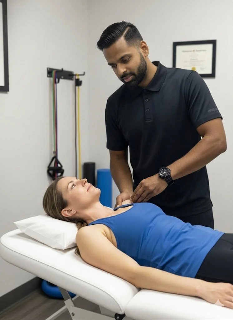
At our 68 Abell Street facility in Toronto, an exhaustive in-person physical assessment is legally and clinically required before beginning therapy. A physiotherapist will utilize specific orthopedic tests, such as the "Empty Can Test" or the "Drop Arm Test," to isolate which of the four S.I.T.S. muscles is compromised. We also assess joint mobility, cervical spine involvement, and functional movement patterns to build a holistic picture of the mechanical failure before obtaining informed patient consent to commence care.
Treatment Modalities at Rehab Mechanics
Rehabilitation requires a precise, structured approach. Passive rest is generally ineffective for tendon injuries, as it leads to further atrophy (muscle wasting) and stiffness. Tendons require mechanical loading to stimulate collagen synthesis and heal.
Soft Tissue Therapy and Down-Regulation
Before aggressive strengthening begins, soft tissue therapy is heavily utilized. When the rotator cuff is injured, larger surrounding muscles—such as the upper trapezius, levator scapulae, and pectoralis major—often go into protective spasm, creating a rigid, elevated shoulder posture. Manual release techniques help to down-regulate the nervous system, reduce this compensatory hypertonicity, and restore the necessary joint space for pain-free movement.
Corrective Exercises: The Progressive Loading Model
"You cannot fire a cannon from a canoe." This clinical adage means that your arm cannot generate healthy strength if the shoulder blade it attaches to is unstable. Our corrective exercise protocols aim to build that stable foundation, generally following a strictly phased progression:
1. Phase One: The Isometric Phase
In the acute phase of injury, movement is often too painful. We introduce isometrics—contracting the rotator cuff muscles against a fixed resistance (like a wall) without actually moving the joint. A typical protocol involves holding a moderate contraction for 30 to 45 seconds for multiple repetitions. This safely introduces a mechanical load to the tendon while creating a profound analgesic (pain-relieving) effect on the localized nervous system.
2. Phase Two: The Isotonic and Concentric Phase
Once pain is manageable, movement is introduced using very light resistance bands or gravity-eliminated weights (such as lying on your side). The focus here is on isolated, controlled contractions to rebuild the foundational strength of the specific injured muscle, as well as engaging the rhomboids and serratus anterior to secure the shoulder blade to the rib cage.
3. Phase Three: The Eccentric Phase
Tendons respond exceptionally well to eccentric loading—the lengthening phase of a muscle contraction. For example, using your healthy arm to pull a resistance band into external rotation, and then using the injured arm to slowly, over a count of four seconds, resist the band as it pulls your arm back inward. This high-tension, controlled lengthening signals the body to align new collagen fibers along the lines of mechanical stress, building robust tensile resilience.
4. Phase Four: The Functional and Proprioceptive Phase
The final stage bridges the gap between basic rehabilitation and real-world demands. This involves integrating the shoulder with full-body kinetic movements, multi-planar overhead presses, and proprioceptive drills (like stabilizing a medicine ball against a wall) to restore full occupational, athletic, and functional capacity.
Progressive Loading Framework
Rehabilitation PhaseExercise ExampleClinical Objective
Phase 1: Isometric
Wall pushes (Internal/External rotation); 45-second holds.
Introduce safe tendon loading; down-regulate pain signals.
Phase 2: Concentric
Side-lying external rotation; Prone I, Y, and T raises.
Rebuild isolated muscular strength and dynamic scapular stability.
Phase 3: Eccentric
Slow-release resistance band rotations (4-second negative).
Stimulate structural collagen alignment and improve tissue resilience.
Phase 4: Functional
Overhead kettlebell carries / Plyometric ball drops.
Restore full occupational or athletic capacity and dynamic joint control.
Author Biography
Written by Sanjay Attwala (BSC, MSC, RPT), Registered Physiotherapist.
Sanjay Attwala manages patient care at Rehab Mechanics (S. Attwala Physiotherapy Professional Corporation) located at 68 Abell Street, Toronto. He is in good standing with the College of Physiotherapists of Ontario (CPO). Learn more about our highly qualified clinical team here.
Medical Disclaimer:
The content provided in this article is for general educational and informational purposes only and does not constitute formal medical advice. Individual physiological responses to physiotherapy vary, and Rehab Mechanics does not guarantee specific treatment outcomes. An in-person assessment is legally and clinically required to develop an individualized treatment plan and obtain informed consent before commencing care.