What is Retrolisthesis and Can Physiotherapy Help Treat It? | Rehab Mechanics Toronto
What is Retrolisthesis and Can Physiotherapy Help Treat It?
Summary for OUR HIGH FLYERS (TL;DR):
Physiotherapy cannot physically push a slipped vertebra back into place, but it is the primary conservative treatment for managing the pain and instability caused by retrolisthesis. By aggressively strengthening the deep core stabilizers (like the multifidus and transversus abdominis), addressing muscular imbalances in the hips, and modifying daily movement patterns, physiotherapy acts as an "internal brace" to prevent further backward slippage and relieve nerve compression.
Key Takeaways:
Primary Symptoms: A deep, localized ache in the lower back that worsens with spinal extension (bending backward) or prolonged standing. If the slipped vertebra pinches a nerve root, pain, tingling, numbness, or weakness may radiate down the buttocks and legs (a condition broadly referred to as sciatica or radiculopathy).
The Pathology: Retrolisthesis occurs when a single vertebra in the spine slips backward relative to the vertebra immediately below it. It is primarily driven by biomechanical failure, often caused by severe degenerative disc disease, arthritis of the facet joints, or acute physical trauma.
Core Modalities: Evidence-based rehabilitation heavily avoids aggressive spinal manipulation at the hypermobile segment. Instead, it relies on strict core stabilization protocols (such as the McGill Big 3), pelvic tilting neuromuscular control, and lower body soft tissue release to reduce compensatory strain.
General Timelines: While the structural skeletal slippage is permanent without surgical fusion, patients who strictly adhere to a stabilization program often see a significant reduction in pain and neurological symptoms within 8 to 12 weeks of targeted physiotherapy, often returning to high-level functional activities.

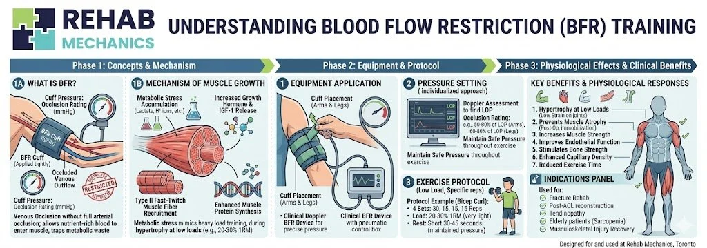
Medical illustration of the lumbar spine comparing normal vertebral alignment with retrolisthesis, where the upper vertebra has translated backward over the lower vertebra, narrowing the intervertebral foramen and crowding the spinal nerve root.
Understanding the Anatomy and Biomechanics of Retrolisthesis
When patients hear the term "slipped disc," they usually envision a herniation—where the soft, jelly-like center of the intervertebral disc pushes out through its tough exterior ring. However, a "slipped bone" is a different mechanical failure altogether.
The human spine is a perfectly stacked column of bones (vertebrae). When this column loses its structural integrity and a vertebra slides forward, it is called anterolisthesis (or spondylolisthesis). When a vertebra slides backward toward the spinal canal, it is diagnosed as retrolisthesis. This backward translation occurs most frequently in the highly mobile segments of the cervical spine (neck) and the weight-bearing segments of the lumbar spine (lower back), specifically at the L3-L4, L4-L5, or L5-S1 levels.
Why Does the Spine Slip Backward? The Mechanics of Instability
Retrolisthesis is fundamentally an issue of mechanical instability and joint failure. Under healthy conditions, the vertebrae are held firmly in place by a complex, redundant system of intervertebral discs (which act as shock absorbers and spacers) and facet joints (which interlock like hinges at the back of the spine to prevent excessive sliding).
The most common drivers of this structural failure include:
Degenerative Disc Disease (DDD): This is the most frequent culprit. As we age, or through repetitive mechanical overload, the discs lose their hydration and height (disc desiccation). A flatter, deflated disc brings the two vertebrae closer together. This loss of height creates "slack" in the longitudinal ligaments holding the spine together, effectively allowing the bone above to shift backward on the bone below.
Facet Joint Osteoarthritis: The facet joints are covered in smooth cartilage. Degeneration of this cartilage strips away the physical "brakes" that keep the vertebrae properly stacked. As the joints wear down, they can subluxate (partially dislocate), facilitating the backward slide.
Trauma and Ligamentous Laxity: High-impact injuries, such as a motor vehicle accident, severe whiplash, or a heavy fall, can rupture the stabilizing ligaments (specifically the anterior and posterior longitudinal ligaments) or fracture the bony stabilizing structures of the spine.
Radiological Grading: How Severe is the Slip?
When diagnosing retrolisthesis, medical professionals utilize radiological imaging to grade the severity of the slip, typically referencing a modified Meyerding grading system. The grade is determined by the percentage that the upper vertebra has slipped backward over the lower vertebra:
Grade 1: $1\%$ to $25\%$ slippage. (This is the most common presentation in a physiotherapy clinic and is highly responsive to conservative care).
Grade 2: $26\%$ to $50\%$ slippage.
Grade 3: $51\%$ to $75\%$ slippage.
Grade 4: $76\%$ to $100\%$ slippage. (Grades 3 and 4 are severe structural failures that frequently require surgical stabilization).
The Neurological Threat: Foraminal Stenosis and Radiculopathy
The primary reason retrolisthesis is so painful is rarely just the shifting bone itself; it is the narrowing of the intervertebral foramen—the small, bony windows on the sides of the spine where the spinal nerves exit the spinal cord and travel down the legs.
When a bone slides backward, it effectively shrinks the size of this window (a condition called foraminal stenosis). Furthermore, the collapsed disc space often causes the ligamentum flavum (a ligament inside the spinal canal) to buckle inward, further crowding the area.
This mechanical crowding pinches the exiting nerve root. Depending on which level of the spine slips, the symptoms vary wildly:
L4 Nerve Impingement: Often causes sharp pain radiating to the front of the thigh, accompanied by weakness in straightening the knee.
L5 Nerve Impingement: Typically refers pain down the side of the leg and into the top of the foot, potentially causing "foot drop" (an inability to lift the big toe or ankle).
S1 Nerve Impingement: Shoots pain down the back of the calf to the heel and sole of the foot, often reducing the Achilles reflex.
Clinical Assessment: Identifying the Instability
At our 68 Abell Street clinic, patients often arrive with a static X-ray or MRI report confirming a Grade 1 or Grade 2 retrolisthesis. However, a static scan taken while lying down inside an MRI tube only tells us what the bone looks like at rest; an in-person physical assessment tells us how the body is functioning around it under gravity.
Dynamic Stability Testing and Directional Preference
A registered physiotherapist will perform specific orthopedic tests to identify your "directional preference." For a patient with retrolisthesis, spinal extension (bending backward or reaching overhead) frequently reproduces their severe, shooting pain because it structurally forces the vertebra further backward, jamming the facet joints and further narrowing the spinal canal. Conversely, spinal flexion (bending forward slightly) often opens the neural foramina, offering temporary relief from the leg pain.
If instability is suspected, a physician may order dynamic flexion-extension X-rays. These are images taken while the patient is actively bending entirely forward and then entirely backward. These dynamic scans allow clinicians to see if the vertebra is actively sliding back and forth during movement, which heavily dictates the aggressiveness of the stabilization protocol.
Neurological Screening and "Red Flags"
We will also conduct a thorough, legally required neurological screen—testing deep tendon reflexes, myotomes (specific muscle strength testing), and dermatomes (skin sensation mapping)—to determine the exact severity of the nerve root compression.
(Clinical Note: Severe, rapidly progressing neurological deficits, such as a sudden loss of bowel/bladder control, profound "saddle" numbness around the groin, or profound leg weakness causing you to stumble, are considered medical red flags (Cauda Equina Syndrome). These indicate absolute spinal cord compression and require immediate emergency medical intervention, not physical therapy.)
Comprehensive Treatment Modalities at Rehab Mechanics
Conservative management of retrolisthesis requires a highly delicate clinical balance: we must mobilize the stiff, compensatory areas of the kinetic chain while aggressively stabilizing the hypermobile (slipped) segment.
1. Advanced Core Stabilization (The Internal Brace)
Because the passive anatomical structures (ligaments, bone, and discs) have failed to hold the spine in place, we must train the active structures (the muscular system) to take over the job. This does not mean doing traditional sit-ups, crunches, or heavy deadlifts, which place massive, dangerous compressive loads and shear forces directly on the unstable discs.
Instead, we utilize evidence-based stabilization protocols, drawing heavily from the McGill Big 3 (developed by Dr. Stuart McGill, a world-renowned spine biomechanist). The goal is to build immense muscular endurance—not necessarily peak strength—in the deep core while keeping the spine in a strictly neutral, pain-free position.
The Modified Curl-up: Replaces the standard crunch. One leg is bent to lock the pelvis in a neutral position, and the hands are placed under the lumbar spine to preserve its natural curve. The movement isolates the transversus abdominis (the deep corset muscle) without flexing the unstable lower back.
The Side Plank: An unparalleled exercise for building endurance in the quadratus lumborum (QL) and lateral abdominal obliques, providing crucial side-to-side stability for the slipping vertebrae.
The Bird-Dog: This movement (extending opposite arm and leg while on all fours) specifically targets the multifidus—a series of tiny, highly complex muscles that interlace directly between the vertebrae. A strong multifidus is the ultimate "internal brace" against backward slippage.
2. Correcting Pelvic Mechanics and Posture
The position of your pelvis directly dictates the curve and shear forces placed on your lower back. Many patients with retrolisthesis suffer from an excessive anterior pelvic tilt (a severe, pronounced arch in the lower back, often associated with "Lower Cross Syndrome").
This excessive arch physically encourages the vertebrae to slide backward down the slope of the bone below it. Physiotherapy focuses heavily on neuromuscular re-education. We teach patients how to actively control their pelvis, often cueing a subtle posterior pelvic tilt (tucking the tailbone slightly) during heavy lifting, prolonged standing, or reaching overhead. This conscious postural adjustment structurally opens the neural windows and decompresses the pinched nerves throughout the day.
3. Soft Tissue Therapy and Joint Mobilization (Above and Below)
A cardinal rule of treating hypermobility (a slipping joint) is that we explicitly avoid aggressive, cracking manipulations at the exact site of the retrolisthesis. Forcing motion into a joint that is already too loose is clinically counterproductive.
Instead, we aggressively treat the joints above and below the injury. If your hips are extremely stiff, or your mid-back (thoracic spine) lacks rotational mobility, your lower back is forced to overcompensate and move excessively to get you through your daily tasks. By using soft tissue therapy to release chronically tight hip flexors (psoas) and manual joint mobilization to restore thoracic extension, we eliminate the mechanical demand being unfairly placed on the unstable lumbar segment.
4. Postural and Ergonomic Management at Home
Rehabilitation must extend beyond the clinic walls. For a patient with an unstable spine, how they sleep and sit dictates how they heal.
Sleeping Posture: Patients with lumbar retrolisthesis often find relief sleeping in a fetal position (on their side with knees pulled toward the chest) with a thick pillow between the knees to prevent the top leg from rotating the spine. If sleeping on the back, placing a large bolster under the knees flattens the lumbar curve, reducing the backward shear force on the slipped bone.
Sitting: Prolonged sitting compresses the discs. We highly recommend utilizing a lumbar roll in office chairs and car seats, and taking standing micro-breaks every 30 minutes to rehydrate the intervertebral discs.
Phase Breakdown for Retrolisthesis Rehabilitation
Rehabilitation Phase
Primary Interventions & Modalities
Clinical Objective
Phase 1: Pain & Inflammation Down-Regulation
Activity modification (strictly avoiding heavy extension), positional relief strategies, soft tissue release of hypertonic lumbar erectors and hamstrings.
Reduce acute nerve root irritation (sciatica) and alleviate severe compensatory muscle spasms in the lower back.
Phase 2: Deep Core Activation
Isolation of the transversus abdominis and multifidus; precise introduction of the McGill Big 3 protocols in static, neutral spine positions.
Create an "internal brace" of muscular stiffness to artificially stabilize the slipping vertebral segment and prevent further micro-trauma.
Phase 3: Hip & Thoracic Mobility
Stretching hip flexors (psoas), targeted manual joint mobilization of the thoracic spine and Sacroiliac (SI) joints.
Reduce the compensatory mechanical load being placed on the unstable lumbar spine by restoring movement upstream and downstream.
Phase 4: Functional Load Integration
Squat and hinge mechanics, anti-rotation exercises (Pallof presses), and heavy lifting with strict neutral spine control.
Ensure the patient can return to occupational lifting or athletics without triggering a neurological relapse or facet joint irritation.
Author BiographyWritten by Sanjay Attwala (BSC, MSC, RPT), Registered Physiotherapist. Sanjay Attwala manages patient care at Rehab Mechanics (S. Attwala Physiotherapy Professional Corporation) located at 68 Abell Street, Toronto. He is in good standing with the College of Physiotherapists of Ontario (CPO). Learn more about our highly qualified clinical team here.
Medical Disclaimer:The content provided in this article is for general educational and informational purposes only and does not constitute formal medical advice. Retrolisthesis is a serious structural condition that requires radiological imaging (X-ray/MRI) for a definitive diagnosis and grading. Individual responses to physiotherapy vary, and Rehab Mechanics does not guarantee specific treatment outcomes. Severe grades of slippage may require surgical intervention. An in-person assessment is legally and clinically required to rule out severe neurological red flags and obtain informed consent before commencing conservative care.