How Does Shockwave Therapy Treat Tennis Elbow? Physiotherapy in Toronto | Rehab Mechanics
How Does Shockwave Therapy Treat Tennis Elbow?
Summary for our “efficient movers” (TL;DR): Extracorporeal Shockwave Therapy (ESWT) treats tennis elbow (lateral epicondylitis) by delivering high-energy acoustic sound waves into the damaged extensor tendon. This creates a controlled micro-trauma that breaks down calcifications, stimulates localized blood flow (angiogenesis), and forces the body to restart a stalled cellular repair cascade. For full resolution, this passive modality must be combined with an active, progressive eccentric loading and heavy slow resistance (HSR) exercise program to rebuild tendon tensile strength.
Key Takeaways:
Primary Symptoms: A sharp, stabbing, or burning pain isolated to the outer bony prominence of the elbow, which frequently radiates down the back of the forearm toward the wrist.
Functional Deficits: This condition is uniquely characterized by a significantly weakened and painful grip. Everyday tasks—such as turning a tight doorknob, pouring a pot of coffee, typing on a keyboard, or even offering a firm handshake—can trigger severe, disproportionate pain.
Core Modalities: Evidence-based management requires a multimodal clinical approach: Shockwave Therapy to aggressively address the cellular tendon degeneration, targeted soft tissue release for the hypertonic forearm muscle bellies, and a strict, phased tendon-loading exercise protocol.
General Timelines: Chronic cases of lateral epicondylitis are notoriously stubborn because tendons, unlike muscles, have an exceptionally poor natural blood supply. Comprehensive rehabilitation typically requires 6 to 12 weeks of strict adherence to a clinical plan before true structural remodeling yields permanent functional pain relief.
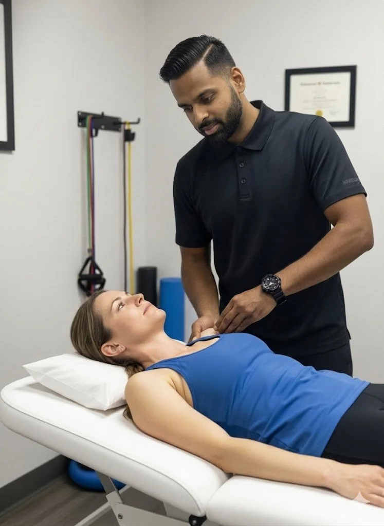
Registered Physiotherapist (Sanjay Attwala) engaging in a variety of cutting edge techniques for different types of tennis elbow injuries.
Understanding the Anatomy and Biomechanics of Lateral Epicondylitis
Despite its colloquial name, "tennis elbow" is a highly prevalent repetitive strain injury. Statistically, it is seen far more frequently in office workers, manual laborers, carpenters, plumbers, and individuals who perform repetitive gripping, lifting, or wrist extension tasks than in actual racquet sports athletes.
Medically classified as lateral epicondylitis or lateral elbow tendinopathy, this condition fundamentally affects the common extensor tendon. This thick band of connective tissue serves as the primary anchor point connecting your forearm extensor muscles to the lateral epicondyle—the small, bony bump on the outside of your elbow joint.
The ECRB: The Core of the Mechanical Failure
The most frequently implicated muscle in this complex is the Extensor Carpi Radialis Brevis (ECRB). To understand why this specific tendon fails, we must look at the biomechanics of the human hand.
When you squeeze your hand to grip an object tightly, your flexor muscles (on the palm side of your forearm) contract. However, if only your flexors contracted, your wrist would curl inward uncontrollably. To maintain a strong, functional grip, the ECRB muscle must simultaneously contract to hold your wrist perfectly straight (in slight extension). Therefore, every time you forcefully grip a tool, type on a keyboard, or hold a heavy pan, the ECRB tendon undergoes immense mechanical tension at the elbow. Over thousands of repetitive cycles, this tension exceeds the tissue's capacity to adapt.
Tendinitis vs. Tendinopathy: The Stalled Healing Cycle
Much like plantar fasciitis or Achilles pain, the suffix "-itis" is often a clinical misnomer for chronic cases of tennis elbow. While early-stage injuries (within the first few weeks) involve acute inflammation (tendinitis), long-term tennis elbow is more accurately classified as a tendinopathy or tendinosis.
When the ECRB muscle is continuously overworked without adequate recovery time, the tendon sustains repetitive microscopic tears. In a healthy state, the body easily repairs these micro-tears overnight. However, chronic overload disrupts this delicate balance, leading to a state of failed biological healing.
In a state of tendinopathy, the structural collagen fibers within the tendon become disorganized and frayed. Instead of lying in neat, strong, parallel lines (like a combed ponytail), the fibers become tangled and weak (like a bowl of spaghetti). Furthermore, the body attempts to heal the area by growing abnormal, highly sensitive nerve endings and fragile micro-blood vessels (a process called neovascularization). Because the common extensor tendon has a notoriously poor natural blood supply at its bony attachment, the body simply struggles to deliver the biological materials necessary to rebuild the tissue. This results in a stalled, chronic pain cycle that will not resolve with simple rest.
Clinical Assessment: Pinpointing the Root Cause
Pain on the outside of the elbow is not universally caused by lateral epicondylitis. Accurate diagnosis is the vital first step to ensuring effective care, as treating the wrong pathology will prolong the injury. At our 68 Abell Street facility in Toronto, a comprehensive in-person physical assessment is clinically and legally required before initiating any treatment plan.
A registered physiotherapist will utilize specific orthopedic and neurological tests to isolate the mechanical failure. This typically involves:
Cozen’s Test: The patient is asked to make a fist, pronate their forearm (palm down), and extend the wrist backward while the physiotherapist applies heavy resistance. A positive test yields sharp pain at the lateral epicondyle.
Maudsley’s Test: The physiotherapist applies resistance exclusively to the middle finger as the patient attempts to lift it. This specifically stresses the extensor digitorum muscle, which shares the common extensor tendon.
Mill's Test: A passive stretch of the extensor tendons, pushing the wrist into full flexion while straightening the elbow, to evaluate tissue extensibility and pain provocation.
Grip Dynamometry: We often measure grip strength with the elbow completely straight versus bent at 90 degrees. In true tennis elbow, grip strength is profoundly weaker and more painful when the elbow is straight due to the maximum stretch placed on the ECRB.
Ruling Out Competing Diagnoses
Crucially, the assessment must evaluate the entire upper kinetic chain to rule out conditions that mimic tennis elbow:
Cervical Radiculopathy: Pinched or irritated nerves in the neck (specifically the C5 or C6 nerve roots) can refer a burning, aching pain straight down the arm, masquerading as elbow pathology.
Radial Tunnel Syndrome: Compression of the radial nerve as it passes through the supinator muscle in the forearm. This causes a deep, aching pain that is typically located slightly further down the arm (into the muscle belly) rather than directly on the bony epicondyle.
Shoulder Dysfunctions: Often, weakness in the rotator cuff or scapular stabilizers forces the elbow to overcompensate during lifting tasks, making the elbow the "victim" of a weak shoulder.
Only after a precise diagnosis is established, and informed patient consent is obtained detailing the risks and benefits of the proposed plan, will therapeutic interventions commence.
Comprehensive Treatment Modalities at Rehab Mechanics
Rehabilitating chronic tennis elbow requires a highly specific, dual-pronged approach: first, we must interrupt the stalled degenerative cycle; second, we must actively rebuild the tendon's tensile capacity to handle daily loads.
Extracorporeal Shockwave Therapy (ESWT)
For chronic lateral epicondylitis that has not responded to basic rest, ice, and stretching (often cases lasting longer than 3 to 6 months), Shockwave Therapy is considered a primary, highly effective, and evidence-based intervention.
The treatment involves a clinical device that generates high-energy acoustic pressure waves. When the applicator is applied directly to the lateral epicondyle, these waves penetrate the skin and create micro-cavitation bubbles within the degenerated tendon tissue. This purposeful, controlled mechanical stimulus achieves three critical physiological outcomes:
Angiogenesis (New Blood Vessels): It stimulates the formation of new micro-blood vessels. This dramatically increases the localized blood flow and nutrient delivery to the avascular (blood-poor) tendon attachment, providing the raw materials needed for cellular repair.
Re-initiating the Healing Cascade: The acoustic micro-trauma forces the body to abandon the stalled chronic state (tendinosis). It essentially "tricks" the localized immune system into restarting the acute inflammatory healing cascade, signaling fibroblasts (repair cells) to lay down new, healthy type-I collagen.
Profound Analgesia: The high-frequency acoustic pulses over-stimulate the local nerve endings, which helps to deplete Substance P—a neurotransmitter responsible for relaying chronic pain signals to the central nervous system. This offers patients significant functional pain relief that outlasts the treatment session.
The Patient Experience: Shockwave therapy is an active, stimulating treatment. Patients typically feel a rapid, pneumatic tapping sensation that can be uncomfortable (often described as a "good, productive ache"). A typical session lasts only 5 to 10 minutes.
Crucial Clinical Note: Because shockwave intentionally restarts the inflammatory process to heal the tendon, patients are strictly advised not to use ice or anti-inflammatory medications (like Ibuprofen/Advil) for 48 hours post-treatment, as these will suppress the exact biological response we are trying to create.
Soft Tissue Therapy and Joint Mobilization
While shockwave targets the tendon attachment directly at the bone, the large muscle bellies of the forearm must also be addressed. When the extensor tendon is painful, the surrounding muscles (like the brachioradialis and supinator) frequently go into a state of hypertonicity (chronic tightness) in a subconscious attempt to guard the joint.
Soft tissue therapy, including manual myofascial release, Instrument-Assisted Soft Tissue Mobilization (IASTM), and active release techniques, are applied directly to the forearm extensors and flexors. By releasing this dense muscular tension, we physically reduce the mechanical "pull" or traction being continuously exerted on the injured, sensitive tendon attachment. Additionally, joint mobilizations (such as Mulligan mobilizations with movement) may be utilized to restore pain-free tracking of the radial head at the elbow joint.
Corrective Exercises: Rebuilding Tendon Resilience
Passive modalities like shockwave therapy and manual release create the optimal biological environment for healing, but active mechanical loading is absolutely required to make the tendon physically stronger.
Tendons do not respond to passive rest; they adapt and strengthen only in response to mechanical load. The cornerstone of late-stage tennis elbow rehabilitation involves a highly structured progression of loading:
1. The Isometric Phase
In the early stages, when the tendon is highly reactive and painful, we utilize isometrics—contracting the extensor muscles against an immovable resistance without moving the wrist joint. This introduces a safe mechanical load that triggers an analgesic (pain-relieving) effect in the cortex of the brain without stretching the damaged fibers.
2. The Eccentric Loading Phase
As pain subsides, we introduce eccentric loading—the slow, controlled lengthening of a muscle while it is under tension. A classic clinical protocol involves using a specialized rubber resistance bar (like a "FlexBar" for the Tyler Twist exercise) or a light dumbbell. The patient uses their uninjured hand to lift the weight into wrist extension, and then uses the injured arm to slowly lower the weight downward over a count of 4 to 5 seconds. This high-tension, slow-release movement signals the body to align the newly synthesized collagen fibers strictly along the lines of mechanical stress.
3. Heavy Slow Resistance (HSR)
Modern physiotherapy research increasingly supports Heavy Slow Resistance training for tendinopathies. This involves performing both the lifting (concentric) and lowering (eccentric) phases of wrist extension using a heavier weight, but at a very slow, deliberate tempo (e.g., 3 seconds up, 3 seconds down). HSR has been shown to be equally as effective as pure eccentrics for collagen remodeling, and often results in higher patient compliance.
Treatment Phase Breakdown for Lateral Epicondylitis
Rehabilitation Phase
Primary Interventions & Modalities
Clinical Objective
Phase 1: Pain Management & Down-Regulation
Isometric holds, Soft Tissue Release (IASTM), Kinesiology Taping, Bracing (counterforce strap), Ergonomic assessment.
Reduce acute tissue reactivity, decrease forearm muscle hypertonicity, and offload the ECRB tendon during essential daily tasks.
Phase 2: Cellular Stimulation
Extracorporeal Shockwave Therapy (ESWT), Manual Joint Mobilizations.
Break down localized calcifications, induce angiogenesis (new blood flow), and restart the stalled biological healing cascade.
Phase 3: Structural Remodeling
Eccentric Wrist Extensions, FlexBar (Tyler Twist) protocols, Heavy Slow Resistance (HSR).
Guide the parallel alignment of new collagen fibers, aggressively rebuild tendon tensile strength, and restore baseline grip capacity.
Phase 4: Kinetic Chain Integration
Rotator cuff strengthening, Scapular stabilization, multi-joint occupational/athletic simulations.
Ensure the shoulder, elbow, and wrist operate cohesively, eliminating upstream weaknesses to prevent future mechanical overload and injury recurrence.
Author BiographyWritten by Sanjay Attwala (BSC, MSC, RPT), Registered Physiotherapist. Sanjay Attwala manages patient care at Rehab Mechanics (S. Attwala Physiotherapy Professional Corporation) located at 68 Abell Street, Toronto. He is in good standing with the College of Physiotherapists of Ontario (CPO). Learn more about our highly qualified clinical team here.
Medical Disclaimer:The content provided in this article is for general educational and informational purposes only and does not constitute formal medical advice. Individual physiological responses to physiotherapy vary significantly, and Rehab Mechanics does not guarantee specific treatment outcomes or timelines. An in-person assessment is legally and clinically required to develop an individualized treatment plan, definitively diagnose the source of pain, and obtain informed consent before commencing care.