
How Does Shockwave Therapy Treat Tennis Elbow? Physiotherapy in Toronto | Rehab Mechanics
How Does Shockwave Therapy Treat Tennis Elbow?
Summary for our “efficient movers” (TL;DR): Extracorporeal Shockwave Therapy (ESWT) treats tennis elbow (lateral epicondylitis) by delivering high-energy acoustic sound waves into the damaged extensor tendon. This creates a controlled micro-trauma that breaks down calcifications, stimulates localized blood flow (angiogenesis), and forces the body to restart a stalled cellular repair cascade. For full resolution, this passive modality must be combined with an active, progressive eccentric loading and heavy slow resistance (HSR) exercise program to rebuild tendon tensile strength.
Key Takeaways:
Primary Symptoms: A sharp, stabbing, or burning pain isolated to the outer bony prominence of the elbow, which frequently radiates down the back of the forearm toward the wrist.
Functional Deficits: This condition is uniquely characterized by a significantly weakened and painful grip. Everyday tasks—such as turning a tight doorknob, pouring a pot of coffee, typing on a keyboard, or even offering a firm handshake—can trigger severe, disproportionate pain.
Core Modalities: Evidence-based management requires a multimodal clinical approach: Shockwave Therapy to aggressively address the cellular tendon degeneration, targeted soft tissue release for the hypertonic forearm muscle bellies, and a strict, phased tendon-loading exercise protocol.
General Timelines: Chronic cases of lateral epicondylitis are notoriously stubborn because tendons, unlike muscles, have an exceptionally poor natural blood supply. Comprehensive rehabilitation typically requires 6 to 12 weeks of strict adherence to a clinical plan before true structural remodeling yields permanent functional pain relief.
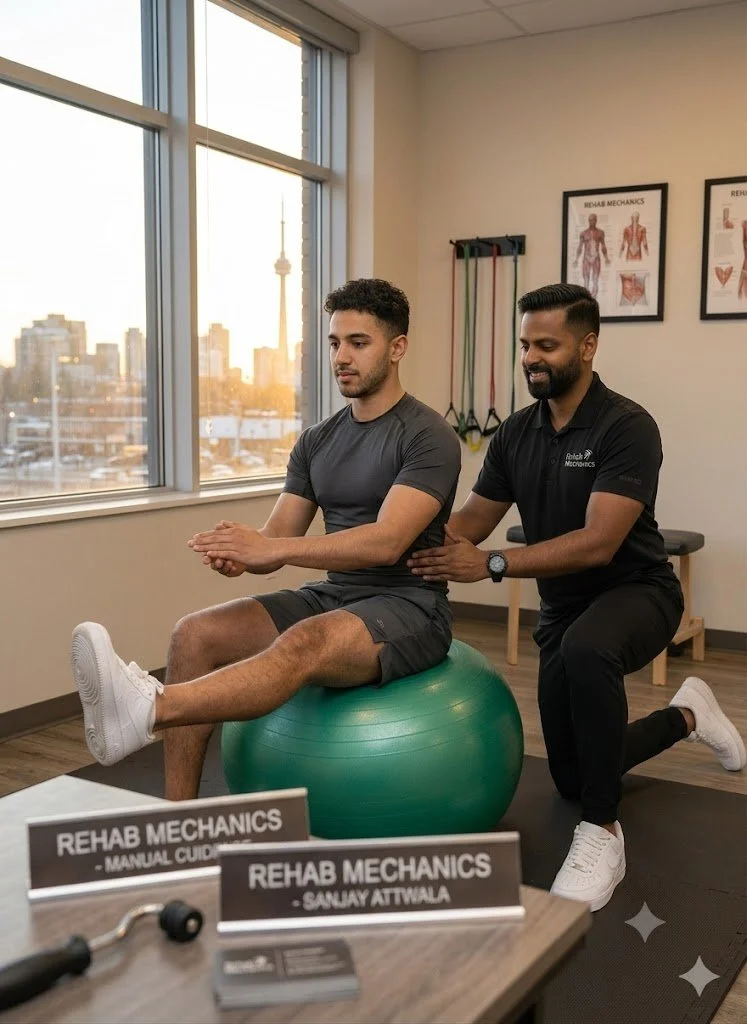
Registered Physiotherapist (Sanjay Attwala) engaging in a variety of cutting edge techniques for different types of tennis elbow injuries.
Understanding the Anatomy and Biomechanics of Lateral Epicondylitis
Despite its colloquial name, "tennis elbow" is a highly prevalent repetitive strain injury. Statistically, it is seen far more frequently in office workers, manual laborers, carpenters, plumbers, and individuals who perform repetitive gripping, lifting, or wrist extension tasks than in actual racquet sports athletes.
Medically classified as lateral epicondylitis or lateral elbow tendinopathy, this condition fundamentally affects the common extensor tendon. This thick band of connective tissue serves as the primary anchor point connecting your forearm extensor muscles to the lateral epicondyle—the small, bony bump on the outside of your elbow joint.
The ECRB: The Core of the Mechanical Failure
The most frequently implicated muscle in this complex is the Extensor Carpi Radialis Brevis (ECRB). To understand why this specific tendon fails, we must look at the biomechanics of the human hand.
When you squeeze your hand to grip an object tightly, your flexor muscles (on the palm side of your forearm) contract. However, if only your flexors contracted, your wrist would curl inward uncontrollably. To maintain a strong, functional grip, the ECRB muscle must simultaneously contract to hold your wrist perfectly straight (in slight extension). Therefore, every time you forcefully grip a tool, type on a keyboard, or hold a heavy pan, the ECRB tendon undergoes immense mechanical tension at the elbow. Over thousands of repetitive cycles, this tension exceeds the tissue's capacity to adapt.
Tendinitis vs. Tendinopathy: The Stalled Healing Cycle
Much like plantar fasciitis or Achilles pain, the suffix "-itis" is often a clinical misnomer for chronic cases of tennis elbow. While early-stage injuries (within the first few weeks) involve acute inflammation (tendinitis), long-term tennis elbow is more accurately classified as a tendinopathy or tendinosis.
When the ECRB muscle is continuously overworked without adequate recovery time, the tendon sustains repetitive microscopic tears. In a healthy state, the body easily repairs these micro-tears overnight. However, chronic overload disrupts this delicate balance, leading to a state of failed biological healing.
In a state of tendinopathy, the structural collagen fibers within the tendon become disorganized and frayed. Instead of lying in neat, strong, parallel lines (like a combed ponytail), the fibers become tangled and weak (like a bowl of spaghetti). Furthermore, the body attempts to heal the area by growing abnormal, highly sensitive nerve endings and fragile micro-blood vessels (a process called neovascularization). Because the common extensor tendon has a notoriously poor natural blood supply at its bony attachment, the body simply struggles to deliver the biological materials necessary to rebuild the tissue. This results in a stalled, chronic pain cycle that will not resolve with simple rest.
Clinical Assessment: Pinpointing the Root Cause
Pain on the outside of the elbow is not universally caused by lateral epicondylitis. Accurate diagnosis is the vital first step to ensuring effective care, as treating the wrong pathology will prolong the injury. At our 68 Abell Street facility in Toronto, a comprehensive in-person physical assessment is clinically and legally required before initiating any treatment plan.
A registered physiotherapist will utilize specific orthopedic and neurological tests to isolate the mechanical failure. This typically involves:
Cozen’s Test: The patient is asked to make a fist, pronate their forearm (palm down), and extend the wrist backward while the physiotherapist applies heavy resistance. A positive test yields sharp pain at the lateral epicondyle.
Maudsley’s Test: The physiotherapist applies resistance exclusively to the middle finger as the patient attempts to lift it. This specifically stresses the extensor digitorum muscle, which shares the common extensor tendon.
Mill's Test: A passive stretch of the extensor tendons, pushing the wrist into full flexion while straightening the elbow, to evaluate tissue extensibility and pain provocation.
Grip Dynamometry: We often measure grip strength with the elbow completely straight versus bent at 90 degrees. In true tennis elbow, grip strength is profoundly weaker and more painful when the elbow is straight due to the maximum stretch placed on the ECRB.
Ruling Out Competing Diagnoses
Crucially, the assessment must evaluate the entire upper kinetic chain to rule out conditions that mimic tennis elbow:
Cervical Radiculopathy: Pinched or irritated nerves in the neck (specifically the C5 or C6 nerve roots) can refer a burning, aching pain straight down the arm, masquerading as elbow pathology.
Radial Tunnel Syndrome: Compression of the radial nerve as it passes through the supinator muscle in the forearm. This causes a deep, aching pain that is typically located slightly further down the arm (into the muscle belly) rather than directly on the bony epicondyle.
Shoulder Dysfunctions: Often, weakness in the rotator cuff or scapular stabilizers forces the elbow to overcompensate during lifting tasks, making the elbow the "victim" of a weak shoulder.
Only after a precise diagnosis is established, and informed patient consent is obtained detailing the risks and benefits of the proposed plan, will therapeutic interventions commence.
Comprehensive Treatment Modalities at Rehab Mechanics
Rehabilitating chronic tennis elbow requires a highly specific, dual-pronged approach: first, we must interrupt the stalled degenerative cycle; second, we must actively rebuild the tendon's tensile capacity to handle daily loads.
Extracorporeal Shockwave Therapy (ESWT)
For chronic lateral epicondylitis that has not responded to basic rest, ice, and stretching (often cases lasting longer than 3 to 6 months), Shockwave Therapy is considered a primary, highly effective, and evidence-based intervention.
The treatment involves a clinical device that generates high-energy acoustic pressure waves. When the applicator is applied directly to the lateral epicondyle, these waves penetrate the skin and create micro-cavitation bubbles within the degenerated tendon tissue. This purposeful, controlled mechanical stimulus achieves three critical physiological outcomes:
Angiogenesis (New Blood Vessels): It stimulates the formation of new micro-blood vessels. This dramatically increases the localized blood flow and nutrient delivery to the avascular (blood-poor) tendon attachment, providing the raw materials needed for cellular repair.
Re-initiating the Healing Cascade: The acoustic micro-trauma forces the body to abandon the stalled chronic state (tendinosis). It essentially "tricks" the localized immune system into restarting the acute inflammatory healing cascade, signaling fibroblasts (repair cells) to lay down new, healthy type-I collagen.
Profound Analgesia: The high-frequency acoustic pulses over-stimulate the local nerve endings, which helps to deplete Substance P—a neurotransmitter responsible for relaying chronic pain signals to the central nervous system. This offers patients significant functional pain relief that outlasts the treatment session.
The Patient Experience: Shockwave therapy is an active, stimulating treatment. Patients typically feel a rapid, pneumatic tapping sensation that can be uncomfortable (often described as a "good, productive ache"). A typical session lasts only 5 to 10 minutes.
Crucial Clinical Note: Because shockwave intentionally restarts the inflammatory process to heal the tendon, patients are strictly advised not to use ice or anti-inflammatory medications (like Ibuprofen/Advil) for 48 hours post-treatment, as these will suppress the exact biological response we are trying to create.
Soft Tissue Therapy and Joint Mobilization
While shockwave targets the tendon attachment directly at the bone, the large muscle bellies of the forearm must also be addressed. When the extensor tendon is painful, the surrounding muscles (like the brachioradialis and supinator) frequently go into a state of hypertonicity (chronic tightness) in a subconscious attempt to guard the joint.
Soft tissue therapy, including manual myofascial release, Instrument-Assisted Soft Tissue Mobilization (IASTM), and active release techniques, are applied directly to the forearm extensors and flexors. By releasing this dense muscular tension, we physically reduce the mechanical "pull" or traction being continuously exerted on the injured, sensitive tendon attachment. Additionally, joint mobilizations (such as Mulligan mobilizations with movement) may be utilized to restore pain-free tracking of the radial head at the elbow joint.
Corrective Exercises: Rebuilding Tendon Resilience
Passive modalities like shockwave therapy and manual release create the optimal biological environment for healing, but active mechanical loading is absolutely required to make the tendon physically stronger.
Tendons do not respond to passive rest; they adapt and strengthen only in response to mechanical load. The cornerstone of late-stage tennis elbow rehabilitation involves a highly structured progression of loading:
1. The Isometric Phase
In the early stages, when the tendon is highly reactive and painful, we utilize isometrics—contracting the extensor muscles against an immovable resistance without moving the wrist joint. This introduces a safe mechanical load that triggers an analgesic (pain-relieving) effect in the cortex of the brain without stretching the damaged fibers.
2. The Eccentric Loading Phase
As pain subsides, we introduce eccentric loading—the slow, controlled lengthening of a muscle while it is under tension. A classic clinical protocol involves using a specialized rubber resistance bar (like a "FlexBar" for the Tyler Twist exercise) or a light dumbbell. The patient uses their uninjured hand to lift the weight into wrist extension, and then uses the injured arm to slowly lower the weight downward over a count of 4 to 5 seconds. This high-tension, slow-release movement signals the body to align the newly synthesized collagen fibers strictly along the lines of mechanical stress.
3. Heavy Slow Resistance (HSR)
Modern physiotherapy research increasingly supports Heavy Slow Resistance training for tendinopathies. This involves performing both the lifting (concentric) and lowering (eccentric) phases of wrist extension using a heavier weight, but at a very slow, deliberate tempo (e.g., 3 seconds up, 3 seconds down). HSR has been shown to be equally as effective as pure eccentrics for collagen remodeling, and often results in higher patient compliance.
Treatment Phase Breakdown for Lateral Epicondylitis
Rehabilitation Phase
Primary Interventions & Modalities
Clinical Objective
Phase 1: Pain Management & Down-Regulation
Isometric holds, Soft Tissue Release (IASTM), Kinesiology Taping, Bracing (counterforce strap), Ergonomic assessment.
Reduce acute tissue reactivity, decrease forearm muscle hypertonicity, and offload the ECRB tendon during essential daily tasks.
Phase 2: Cellular Stimulation
Extracorporeal Shockwave Therapy (ESWT), Manual Joint Mobilizations.
Break down localized calcifications, induce angiogenesis (new blood flow), and restart the stalled biological healing cascade.
Phase 3: Structural Remodeling
Eccentric Wrist Extensions, FlexBar (Tyler Twist) protocols, Heavy Slow Resistance (HSR).
Guide the parallel alignment of new collagen fibers, aggressively rebuild tendon tensile strength, and restore baseline grip capacity.
Phase 4: Kinetic Chain Integration
Rotator cuff strengthening, Scapular stabilization, multi-joint occupational/athletic simulations.
Ensure the shoulder, elbow, and wrist operate cohesively, eliminating upstream weaknesses to prevent future mechanical overload and injury recurrence.
Author BiographyWritten by Sanjay Attwala (BSC, MSC, RPT), Registered Physiotherapist. Sanjay Attwala manages patient care at Rehab Mechanics (S. Attwala Physiotherapy Professional Corporation) located at 68 Abell Street, Toronto. He is in good standing with the College of Physiotherapists of Ontario (CPO). Learn more about our highly qualified clinical team here.
Medical Disclaimer:The content provided in this article is for general educational and informational purposes only and does not constitute formal medical advice. Individual physiological responses to physiotherapy vary significantly, and Rehab Mechanics does not guarantee specific treatment outcomes or timelines. An in-person assessment is legally and clinically required to develop an individualized treatment plan, definitively diagnose the source of pain, and obtain informed consent before commencing care.

What Are the Best Corrective Exercises for a Rotator Cuff Injury? Physiotherapy in Toronto | Rehab Mechanics
What Are the Best Corrective Exercises for a Rotator Cuff Injury?
TL;DR
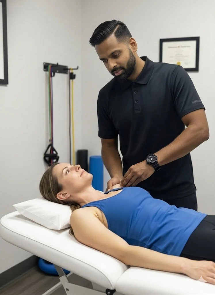
The most effective corrective exercises for a rotator cuff injury focus on progressively loading the tendon to restore tensile strength. Rehabilitation must transition from low-impact isometric holds designed to reduce pain, through to functional isotonic movements that restore full dynamic stability. Comprehensive care addresses not just the rotator cuff, but the stabilizing muscles of the entire shoulder blade.
Key Takeaways:
Primary Symptoms: A deep, dull ache in the shoulder joint, catching or pinching sensations when lifting the arm overhead, significant weakness during outward rotation, and pain that disrupts sleep (particularly when lying on the affected shoulder).
Core Modalities: Evidence-based rehabilitation combines pain-relieving manual soft tissue therapy, scapular stabilization, and a strictly phased, high-load corrective exercise program.
General Timelines: Depending on the specific pathology—whether acute tendinitis, chronic degenerative tendinopathy, or a partial-thickness tear—rehabilitation typically spans from 6 to 12 weeks of consistent clinical adherence before true structural remodeling occurs.
(Visual Asset Placeholder: Anatomical diagram of the shoulder joint)
Alt Text: Medical illustration of the human shoulder highlighting the four S.I.T.S. muscles of the rotator cuff—supraspinatus, infraspinatus, teres minor, and subscapularis—surrounding the glenohumeral joint and scapula.
Understanding the Anatomy of a Rotator Cuff Injury
The human shoulder is uniquely designed to prioritize mobility over stability. It is often compared to a golf ball sitting on a small tee. To keep the "ball" (the head of the humerus) centered on the "tee" (the glenoid fossa) during movement, the body relies on a dynamic group of four muscles and their accompanying tendons, collectively known as the rotator cuff.
Clinically, these are referred to as the S.I.T.S. muscles:
Supraspinatus: Responsible for initiating the lifting (abduction) of the arm away from the body. This is the most frequently injured tendon in the rotator cuff complex.
Infraspinatus: The primary muscle responsible for externally rotating the arm.
Teres Minor: Assists the infraspinatus with external rotation and joint stabilization.
Subscapularis: Located on the front of the shoulder blade, this powerful muscle is responsible for internal rotation.
Rotator cuff injuries frequently manifest as either acute tears (often from a sudden trauma or fall) or degenerative tendinopathy (the gradual wearing down of the tendon from overuse and poor biomechanics).
Impingement Syndrome and Scapular Dyskinesis
A rotator cuff rarely fails in isolation. Often, injuries are the result of poor structural biomechanics over a long period. For example, individuals with "upper cross syndrome"—characterized by a forward head posture and rounded shoulders, common in office workers—experience a narrowing of the subacromial space (the gap under the shoulder bone where the supraspinatus tendon travels).
When the shoulder blade (scapula) sits in an optimal position, this tendon glides smoothly. However, when the shoulder is chronically rounded forward, the bone repeatedly pinches or "impinges" the tendon against the acromion with every overhead movement. This repetitive micro-trauma leads to inflammation, cellular degeneration, and eventual tearing. Therefore, treating the rotator cuff always requires treating the posture and stability of the entire shoulder blade (a concept known as correcting scapular dyskinesis).
Clinical Assessment: Diagnosing the Root Cause
Because shoulder pain can originate from multiple sources—including referred nerve pain from the cervical spine (neck) or a frozen shoulder (adhesive capsulitis)—accurate diagnosis is the vital first step.
At our 68 Abell Street facility in Toronto, an exhaustive in-person physical assessment is legally and clinically required before beginning therapy. A physiotherapist will utilize specific orthopedic tests, such as the "Empty Can Test" or the "Drop Arm Test," to isolate which of the four S.I.T.S. muscles is compromised. We also assess joint mobility, cervical spine involvement, and functional movement patterns to build a holistic picture of the mechanical failure before obtaining informed patient consent to commence care.
Treatment Modalities at Rehab Mechanics
Rehabilitation requires a precise, structured approach. Passive rest is generally ineffective for tendon injuries, as it leads to further atrophy (muscle wasting) and stiffness. Tendons require mechanical loading to stimulate collagen synthesis and heal.
Soft Tissue Therapy and Down-Regulation
Before aggressive strengthening begins, soft tissue therapy is heavily utilized. When the rotator cuff is injured, larger surrounding muscles—such as the upper trapezius, levator scapulae, and pectoralis major—often go into protective spasm, creating a rigid, elevated shoulder posture. Manual release techniques help to down-regulate the nervous system, reduce this compensatory hypertonicity, and restore the necessary joint space for pain-free movement.
Corrective Exercises: The Progressive Loading Model
"You cannot fire a cannon from a canoe." This clinical adage means that your arm cannot generate healthy strength if the shoulder blade it attaches to is unstable. Our corrective exercise protocols aim to build that stable foundation, generally following a strictly phased progression:
1. Phase One: The Isometric Phase
In the acute phase of injury, movement is often too painful. We introduce isometrics—contracting the rotator cuff muscles against a fixed resistance (like a wall) without actually moving the joint. A typical protocol involves holding a moderate contraction for 30 to 45 seconds for multiple repetitions. This safely introduces a mechanical load to the tendon while creating a profound analgesic (pain-relieving) effect on the localized nervous system.
2. Phase Two: The Isotonic and Concentric Phase
Once pain is manageable, movement is introduced using very light resistance bands or gravity-eliminated weights (such as lying on your side). The focus here is on isolated, controlled contractions to rebuild the foundational strength of the specific injured muscle, as well as engaging the rhomboids and serratus anterior to secure the shoulder blade to the rib cage.
3. Phase Three: The Eccentric Phase
Tendons respond exceptionally well to eccentric loading—the lengthening phase of a muscle contraction. For example, using your healthy arm to pull a resistance band into external rotation, and then using the injured arm to slowly, over a count of four seconds, resist the band as it pulls your arm back inward. This high-tension, controlled lengthening signals the body to align new collagen fibers along the lines of mechanical stress, building robust tensile resilience.
4. Phase Four: The Functional and Proprioceptive Phase
The final stage bridges the gap between basic rehabilitation and real-world demands. This involves integrating the shoulder with full-body kinetic movements, multi-planar overhead presses, and proprioceptive drills (like stabilizing a medicine ball against a wall) to restore full occupational, athletic, and functional capacity.
Progressive Loading Framework
Rehabilitation PhaseExercise ExampleClinical Objective
Phase 1: Isometric
Wall pushes (Internal/External rotation); 45-second holds.
Introduce safe tendon loading; down-regulate pain signals.
Phase 2: Concentric
Side-lying external rotation; Prone I, Y, and T raises.
Rebuild isolated muscular strength and dynamic scapular stability.
Phase 3: Eccentric
Slow-release resistance band rotations (4-second negative).
Stimulate structural collagen alignment and improve tissue resilience.
Phase 4: Functional
Overhead kettlebell carries / Plyometric ball drops.
Restore full occupational or athletic capacity and dynamic joint control.
Author Biography
Written by Sanjay Attwala (BSC, MSC, RPT), Registered Physiotherapist.
Sanjay Attwala manages patient care at Rehab Mechanics (S. Attwala Physiotherapy Professional Corporation) located at 68 Abell Street, Toronto. He is in good standing with the College of Physiotherapists of Ontario (CPO). Learn more about our highly qualified clinical team here.
Medical Disclaimer:
The content provided in this article is for general educational and informational purposes only and does not constitute formal medical advice. Individual physiological responses to physiotherapy vary, and Rehab Mechanics does not guarantee specific treatment outcomes. An in-person assessment is legally and clinically required to develop an individualized treatment plan and obtain informed consent before commencing care.
How to Treat Plantar Fasciitis with Physiotherapy? Physiotherapy in Toronto | Rehab Mechanics
How to Treat Plantar Fasciitis with Physiotherapy?
Physiotherapy treats plantar fasciitis by reducing fascial tension, correcting lower extremity biomechanics, and stimulating tissue repair through a combination of targeted clinical modalities. Comprehensive rehabilitation addresses the root cause of the mechanical overload rather than just masking the localized pain.
Key Takeaways:
Primary Symptoms: Sharp, localized heel pain that is most severe during the first steps in the morning or upon weight-bearing after prolonged periods of rest. Pain often presents as a stabbing sensation at the bottom of the heel.
Core Modalities: Evidence-based treatment typically involves a combination of custom orthotics, extracorporeal shockwave therapy, targeted soft tissue release, therapeutic taping, and progressive corrective exercises.
General Timelines: Recovery timelines vary significantly based on individual physiological responses, the chronicity of the injury, and adherence to rehabilitation protocols. While immediate symptom relief can occur early in treatment, true structural remodeling of the fascial tissue often takes several weeks to months of consistent care.
(Visual Asset Placeholder: Clinical diagram of the foot's fascial band) Alt Text: Anatomical diagram of the human foot highlighting the plantar fascia band connecting the heel bone to the toes, illustrating the Windlass mechanism and the common site of inflammation at the medial calcaneal tubercle in plantar fasciitis.
Understanding the Anatomy of Plantar Fasciitis
The plantar fascia is a thick, web-like band of fibrous connective tissue (aponeurosis) that runs across the bottom of your foot. It originates at the medial tubercle of the heel bone (calcaneus) and fans out to connect to the base of your toes. Functionally, it acts as a dynamic shock absorber and is the primary support structure for the longitudinal arch of your foot, bearing up to 14% of the total load of the foot during the walking cycle.
Plantar fasciitis occurs when excessive mechanical stress or repetitive strain leads to microscopic tearing and subsequent inflammation of this tissue, most commonly right at its attachment point on the heel bone. It is one of the most frequent repetitive strain injuries evaluated at our Queen West clinic.
Fasciitis vs. Fasciosis: The Chronic Degeneration Cycle
While the suffix "-itis" implies acute inflammation, current medical research indicates that chronic cases (lasting longer than a few weeks) are more accurately described as plantar fasciosis. Fasciosis involves non-inflammatory structural degeneration. In these persistent cases, the collagen fibers that make up the fascia become disorganized, fragmented, and lose their vascularity (blood supply). Understanding this distinction is crucial because treating chronic cellular degeneration requires active mechanical loading and biological stimulation, rather than simple rest and anti-inflammatory medication.
Risk Factors and the Kinetic Chain
Plantar fasciitis rarely develops in isolation. It is typically the result of compounding biomechanical flaws or environmental factors, including:
Foot Mechanics: Both flat feet (pes planus) and rigid, high arches (pes cavus) alter how weight is distributed, increasing tension on the fascia.
Occupational Hazards: Service industry workers, nurses, or tradespeople who stand for prolonged hours on unyielding surfaces (like concrete) face continuous micro-trauma.
Activity Spikes: Distance runners or athletes who rapidly increase their training volume or change their running surface frequently overload the tissue before it can adapt.
Upstream Restrictions: Deficiencies in ankle mobility or weakness in the gluteal muscles can force the foot to compensate, placing an unnatural burden on the medial arch.
The Biomechanics of "First Step" Morning Pain
A hallmark characteristic of plantar fasciitis is severe pain during the first few steps in the morning. This phenomenon is biomechanically driven. During sleep, the foot naturally rests in a slightly pointed (plantarflexed) position. This allows the inflamed or damaged plantar fascia to artificially shorten and attempt to heal in a contracted state overnight.
When a patient takes their first step out of bed, the sudden upward bending (dorsiflexion) of the foot and toes forcefully stretches this newly healed, contracted tissue. This rapid stretch re-tears the micro-fibers, triggering an acute pain response. As the patient walks and the tissue warms up, it becomes more pliable, and the sharp pain often dulls to a deep ache. However, as tissue fatigue sets in toward the end of the day, a dull, throbbing pain frequently returns.
Clinical Assessment: Moving Beyond a Basic Diagnosis
Effective rehabilitation begins with diagnostic accuracy. Heel pain is not always plantar fasciitis. At our 68 Abell Street facility, care begins with an exhaustive in-person physical assessment. This includes a comprehensive gait analysis, joint mobility testing, and load-testing to rule out competing diagnoses.
Common conditions that mimic plantar fasciitis include:
Fat Pad Atrophy: The degradation of the natural shock-absorbing fat cushion under the heel bone, often presenting as a deep bruise sensation.
Calcaneal Stress Fractures: Micro-fractures in the heel bone caused by repetitive impact.
Tarsal Tunnel Syndrome: The compression of the posterior tibial nerve, which can cause shooting pain, numbness, or tingling down into the heel and arch.
Only after a precise mechanical diagnosis is confirmed, and informed patient consent is obtained, is an individualized treatment plan initiated.
Treatment Modalities at Rehab Mechanics
Rehabilitation for plantar fasciitis is a phased, multimodal process. Passive treatments are utilized to manage acute pain, while active loading is introduced to rebuild long-term tissue resilience.
Soft Tissue Therapy and The Kinetic Chain
Manual soft tissue therapy focuses on releasing tension not just in the plantar fascia itself, but throughout the entire posterior kinetic chain. The human body operates on a system of interconnected tension. The calf muscles (gastrocnemius and soleus) merge into the Achilles tendon, which attaches to the back of the heel bone. If the calf muscles are chronically tight, they exert an upward pulling force on the heel bone, which in turn stretches and strains the plantar fascia attached to the bottom of the same bone.
By utilizing manual release techniques, deep tissue mobilization, or Instrument-Assisted Soft Tissue Mobilization (IASTM) on the calf complex, physiotherapists can reduce this upstream tension. This down-regulates the local nervous system and directly decreases the mechanical pulling force exerted on the injured heel.
Extracorporeal Shockwave Therapy
Shockwave therapy is a highly effective, evidence-based modality frequently utilized for chronic tendinopathies and stubborn fascial issues (fasciosis) that have failed to respond to conservative stretching.
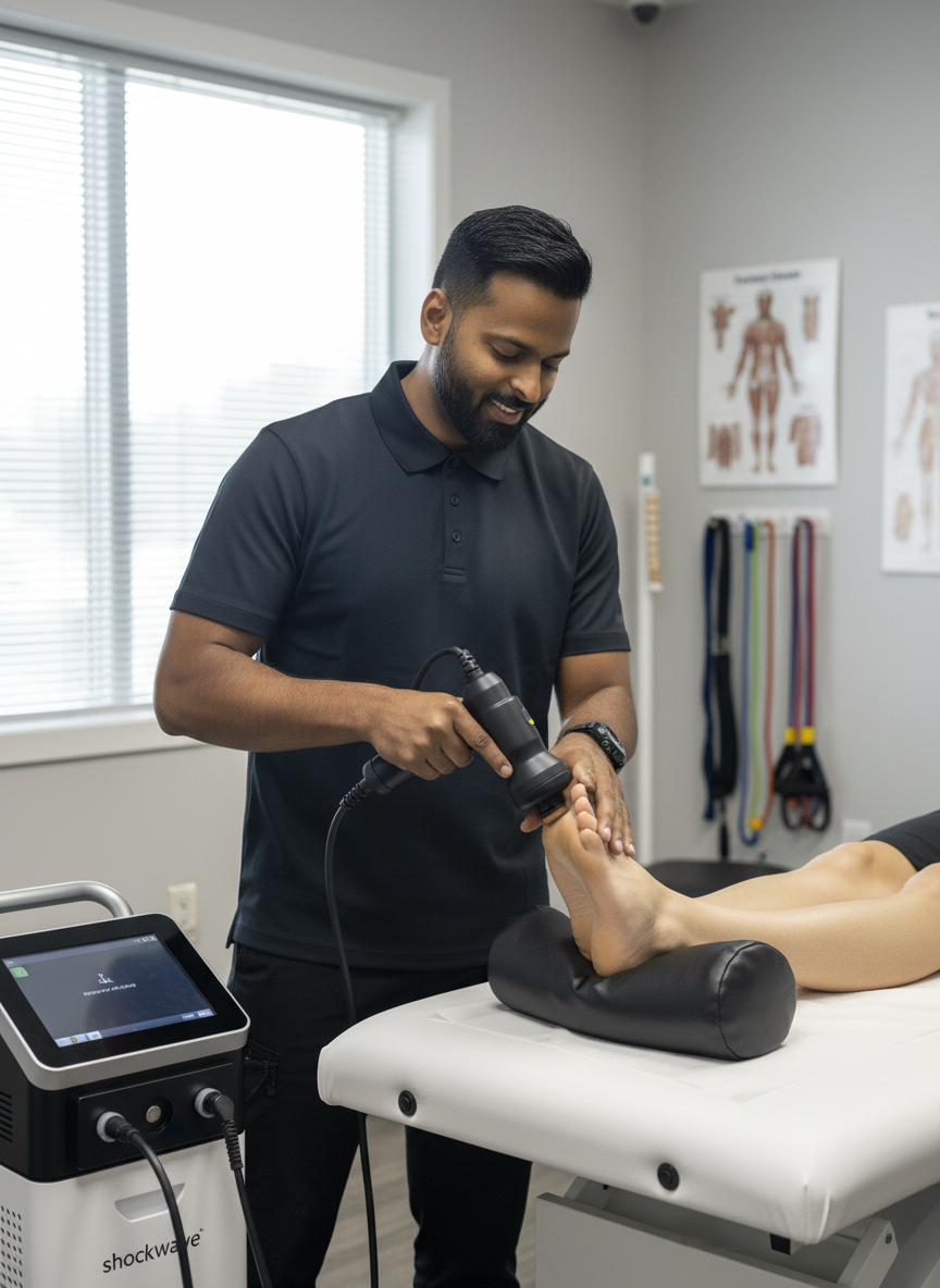
The treatment involves delivering high-energy acoustic sound waves directly into the injured tissue via a specialized clinical device. These waves create controlled micro-trauma within the fascia. This mechanical stimulus disrupts chronic, stalled healing cycles and triggers a renewed biological repair cascade. Specifically, it stimulates localized blood flow and the formation of new micro-blood vessels (angiogenesis) in a tissue that has poor natural blood supply. Furthermore, the acoustic pulses help to deplete Substance P—a neurotransmitter responsible for relaying pain signals—offering patients functional relief as the tissue rebuilds.
Custom Orthotics and Biomechanical Offloading
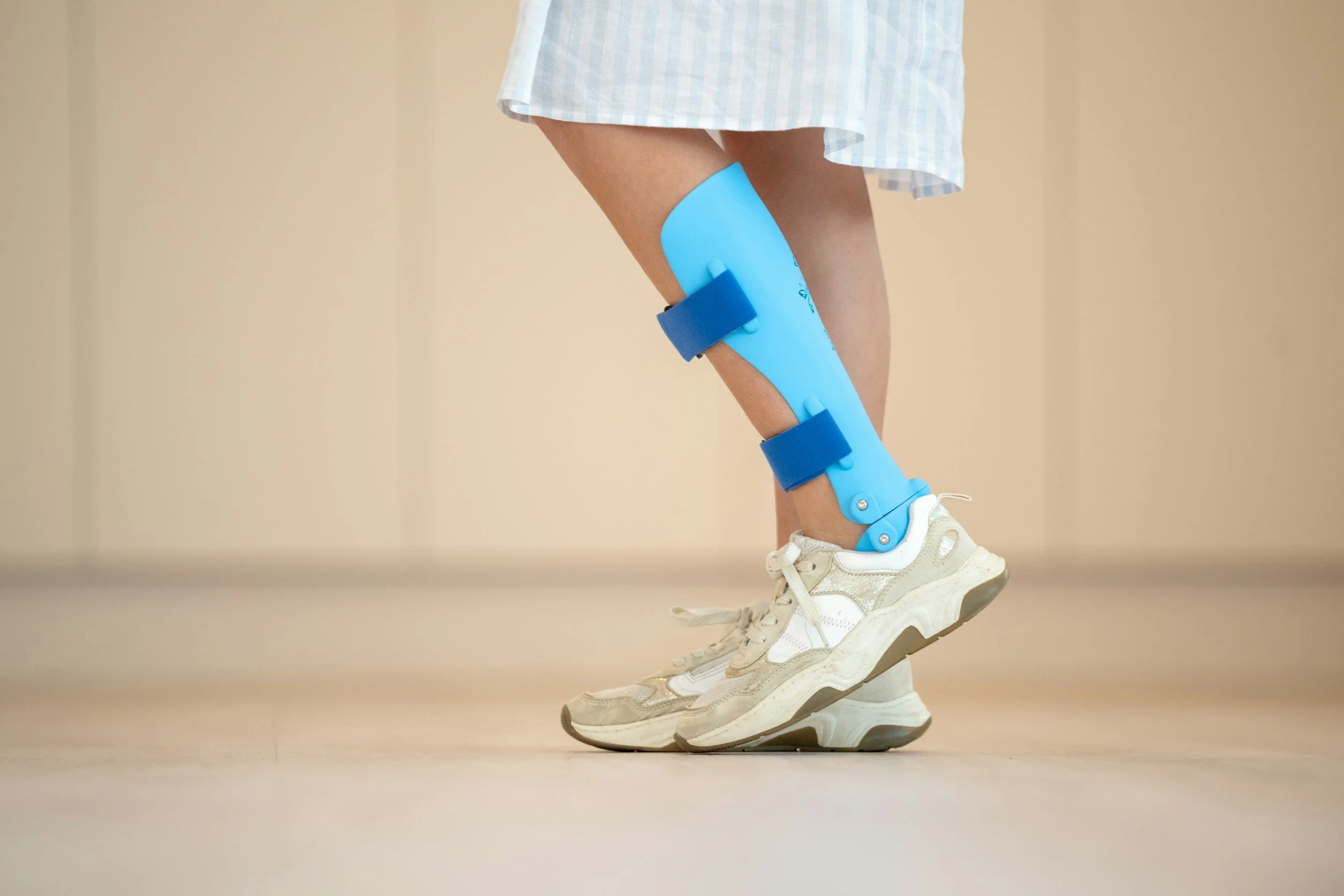
For patients whose plantar fasciitis is fundamentally driven by structural biomechanical issues—such as excessive pronation (inward rolling of the foot) or unusually high, rigid arches—custom orthotics provide necessary mechanical support.
Unlike generic, over-the-counter gel inserts which merely compress under body weight, custom orthotics are prescribed medical devices cast to the exact contours of the patient's foot while held in a neutral, biomechanically optimal position. By purposefully redistributing the daily load across the entire surface area of the foot and correcting inward collapse during the walking cycle, orthotics successfully reduce the daily mechanical strain on the healing fascia, allowing it the physiological space it requires to heal.
Taping and Home Management
Between clinical visits, managing daily loads is critical. Our physiotherapists frequently utilize specialized taping techniques, such as low-dye taping, to artificially support the longitudinal arch and compress the heel pad. This temporary strapping acts as an external ligament, reducing the stretch placed on the fascia during essential daily walking.
Patients are also educated on home-management strategies, including rolling the arch of the foot over a frozen water bottle to manage acute inflammatory flare-ups, and the strict avoidance of walking barefoot on hard surfaces, particularly first thing in the morning.
Corrective Exercise Protocols: High-Load Strength Training
Passive treatments alone are rarely sufficient for long-term resolution. The plantar fascia must be physically loaded to build tensile strength and align new collagen fibers correctly.
A cornerstone of modern evidence-based physiotherapy for this condition involves high-load strength training. By placing a rolled towel under the toes to engage the Windlass Mechanism (a natural tightening of the fascia when the toes are bent upward) and performing slow, heavily loaded calf raises, the tissue is subjected to high-load eccentric strengthening.
A typical protocol involves moving very slowly: 3 seconds raising up, a 2-second hold at the top, and 3 seconds lowering down. This slow, controlled tension under heavy load signals the body to lay down stronger, more resilient connective tissue capable of handling the high-impact demands of running, jumping, and daily occupational life.
Comparing Interventions for Plantar Fasciitis
Treatment Modality
Primary Mechanism of Action
Clinical Application
Soft Tissue Therapy
Reduces muscular and fascial tension in the interconnected posterior chain (calves/Achilles).
Immediate symptom management, improving ankle mobility, and reducing "pull" on the heel.
Shockwave Therapy
Stimulates cellular metabolism, breaks down calcifications, and induces local angiogenesis (blood flow).
Chronic, stubborn fascial pain (lasting > 3 months) that resists conservative stretching.
Custom Orthotics
Mechanically corrects foot posture, prevents excessive pronation, and redistributes weight-bearing load.
Long-term biomechanical management and off-loading of the medial arch.
Clinical Taping
Provides temporary structural support to the longitudinal arch.
Acute pain management during essential daily weight-bearing activities.
Corrective Exercises
Increases load tolerance and guides the alignment of new collagen fibers through eccentric loading.
Long-term functional rehabilitation, rebuilding tensile strength, and injury prevention.
Author BiographyWritten by Mr. Sanjay Attwala (BSC, MSC, RPT), Registered Physiotherapist. Sanjay Attwala manages patient care at Rehab Mechanics (S. Attwala Physiotherapy Professional Corporation) located at 68 Abell Street, Toronto. He is in good standing with the College of Physiotherapists of Ontario (CPO). Learn more about our clinical team here.
Medical Disclaimer:The content provided in this article is for general educational and informational purposes only and does not constitute formal medical advice. Individual physiological responses to physiotherapy vary, and Rehab Mechanics does not guarantee specific treatment outcomes. An in-person assessment is legally and clinically required to develop an individualized treatment plan and obtain informed consent before commencing care.
Do I Absolutely Need Surgery to Fix a Torn Meniscus?
No. The vast majority of degenerative meniscus tears heal entirely without surgery. An aggressive, non-operative physiotherapy protocol of joint unloading, quadriceps strengthening, and neuromuscular retraining stabilizes the knee, permanently eliminates catching pain, and prevents early-onset osteoarthritis.
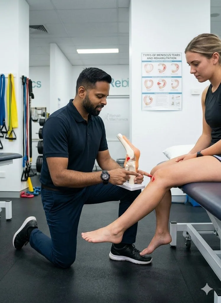
The Misunderstanding of Knee Cartilage Injuries
An MRI reading of a "torn meniscus" strikes fear into the hearts of active Torontonians. Whether you twisted your knee playing in a recreational soccer league at Lamport Stadium, or simply felt a sharp pop while crouching down to pick up a box in your Queen West apartment, the immediate assumption is that a torn tissue must be surgically cut out or sewn back together.
For decades, orthopedic surgeons routinely performed arthroscopic surgeries to "clean up" torn menisci. However, modern medical imaging and rigorous clinical trials have completely flipped this narrative.
Unless your knee is physically, immovably locked in place, rushing into surgery is often highly detrimental. Removing cartilage rapidly accelerates joint degeneration. At Rehab Mechanics, we prioritize identifying the true biomechanical cause of your knee pain, utilizing advanced conservative rehabilitation to stabilize the joint and save your natural cartilage.
Structural Analysis of the Meniscus
To understand why conservative physiotherapy is the international standard of care, we must perform a detailed biomechanical analysis of the knee joint and its internal shock absorbers.
The Anatomy of the Knee's Shock Absorbers
The knee is a massive hinge joint where your thigh bone (femur) rests on top of your shin bone (tibia).
The C-Shaped Cushions: Sandwiched between these two bones are two pieces of tough, rubbery fibrocartilage known as the medial (inside) and lateral (outside) menisci.
Mechanical Function: They act as crucial shock absorbers, distributing your body weight evenly across the joint surface and preventing the bones from grinding directly against one another.
The Blood Supply Dilemma (Red Zone vs. White Zone)
The location of the tear dictates the healing potential.
The Red-Red Zone: The outer third of the meniscus has a rich blood supply. Tears here can physically heal and scar back together with proper load management.
The White-White Zone: The inner two-thirds lack a direct blood supply. Tears here will never "knit" back together. However, they do not need to. Physiotherapy trains the surrounding muscles to absorb the shock, rendering the tear entirely asymptomatic.
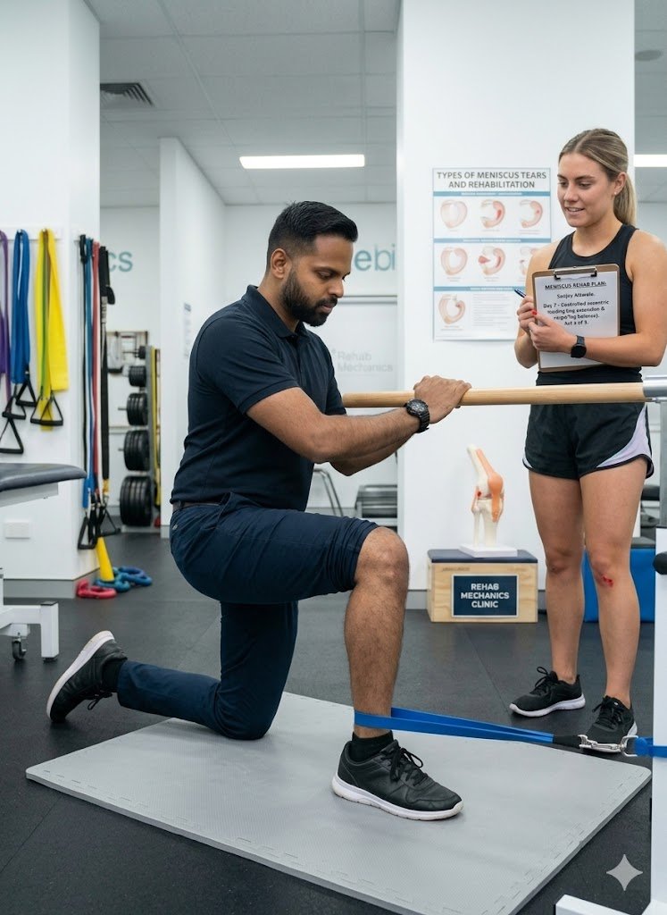
Mechanisms of Injury: Acute vs. Degenerative
Not all tears are created equal, and they demand very different clinical approaches.
Acute Traumatic Tears
The Cause: High-velocity twisting. Planting your foot firmly on the ground and violently rotating your torso (common in basketball, skiing, or slipping on Toronto ice).
The Pathology: These often result in large "bucket-handle" tears that can flip into the joint space, sometimes requiring surgical intervention if they cause a hard mechanical lock.
Chronic Degenerative Tears
The Cause: Wear and tear over decades. As we age, the cartilage naturally dries out and frays.
The Reality: The vast majority of meniscus tears in adults over 35 are degenerative. Studies show that thousands of people walking the streets of Toronto have torn menisci right now and experience zero pain. The tear is a symptom of joint overload, not the root cause.
Identifying the Clinical Red Flags
Meniscus tears present with a highly specific set of mechanical symptoms that differ greatly from standard runner's knee or a basic ligament sprain.
The "Catching" Sensation: A sharp, localized pain when you bend or straighten the knee, feeling as though something is momentarily stuck inside the joint hinge.
Joint Line Tenderness: Pressing your finger precisely into the soft space between the bones on the side of your knee produces exquisite pain.
Delayed Effusion: Unlike an ACL tear which swells instantly, a meniscus tear often causes the knee to slowly puff up with fluid 24 to 48 hours after the initial tweak.
Deep Flexion Pain: An inability to perform a deep squat or kneel on the floor without a sharp pinching sensation in the back of the knee.
The Physiotherapy Protocol: Bulletproofing the Knee
Our clinical approach at Rehab Mechanics focuses strictly on mechanical decompression, reducing joint effusion, and building massive structural support around the damaged cartilage.
1. Joint Decompression and Effusion Management
We cannot build strength if the joint is massively swollen and chemically irritated.
Manual Therapy: We use targeted soft tissue release on the calf, hamstring, and IT band to reduce the compressive forces pulling the knee joint tightly together.
Lymphatic Drainage: Utilizing manual techniques and specific modalities to push the stagnant joint fluid (effusion) out of the knee capsule, instantly reducing the feeling of pressure and restoring range of motion.
2. Neuromuscular Quadriceps Reactivation
Swelling inside the knee triggers a reflex that actively shuts down your quadriceps muscle (Arthrogenic Muscle Inhibition).
Isometric Activation: We utilize pain-free isometric holds and Neuromuscular Electrical Stimulation (NMES) to force the quadriceps to fire, overriding the brain's shutdown signal.
VMO Targeting: Specific focus is placed on the vastus medialis oblique (the teardrop muscle on the inside of the thigh) to ensure the kneecap tracks perfectly, taking pressure off the medial meniscus.
3. Closed Kinetic Chain Strength and Proprioception
Once the swelling is gone, we must teach the leg how to absorb gravity without relying on the damaged cartilage.
Gluteal Stabilization: Strengthening the side glutes (gluteus medius) prevents the knee from caving inward (valgus collapse) during walking or running, which is the primary movement that crushes the meniscus.
Proprioceptive Retraining: Utilizing balance boards and uneven surfaces to retrain the micro-stabilizers in the knee, ensuring the joint reacts instantly to sudden changes in direction without twisting dangerously.
Primary Source Proof
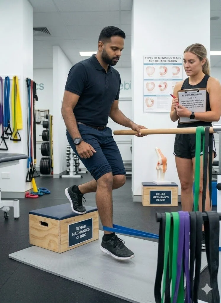
Massive, landmark orthopedic trials (including the ESCAPE trial) have definitively proven that for degenerative meniscus tears, structured, supervised physical therapy yields pain relief and functional outcomes that are identical to arthroscopic partial meniscectomy surgery, without subjecting the patient to surgical risks or accelerated osteoarthritis.
[PDF Action Button] Download Clinical Evidence: Physical Therapy versus Arthroscopic Partial Meniscectomy for Meniscal Tears
Save Your Cartilage Today
You do not have to accept an invasive surgery to fix a clicking, painful knee. By aggressively strengthening the muscular scaffolding around the joint, your body can adapt, compensate, and completely resolve the pain of a torn meniscus.
Book your comprehensive knee assessment today. We will determine the exact mechanical nature of your tear and build a customized roadmap to recovery. We are conveniently located inside the Prime Medical Centre at 68 Abell Street, easily accessible in Toronto Queen West.
Contact us to schedule your appointment:
Email: info@rehabmechanics.com
Phone: (416) 533-3900
About the Author
Mr. Sanjay Attwala (B.Sc., M.Sc., RPT) is a Registered Physiotherapist, clinical director, and the founder of Rehab Mechanics in Toronto. With over 15 years of registered clinical practice and a deep specialization in complex musculoskeletal rehabilitation, Sanjay synthesizes rigorous international academic training with advanced evidence-based therapeutics to guide his clinical practice and patient education initiatives.
Academic Background & Credentials
Master of Science (M.Sc.) in Physiotherapy – University of Keele, United Kingdom (2010).
Bachelor of Science (B.Sc.) – University of Waterloo, Ontario, Canada.
Registered Physiotherapist (RPT) – Regulated health professional in excellent standing with the College of Physiotherapists of Ontario (CPO).
Corporate Entity – Operating officially under the S. Attwala Physiotherapy Professional Corporation with a DBA of Rehab Mechanics.
Clinical Expertise & Philosophy Sanjay’s clinical approach rejects passive symptom management in favor of identifying underlying biomechanical root causes. His diverse expertise spans advanced manual therapies, personalized corrective exercise prescription, and modern physical modalities. At the Rehab Mechanics Toronto Queen West clinic, he routinely diagnoses and treats complex conditions including:
Spinal & Discogenic Pathology – Cervical, thoracic, and lumbar disc injuries, sciatica, and sacroiliac joint (SIJ) dysfunction.
Upper & Lower Extremity Injuries – Rotator cuff tears, frozen shoulder, tennis/golfer’s elbow, carpal tunnel syndrome, and complex ankle/foot pathologies.
Perinatal & Pelvic Health Rehabilitation – Specialized assessment and rehabilitation protocols tailored specifically for women during pregnancy and the post-partum period, addressing pelvic girdle pain, diastasis recti, and core stabilization.
Specialized Rehabilitation – Pelvic health therapy, TMJ dysfunction, post-surgical rehabilitation (including Total Hip and Total Knee Replacements), and custom orthotics dispensing.
Shockwave Therapy: with advanced cutting edge technological devices to suit your needs.
Interdisciplinary Practice & Patient Care Sanjay practices an integrated model of healthcare, working closely alongside medical doctors inside the Prime Medical Centre on Abell Street to streamline patient recovery pathways. He maintains a human-centric, communication-first clinical framework, ensuring that care remains fully customized rather than automated.
His clinical caseload encompasses a broad operational spectrum under Ontario's regulatory frameworks, including:
Motor Vehicle Accident (MVA) Claims – Rehabilitation navigating Ontario’s statutory accident benefits schedule.
Workplace Safety and Insurance Board (WSIB) – Occupational injury management and return-to-work screening.
Extended Health Care (EHC) & Private Practice – Multi-tier insurance coordination and long-term athletic development plans.
Commitment to Research & Community Outside of his clinical caseload at Rehab Mechanics and his additional practice affiliations in Etobicoke, Sanjay is an active health writer and community educator. He translates contemporary peer-reviewed medical research into accessible, actionable guidance on his professional blog. As a dedicated father and husband, he mirrors his professional advice in his personal life, focusing on structural mobility, cross-training, and longevity to help his family and his community thrive. Naturally he takes he a keen interest in rehabilitation for women who are pregnant and post-partum.
Disclaimer: The information provided on this blog is intended for educational and informational purposes only. It does not constitute medical advice, diagnosis, or a treatment plan. Always seek the direct advice of a Registered Physiotherapist, physician, or other qualified health provider regarding any medical condition or physical rehabilitation routine.
Your Achilles heel could be too much rest and not enough physio
Can Physiotherapy Cure Achilles Tendonitis Without Months of Rest?
Yes. Physiotherapy cures Achilles tendonitis without complete rest. Utilizing progressive heavy slow resistance (HSR) training, localized shockwave therapy, and biomechanical correction, physiotherapy regenerates degenerated tendon fibers, restores ankle mobility, and builds load capacity faster than passive immobilization.
The Danger of Ignoring Heel Pain in an Active City
Toronto possesses a massive, highly competitive, and deeply passionate running culture. Whether you are training through the winter for the Toronto Waterfront Marathon, doing sprint intervals along the Martin Goodman Trail, playing weekend basketball in Trinity Bellwoods, or simply power-walking your daily commute through Queen West, your feet and ankles take a massive, repetitive beating on unforgiving concrete.
When a dull, stiff ache develops at the back of your heel, the immediate reaction of most urban athletes is to try and push through it. You might skip a run, ice the heel for a day, take a few anti-inflammatories, and assume it will fade. When the pain finally becomes too sharp to ignore—often forcing a visible limp—the traditional medical advice provided at generic walk-in clinics is typically to stop all activity, rest for six to eight weeks, and wait.
The "Wait and See" Fallacy
However, complete rest is the absolute worst thing you can do for a chronic tendon injury. Tendons are mechanical tissues; they require mechanical load to heal and maintain their structural integrity.
Passive rest simply causes the healthy parts of the tendon to weaken and atrophy. While resting might temporarily reduce the acute inflammation and pain, it guarantees that the tendon will be entirely unprepared to handle the force of your body weight the moment you step back onto the pavement, resulting in a frustrating, immediate relapse.
The Psychological Toll of the Sidelines
For many Torontonians, running or recreational sports are not just physical activities; they are primary stress relievers and social outlets. Being sidelined for months creates intense frustration, anxiety about losing cardiovascular fitness, and fear that the pain will become a permanent, chronic condition. At Rehab Mechanics, our specialized foot and ankle protocols treat Achilles pain by actively rebuilding the tissue, keeping you moving, modifying your load, and keeping you engaged in your active lifestyle while you heal.
Structural Analysis of the Achilles Tendon
To effectively cure this condition, we must move past outdated terms like "tendonitis" and understand the precise cellular failure happening at the back of your ankle. Tendons do not behave like muscles, and they cannot be treated like muscles.
The Anatomy of the Ultimate Spring
The Achilles tendon is the thickest and strongest tendon in the human body. It acts as the vital bridge connecting your powerful calf muscles (the superficial gastrocnemius and the deep soleus) directly into your heel bone (the calcaneus).
The Energy Storage Mechanism: The Achilles is a biological spring. When you run, jump, or hop, the Achilles stretches like a massive, high-tension elastic band, storing kinetic energy. When you push off your toes, it violently snaps back, releasing that stored energy to propel your entire body weight forward.
The Paratenon Sheath: Unlike some tendons that sit inside a synovial sheath, the Achilles is surrounded by a thin layer of highly vascularized tissue called the paratenon. Friction between the tendon and this sheath is often the first source of acute pain.
The Vascular Weakness: Despite its massive tensile strength, the Achilles has a notoriously poor blood supply, particularly in the "mid-portion" region about 2 to 6 centimeters above the heel attachment. This lack of robust blood flow makes it highly susceptible to chronic, failed healing.
Tendonitis vs. Tendinopathy (The Degeneration Cycle)
Most patients diagnosed with Achilles "tendonitis" do not actually have active inflammation. Tendinopathy exists on a specific continuum (known in orthopedic science as the Cook and Purdam continuum).
1. The Reactive Phase (Acute Tendonitis)
Initially, after a massive, sudden spike in training volume, the tendon and its sheath may swell and become acutely inflamed. The tendon swells by drawing in water to become thicker and stiffer in an attempt to handle the sudden overload. This is true tendonitis, and it is highly painful but easily reversible with brief load modification.
2. Tendon Dysrepair
If the extreme load continues without proper recovery days, the tendon cells (tenocytes) become overworked and exhausted. The collagen matrix begins to separate, and the internal structure starts to break down microscopically.
3. The Degenerative Phase (Chronic Tendinosis)
The cells enter a state of panic. Instead of laying down strong, perfectly parallel Type I collagen fibers, the body patches the micro-tears with weak, disorganized, chaotic Type III scar tissue.
The tendon physically thickens, forms painful, hardened nodules (bumps you can feel), and structurally degenerates. Medical imaging will show a dark, swollen mass of dysfunctional tissue. At this stage, anti-inflammatory medications are entirely useless because there is no inflammation left; there is only cellular decay.
Mid-Portion vs. Insertional Tendinopathy
Locating the exact epicenter of your pain is clinically vital, as the treatment protocol changes drastically depending on the location.
Mid-Portion Tendinopathy: The pain is located 2 to 6 cm above the heel bone. It is usually caused by excessive spring loading (running/jumping). It responds exceptionally well to heavy drop-stretching (eccentric work).
Insertional Tendinopathy: The pain is located exactly where the tendon attaches to the heel bone. This is often complicated by a bone spur (Haglund's deformity) or an inflamed bursa sac. This specific variation reacts terribly to deep stretching, requiring a highly modified, flat-ground strengthening protocol.
Common Biomechanical Triggers
Why did the tendon fail in the first place? It is rarely just from "running too much." It is almost always a combination of environmental load and internal biomechanics.
Training Load Errors: This accounts for 80% of Achilles injuries. Doing "too much, too soon, too fast." Transitioning rapidly from running on a soft indoor treadmill all winter to pounding cold outdoor concrete in the spring without an adjustment period is a massive trigger.
Ankle Dorsiflexion Restriction: If your ankle joint is incredibly stiff and cannot bend forward deeply (often due to previous sprains), the Achilles is forced to absorb a massive, unnatural shearing force with every single step, rather than a smooth, linear stretch.
Soleus Muscle Weakness: The soleus (the deep calf muscle) handles up to 8 times your body weight when running. If it is weak, it cannot absorb the shock, transferring 100% of the destructive force directly into the tendon.
Footwear Alterations: Transitioning too quickly from high-cushion running shoes with a massive "heel drop" to trendy "zero-drop" minimalist shoes aggressively alters the pulling angle of the Achilles, forcing it to stretch further than it is adapted to.
Identifying the Clinical Red Flags
Achilles tendinopathy presents with a highly specific, localized set of symptoms that differentiate it from generic calf cramps or plantar fasciitis.
The Morning Hobble: The absolute hallmark symptom. The first few steps out of bed in the morning are agonizingly stiff and painful, forcing you to limp to the bathroom. As the tendon warms up, it becomes pliable again.
The "Warm-Up" Effect (The Trickster): The pain often decreases after 10 minutes of walking or light jogging. This leads athletes to falsely believe the injury is minor and they can train through it. However, the pain returns violently and throbs intensely a few hours after the activity stops.
Post-Rest Stiffness: Beyond the morning, the tendon will drastically stiffen up after sitting at a desk for three hours. Standing up to walk to the printer causes a sharp, grabbing pain.
The Pinch Test: Squeezing the tendon tightly between your thumb and index finger causes sharp, localized pain, and you may physically feel a thick, hardened "bump" or crepitus (a crunchy, squeaky feeling) on the tendon.
The Physiotherapy Protocol: Active Tissue Regeneration
At Rehab Mechanics, we reject passive treatments. Ultrasound, resting in walking boots, and steroid injections (which carry a massive risk of actually rupturing the tendon) are not the answer. Tendons are mechanical structures; they must be fixed with progressive, heavy mechanical load.
1. Advanced Modalities (Neovascularization)
Because the degenerated tendon lacks blood flow, our immediate goal is to stimulate cellular repair at the microscopic level.
Extracorporeal Shockwave Therapy (ESWT): We use high-energy acoustic sound waves to physically shatter the disorganized scar tissue. More importantly, this mechanical stimulus triggers mechanotransduction, forcing the body to grow brand-new capillary blood vessels (neovascularization) directly into the deadened, chronic tendon fibers, flooding the area with healing nutrients.
2. Heavy Slow Resistance (HSR) Training
This is the absolute core of a permanent cure. We must systematically load the tendon to teach the body how to lay down new, perfectly parallel collagen fibers.
Phase 1: Isometric Loading (Pain Relief): We begin with heavy, static holds. You might hold a double-leg calf raise halfway up for 45 seconds. Isometrics are scientifically proven to immediately down-regulate tendon pain and safely engage the muscle without irritating the joint through movement.
Phase 2: Eccentric and Concentric Isotonics: We meticulously guide you through Heavy Slow Resistance protocols. Using a leg press or weighted calf machine, you perform very slow, heavy repetitions (3 seconds up, 3 seconds down). This heavy tension is the most potent stimulus for tendon regeneration, far outperforming bodyweight exercises.
Phase 3: Plyometric and Energy Storage Phase: You cannot return to running just because you can do a calf raise. Running is jumping. We must re-train the tendon's spring capacity using pogo jumps, skipping, and rapid drop-jumps to ensure the Achilles can handle rapid, violent force absorption.
3. Biomechanical Correction and Manual Therapy
We must fix the environmental factors that caused the overload to prevent a recurrence.
Joint Mobilization: Our Registered Physiotherapists apply deep, manual orthopedic glides to the talocrural (ankle) joint to restore full dorsiflexion, instantly taking the mechanical shearing strain off the Achilles.
Deep Myofascial Release: Stripping out the knotted trigger points in the gastrocnemius and soleus to reduce the constant, resting tension pulling on the heel.
Custom Orthotics: If severe overpronation (flat feet) is driving a whipping motion through the tendon, we will cast and dispense custom-molded orthotics to correct your foot posture and alter the biomechanical pull on the heel.
4. Return to Run (RTR) Programming
We do not just tell you to "try running and see how it feels." We utilize specific clinical criteria.
Hop Testing: You must be able to perform 20 single-leg hops with equal height and speed to your uninjured side without pain.
Cadence Manipulation: We may use a metronome to increase your running step rate (cadence) by 5-10%. Taking slightly shorter, faster steps drastically reduces the heavy, bounding load on the Achilles tendon.
Graduated Loading: We implement specific run/walk intervals, ensuring your tendon adapts to the pavement slowly, avoiding the "boom and bust" cycle of reinjury.
Primary Source Proof
Decades of peer-reviewed sports medicine research conclusively demonstrate that a progressive, heavy-load exercise protocol (specifically Heavy Slow Resistance training), often combined with extracorporeal shockwave therapy, is the international gold standard for completely resolving chronic Achilles tendinopathy.
[PDF Action Button] Download Clinical Evidence: The Efficacy of Heavy Slow Resistance Training in the Management of Achilles Tendinopathy
Rebuild Your Load Capacity Today
You do not have to give up running, abandon your athletic goals, or accept chronic, limping heel stiffness as a permanent reality. Expert, targeted physical rehabilitation can reverse the cellular degeneration of your tendon, rebuild your lower leg strength, and get you back to your active urban lifestyle without pain.
Book your comprehensive foot and ankle assessment today. We are conveniently located inside the Prime Medical Centre at 68 Abell Street, easily accessible in Toronto Queen West.
Contact us to schedule your appointment:
Email: info@rehabmechanics.com
Phone: (416) 533-3900
About the Author
Mr. Sanjay Attwala (B.Sc., M.Sc., RPT) is a Registered Physiotherapist, clinical director, and the founder of Rehab Mechanics in Toronto. With over 15 years of registered clinical practice and a deep specialization in complex musculoskeletal rehabilitation, Sanjay synthesizes rigorous international academic training with advanced evidence-based therapeutics to guide his clinical practice and patient education initiatives.
Academic Background & Credentials
Master of Science (M.Sc.) in Physiotherapy – University of Keele, United Kingdom (2010).
Bachelor of Science (B.Sc.) – University of Waterloo, Ontario, Canada.
Registered Physiotherapist (RPT) – Regulated health professional in excellent standing with the College of Physiotherapists of Ontario (CPO).
Corporate Entity – Operating officially under the S. Attwala Physiotherapy Professional Corporation with a DBA of Rehab Mechanics.
Clinical Expertise & Philosophy
Sanjay’s clinical approach rejects passive symptom management in favor of identifying underlying biomechanical root causes. His diverse expertise spans advanced manual therapies, personalized corrective exercise prescription, and modern physical modalities. At the Rehab Mechanics Toronto Queen West clinic, he routinely diagnoses and treats complex conditions including:
Spinal & Discogenic Pathology – Cervical, thoracic, and lumbar disc injuries, sciatica, and sacroiliac joint (SIJ) dysfunction.
Upper & Lower Extremity Injuries – Rotator cuff tears, frozen shoulder, tennis/golfer’s elbow, carpal tunnel syndrome, and complex ankle/foot pathologies.
Perinatal & Pelvic Health Rehabilitation – Specialized assessment and rehabilitation protocols tailored specifically for women during pregnancy and the post-partum period, addressing pelvic girdle pain, diastasis recti, and core stabilization.
Specialized Rehabilitation – Pelvic health therapy, TMJ dysfunction, post-surgical rehabilitation (including Total Hip and Total Knee Replacements), and custom orthotics dispensing.
Shockwave Therapy: with advanced cutting edge technological devices to suit your needs.
Interdisciplinary Practice & Patient Care
Sanjay practices an integrated model of healthcare, working closely alongside medical doctors inside the Prime Medical Centre on Abell Street to streamline patient recovery pathways. He maintains a human-centric, communication-first clinical framework, ensuring that care remains fully customized rather than automated.
His clinical caseload encompasses a broad operational spectrum under Ontario's regulatory frameworks, including:
Motor Vehicle Accident (MVA) Claims – Rehabilitation navigating Ontario’s statutory accident benefits schedule.
Workplace Safety and Insurance Board (WSIB) – Occupational injury management and return-to-work screening.
Extended Health Care (EHC) & Private Practice – Multi-tier insurance coordination and long-term athletic development plans.
Commitment to Research & Community
Outside of his clinical caseload at Rehab Mechanics and his additional practice affiliations in Etobicoke, Sanjay is an active health writer and community educator. He translates contemporary peer-reviewed medical research into accessible, actionable guidance on his professional blog. As a dedicated father and husband, he mirrors his professional advice in his personal life, focusing on structural mobility, cross-training, and longevity to help his family and his community thrive. Naturally he takes he a keen interest in rehabilitation for women who are pregnant and post-partum.
Disclaimer: The information provided on this blog is intended for educational and informational purposes only. It does not constitute medical advice, diagnosis, or a treatment plan. Always seek the direct advice of a Registered Physiotherapist, physician, or other qualified health provider regarding any medical condition or physical rehabilitation routine.
Sacroiliac joint dysfunction
Is My Lingering Lower Back Pain Actually a Sacroiliac Joint Dysfunction?
Yes. Lingering, one-sided lower back pain is frequently misdiagnosed sacroiliac joint (SIJ) dysfunction. Specialized physiotherapy utilizes precise joint mobilization, targeted pelvic stabilization exercises, and muscle energy techniques to correct pelvic asymmetry, instantly relieving unilateral back pain and sharp sciatic-like symptoms.
The Diagnostic Confusion of Lower Back Pain
If you have been struggling with lower back pain that just will not go away despite months of standard core exercises, generic yoga stretching, and multiple trips to various practitioners, you are likely treating the wrong structural tissue.
Many urban professionals in Toronto spend countless hours sitting in ergonomic chairs during hybrid work weeks, enduring long commutes on the Gardiner Expressway, or standing on hard concrete floors. When a deep, nagging ache develops in the lower back, the immediate assumption is often a slipped lumbar disc, a pinched sciatic nerve, or a pulled lumbar muscle.
Patients frequently spend hundreds of dollars on generic massage therapy or chiropractic "adjustments" that provide only a few hours of relief before the exact same pain returns. This relentless cycle of temporary relief and immediate relapse takes a massive psychological toll, leading to deep frustration, anxiety about movement, and an avoidance of the active city lifestyle you love.
However, if your pain is heavily concentrated on one side, located very low in the back (right over the bony "dimples" just above your glutes), and sharply spikes when you transition from sitting to standing or when climbing the stairs at the TTC subway station, you are likely dealing with Sacroiliac Joint (SIJ) Dysfunction.
Because the SI joint shares nerve pathways with the lower lumbar spine and hips, it is an incredibly common—and frequently overlooked—imitator of sciatica. At Rehab Mechanics in Queen West, we specialize in differential diagnosis. We do not just treat generic "back pain" with hot packs and generic stretches; we isolate the specific biomechanical failure in your pelvic girdle to provide immediate, targeted relief and long-term structural resilience.
Structural Analysis of the Sacroiliac Joint
To understand why generic back stretches completely fail to fix SI joint pain—and can sometimes even make it worse—we must perform a highly detailed anatomical analysis of the pelvic ring and the complex mechanics of how your body transfers weight against gravity.
The Anatomy of the Pelvic Keystone
Your pelvis is the absolute foundation of your skeletal structure. It is composed of two large iliac bones (your hip bones) and the sacrum (the shield-shaped, triangular bone at the absolute base of your spine). The sacroiliac joints are the two vertical connection points where the sacrum meets the iliac bones on either side.
Think of the sacrum as the keystone in a Roman arch. It locks the two sides of the pelvis together, allowing your upper body to rest securely on top of your lower body.
The Shock Absorbers: Unlike your knee, hip, or shoulder, the SI joint is not designed for massive, sweeping movements. It is a highly stable, exceptionally rigid joint designed to absorb the massive, repeated shock of your upper body weight and transfer it safely down into your legs every single time your heel strikes the pavement.
Micro-Movements (Nutation and Counternutation): The SI joint only moves about 2 to 4 millimeters. This tiny nodding motion of the sacrum—called nutation (nodding forward) and counternutation (nodding backward)—is just enough to dissipate impact forces safely. When this micro-movement is lost, or becomes excessive, dysfunction begins.
The Dual Stabilization System (Form and Force Closure)
Because the SI joint bears so much weight, it requires a massive, two-part stabilization system to prevent it from collapsing.
1. Form Closure (Bones and Ligaments)
This is the passive stability of the joint. The SI joint is held together by its natural, interlocking puzzle-piece shape (the articular surfaces have ridges and depressions that lock together).
The Ligamentous Network: It is further reinforced by a network of some of the thickest, strongest, and most dense ligaments in the human body, including the sacrospinous, sacrotuberous, and interosseous sacroiliac ligaments. When these ligaments are healthy, the joint is tightly bound.
2. Force Closure (Muscles and Fascia)
This is the active, dynamic stability of the joint. Because the ligaments cannot hold the joint together during heavy athletic movement on their own, your muscles must act as an active compression system.
The Myofascial Slings: The SI joint is violently compressed and stabilized by massive "X" patterns of muscles crossing your back and front, known as myofascial slings.
The Posterior Oblique Sling: For example, your right latissimus dorsi (back muscle) connects through the thick thoracolumbar fascia directly into your left gluteus maximus. When you walk, these opposite muscles fire simultaneously, pulling the fascia taut and squeezing the SI joints together for safe weight transfer.
Mechanisms of SIJ Dysfunction
Pain occurs when the delicate, precise balance of mobility and stability in this joint is disrupted. This biomechanical failure usually falls into two highly specific, contrasting mechanical categories:
1. SIJ Hypermobility (Too Much Movement)
The Cause: This is incredibly common in young women, often triggered by the hormonal ligament laxity during pregnancy (the release of the hormone relaxin softens the pelvic ligaments to prepare for childbirth). It can also be caused by a severe fall directly onto the buttocks on winter ice, or repetitive high-impact rotational sports like golf, tennis, or hockey.
The Pathology: The strong "Form Closure" ligaments are permanently overstretched or micro-torn. The joint moves too much (perhaps 5 or 6 millimeters instead of 2), causing a painful, inflammatory shearing force.
The Muscular Response: The surrounding muscles (specifically the piriformis, hamstrings, and lower back erectors) go into a massive, chronic, exhausting spasm to try and hold the unstable joint together. Stretching these tight muscles actually makes the hypermobility worse, which is why yoga often aggravates this specific condition.
2. SIJ Hypomobility (Too Little Movement)
The Cause: Usually the result of a sedentary desk lifestyle, chronically carrying a heavy laptop bag on one shoulder, having an undiagnosed leg-length discrepancy, or poor postural habits (like always shifting your weight entirely to your right leg while standing in line at a coffee shop).
The Pathology: The joint becomes rigidly locked or slightly rotated out of its normal alignment (known clinically as an upslip, outflare, or anterior torsion). This physical locking jams the cartilaginous joint surfaces violently together, creating sharp, localized inflammation and completely shutting down the natural shock-absorbing micro-movements.
Identifying the Clinical Red Flags
Accurately differentiating SIJ pain from a lumbar disc bulge or true sciatic nerve compression is the most critical step for recovery. Treating a locked pelvis like a herniated disc will yield zero results. If you experience the following highly specific symptoms, the SI joint is the primary structural suspect:
The "Dimple" Pain (Fortin Finger Test): You can point with one single finger directly to the PSIS (the bony dimple on your lower back) as the absolute epicenter of the pain. The pain rarely crosses above the beltline.
Transitional Catching: A sharp, breathtaking, stabbing jolt of pain when altering your posture. This includes rolling over in bed at night, getting out of a low car seat, or standing up after sitting at a desk for an hour.
Unilateral Leg Pain (Pseudosciatica): A deep, heavy ache that radiates into your groin, the front of your thigh, or down the back of your leg. However, unlike true sciatica from a crushed spinal nerve, SI joint referred pain rarely travels below the knee into the calf or foot.
The One-Legged Test: Standing on one leg to put on pants, socks, or shoes is intensely painful, feels structurally weak, or causes a sharp buckling sensation in the hip.
Sitting Intolerance: Sitting on hard surfaces for long periods causes a deep, burning ache in the base of one buttock cheek, forcing you to constantly shift your weight to the opposite side.
The Physiotherapy Intervention: Restoring Pelvic Symmetry
At Rehab Mechanics, we utilize a highly structured, multi-phase clinical pathway to unlock, realign, and permanently stabilize the sacroiliac joint. We do not guess at the source of your pain; we systematically test it.
1. Advanced Diagnostics and Provocation Testing
We use a specialized diagnostic protocol known as a "Cluster Test." We perform a series of five specific orthopedic provocation tests (such as Gaenslen’s Test, the Thigh Thrust, the Sacral Thrust, and the Distraction/Compression Tests). If three out of the five tests accurately reproduce your familiar pain, we have definitively confirmed the SI joint as the pain generator before beginning any manual treatment.
2. Muscle Energy Techniques (MET) and Joint Mobilization
If our assessment reveals that the joint is locked (hypomobile) or rotated out of position, we must realign it mechanically before we can strengthen it.
Patient-Assisted Adjustments (MET): We use Muscle Energy Techniques based on the principle of post-isometric relaxation. Instead of aggressively "cracking" the joint, we place you in a specific position and have you gently push your leg against our manual resistance. This uses your own internal muscular force to smoothly and painlessly rotate the pelvic bones back into symmetrical, neutral alignment.
Grade III/IV Mobilizations: Applying specific, targeted, hands-on manual pressure to the sacrum and ilium to free up restricted connective tissue, break down fascial adhesions, and restore the vital micro-gliding (nutation) motion of the joint.
3. Neuromuscular Core and Gluteal Stabilization
If the joint is hypermobile, or once we have successfully restored alignment to a locked joint, we must aggressively build the muscular "Force Closure" to keep it in place permanently. A passive adjustment is useless if the muscles cannot hold the correction.
Transverse Abdominis Activation: We teach you how to fire your deepest core muscle, the transverse abdominis, which acts as a biological weight belt. When contracted, it violently compresses the SI joints together for immediate stability.
Myofascial Sling Training: We move beyond basic crunches. We prescribe heavy, unilateral (one-sided) exercises like Bulgarian split squats, heavy sled pushes, and asymmetrical farmer's carries. These exercises specifically target the Posterior Oblique Sling (lats and glutes) to build the exact muscle groups responsible for locking the pelvis down during walking and running.
4. Temporary External Support and Ergonomics
While the muscles are rebuilding their strength, we must protect the joint from further mechanical irritation during your daily life.
Sacroiliac Belting: For highly hypermobile patients (especially post-partum mothers), we may properly fit and dispense a specialized, rigid SI joint belt. This belt straps tightly around the bony pelvis, mechanically locking the joints in place to instantly eliminate the painful shearing forces when walking, allowing the inflamed ligaments a chance to scar down and heal.
Sleep and Sitting Ergonomics: We provide actionable lifestyle coaching to prevent nighttime flare-ups. This includes teaching you how to properly prop firm pillows between your knees and ankles to keep the pelvis entirely neutral during side-sleeping, and adjusting your office chair to prevent anterior pelvic tilting.
Primary Source Proof
Clinical guidelines in orthopedic physiotherapy strongly indicate that a combination of manual joint mobilization, specific pelvic stabilization exercises, and accurate differential diagnosis provides the most effective long-term resolution for sacroiliac joint dysfunction, drastically outperforming isolated rest or non-specific lower back stretching.
Download Clinical Evidence: The Efficacy of Manual Therapy and Stabilization Exercises in Sacroiliac Joint Dysfunction
Stop Chasing the Wrong Pain
You do not have to live with sharp, transitional back pain, assume you have a permanent slipped disc, or rely on daily pain medications just to get through your workday. Stop aggressively stretching a joint that actually requires stabilization.
Expert, targeted physical rehabilitation can identify the true biomechanical source of your pelvic pain, correct the mechanical alignment of your keystone joint, and restore your ability to move freely and confidently through the city.
Book your comprehensive spinal and pelvic assessment today. We are conveniently located inside the Prime Medical Centre at 68 Abell Street, providing a modern, easily accessible, ground-floor environment right in the heart of Toronto Queen West.
Contact us to schedule your appointment:
Email: info@rehabmechanics.com
Phone: (416) 533-3900
About the Author
Mr. Sanjay Attwala (B.Sc., M.Sc., RPT) is a Registered Physiotherapist, clinical director, and the founder of Rehab Mechanics in Toronto. With over 15 years of registered clinical practice and a deep specialization in complex musculoskeletal rehabilitation, Sanjay synthesizes rigorous international academic training with advanced evidence-based therapeutics to guide his clinical practice and patient education initiatives.
Academic Background & Credentials
Master of Science (M.Sc.) in Physiotherapy – University of Keele, United Kingdom (2010).
Bachelor of Science (B.Sc.) – University of Waterloo, Ontario, Canada.
Registered Physiotherapist (RPT) – Regulated health professional in excellent standing with the College of Physiotherapists of Ontario (CPO).
Corporate Entity – Operating officially under the S. Attwala Physiotherapy Professional Corporation with a DBA of Rehab Mechanics.
Clinical Expertise & Philosophy
Sanjay’s clinical approach rejects passive symptom management in favor of identifying underlying biomechanical root causes. His diverse expertise spans advanced manual therapies, personalized corrective exercise prescription, and modern physical modalities. At the Rehab Mechanics Toronto Queen West clinic, he routinely diagnoses and treats complex conditions including:
Spinal & Discogenic Pathology – Cervical, thoracic, and lumbar disc injuries, sciatica, and sacroiliac joint (SIJ) dysfunction.
Upper & Lower Extremity Injuries – Rotator cuff tears, frozen shoulder, tennis/golfer’s elbow, carpal tunnel syndrome, and complex ankle/foot pathologies.
Perinatal & Pelvic Health Rehabilitation – Specialized assessment and rehabilitation protocols tailored specifically for women during pregnancy and the post-partum period, addressing pelvic girdle pain, diastasis recti, and core stabilization.
Specialized Rehabilitation – Pelvic health therapy, TMJ dysfunction, post-surgical rehabilitation (including Total Hip and Total Knee Replacements), and custom orthotics dispensing.
Shockwave Therapy: with advanced cutting edge technological devices to suit your needs.
Interdisciplinary Practice & Patient Care
Sanjay practices an integrated model of healthcare, working closely alongside medical doctors inside the Prime Medical Centre on Abell Street to streamline patient recovery pathways. He maintains a human-centric, communication-first clinical framework, ensuring that care remains fully customized rather than automated.
His clinical caseload encompasses a broad operational spectrum under Ontario's regulatory frameworks, including:
Motor Vehicle Accident (MVA) Claims – Rehabilitation navigating Ontario’s statutory accident benefits schedule.
Workplace Safety and Insurance Board (WSIB) – Occupational injury management and return-to-work screening.
Extended Health Care (EHC) & Private Practice – Multi-tier insurance coordination and long-term athletic development plans.
Commitment to Research & Community
Outside of his clinical caseload at Rehab Mechanics and his additional practice affiliations in Etobicoke, Sanjay is an active health writer and community educator. He translates contemporary peer-reviewed medical research into accessible, actionable guidance on his professional blog. As a dedicated father and husband, he mirrors his professional advice in his personal life, focusing on structural mobility, cross-training, and longevity to help his family and his community thrive. Naturally he takes he a keen interest in rehabilitation for women who are pregnant and post-partum.
Disclaimer: The information provided on this blog is intended for educational and informational purposes only. It does not constitute medical advice, diagnosis, or a treatment plan. Always seek the direct advice of a Registered Physiotherapist, physician, or other qualified health provider regarding any medical condition or physical rehabilitation routine.
Physio after total hip replacement
Is Physiotherapy Strictly Necessary After a Total Hip Replacement?
Yes. Post-operative physiotherapy is strictly necessary following a total hip replacement. Immediate, supervised rehabilitation prevents dangerous scar tissue formation, rebuilds atrophied gluteal muscles, restores safe gait mechanics, and guarantees you regain full, pain-free mobility without risking surgical dislocation.
The Reality of Hip Surgery Recovery
For years, you may have suffered through the agonizing, bone-on-bone grinding of hip osteoarthritis. The inability to walk your dog through Trinity Bellwoods, the sharp pain when getting out of a low car, and the chronic limping likely drove you to make the major medical decision to undergo a Total Hip Arthroplasty (THA).
The surgery itself is a remarkable feat of modern orthopedic engineering. The surgeon successfully removes the degenerated bone and installs a state-of-the-art titanium and ceramic implant. However, walking out of the hospital does not mean you are healed; it means the hardware is installed.
Your body's soft tissues—the muscles, tendons, and ligaments that control the leg—have been severely traumatized by the surgical incision. Furthermore, they have likely spent years compensating and atrophying prior to the surgery. At Rehab Mechanics in Toronto Queen West, our specialized post-surgical rehabilitation programs act as the crucial bridge between your hospital discharge and your return to a vibrant, independent urban lifestyle.
Structural Analysis of a Total Hip Arthroplasty
To understand the absolute necessity of our precise physiotherapy protocols, we must perform a biomechanical analysis of what actually happened during your surgery.
The Surgical Intervention
The hip is a deep ball-and-socket joint. To replace it, significant structural alteration occurs.
The Bone Resection: The arthritic head of the femur (the ball) is sawed off. A titanium stem is hammered down into the hollow center of your thigh bone.
The Acetabular Reaming: The damaged socket in your pelvis is ground away using a surgical reamer, and a new metal cup with a medical-grade plastic liner is pressed into place.
Surgical Approaches and Soft Tissue Trauma
The way the surgeon accesses the joint dictates your rehabilitation requirements.
The Posterior Approach
This is the traditional and most common method. The incision is made at the back of the hip.
The Trauma: The surgeon must physically cut through the gluteus maximus and detach several deep external rotator muscles to reach the joint.
The Dislocation Risk: Because the posterior supporting structures are severed, you are at a high risk of the new joint popping out the back (dislocation) if you bend your hip past 90 degrees or cross your legs.
The Anterior Approach
A newer technique where the incision is made at the front of the hip.
The Trauma: The surgeon works between muscle fibers rather than cutting them, often leading to a faster initial recovery.
The Trade-off: However, this approach places heavy tension on the hip flexors and the lateral femoral cutaneous nerve, requiring highly specific soft tissue therapy to prevent chronic front-of-thigh pain.
The Threat of the Trendelenburg Gait
Without intensive physiotherapy, patients frequently develop a permanent, severely altered walking pattern known clinically as a Trendelenburg gait.
Gluteus Medius Shutdown: The side glute muscle (gluteus medius) is responsible for keeping your pelvis level when you stand on one leg during the walking cycle.
The "Hip Drop": Due to surgical trauma and pain inhibition, this muscle completely shuts down. When you step onto the operated leg, your pelvis violently drops on the opposite side, forcing you to throw your torso over the surgical leg to compensate.
Long-Term Consequences: This severe limping places catastrophic stress on your lower back and opposite knee, creating entirely new chronic pain syndromes.
The Physiotherapy Protocol: Restoring Your Foundation
Our post-surgical clinical pathway is aggressive yet heavily structured, ensuring we safely rebuild your strength without violating the surgeon's precautions.
Phase 1: Acute Healing and Precaution Management (Weeks 1-4)
Immediately following hospital discharge, our goal is joint protection and fundamental activation.
Strict Precaution Adherence: We drill your specific hip precautions into your daily routine, teaching you exactly how to safely get out of bed, use the toilet, and enter a vehicle without risking a disastrous hip dislocation.
Isometric Reactivation: Using very low-level isometric contractions (like glute squeezes and quad sets) to force the brain to re-establish an electrical connection with the traumatized muscles without actually moving the joint.
Edema Management: Utilizing manual lymphatic drainage techniques and targeted ankle pumps to push the massive post-operative blood pooling and swelling out of the lower leg to prevent deep vein thrombosis (DVT).
Phase 2: Closed Kinetic Chain Loading (Weeks 4-8)
Once the incision is healed and the joint capsule is stable, we must teach the leg to bear weight again.
Gait Retraining: We systematically transition you from a walker, to a cane, to independent walking. We use mirror feedback and tactile cueing to actively break the habit of the Trendelenburg limp, forcing the gluteus medius to fire.
Functional Strengthening: Introducing controlled mini-squats, specialized step-ups, and targeted resistance band work to rebuild the structural integrity of the pelvic girdle.
Phase 3: Advanced Work Hardening and Balance (Weeks 8-12+)
Proprioceptive Recalibration: Your biological joint had millions of nerve endings that told your brain where your leg was in space. The titanium joint has none. We utilize balance boards and uneven surface training to force your muscles to compensate and develop high-level joint awareness, drastically reducing your risk of future falls.
Return to Activity: Tailoring the final phase of rehab to your specific goals—whether that means safely returning to the golf course, hiking, or managing a strenuous physical occupation.
Primary Source Proof
Decades of peer-reviewed orthopedic literature unanimously confirm that supervised, protocol-driven physical therapy following total hip arthroplasty yields dramatically superior long-term outcomes in walking speed, pain reduction, and muscular endurance compared to self-directed home exercise.
[PDF Action Button] Download Clinical Evidence: The Efficacy of Supervised Physiotherapy Following Total Hip Arthroplasty
Do Not Compromise Your Surgical Investment
You endured the pain of surgery to get your life back; do not let inadequate rehabilitation compromise the result. If scar tissue sets in or your glutes remain atrophied, you will trade hip pain for a permanent, debilitating limp.
Book your specialized post-surgical assessment today. We are conveniently located inside the Prime Medical Centre at 68 Abell Street, providing an accessible, ground-floor environment for immediate post-op patients in downtown Toronto.
Contact us to schedule your appointment:
Email: info@rehabmechanics.com
Phone: (416) 533-3900
About the Author
Mr. Sanjay Attwala (B.Sc., M.Sc., RPT) is a Registered Physiotherapist, clinical director, and the founder of Rehab Mechanics in Toronto. With over 15 years of registered clinical practice and a deep specialization in complex musculoskeletal rehabilitation, Sanjay synthesizes rigorous international academic training with advanced evidence-based therapeutics to guide his clinical practice and patient education initiatives.
Academic Background & Credentials
Master of Science (M.Sc.) in Physiotherapy – University of Keele, United Kingdom (2010).
Bachelor of Science (B.Sc.) – University of Waterloo, Ontario, Canada.
Registered Physiotherapist (RPT) – Regulated health professional in excellent standing with the College of Physiotherapists of Ontario (CPO).
Corporate Entity – Operating officially under the S. Attwala Physiotherapy Professional Corporation with a DBA of Rehab Mechanics.
Clinical Expertise & Philosophy
Sanjay’s clinical approach rejects passive symptom management in favor of identifying underlying biomechanical root causes. His diverse expertise spans advanced manual therapies, personalized corrective exercise prescription, and modern physical modalities. At the Rehab Mechanics Toronto Queen West clinic, he routinely diagnoses and treats complex conditions including:
Spinal & Discogenic Pathology – Cervical, thoracic, and lumbar disc injuries, sciatica, and sacroiliac joint (SIJ) dysfunction.
Upper & Lower Extremity Injuries – Rotator cuff tears, frozen shoulder, tennis/golfer’s elbow, carpal tunnel syndrome, and complex ankle/foot pathologies.
Perinatal & Pelvic Health Rehabilitation – Specialized assessment and rehabilitation protocols tailored specifically for women during pregnancy and the post-partum period, addressing pelvic girdle pain, diastasis recti, and core stabilization.
Specialized Rehabilitation – Pelvic health therapy, TMJ dysfunction, post-surgical rehabilitation (including Total Hip and Total Knee Replacements), and custom orthotics dispensing.
Shockwave Therapy: with advanced cutting edge technological devices to suit your needs.
Interdisciplinary Practice & Patient Care
Sanjay practices an integrated model of healthcare, working closely alongside medical doctors inside the Prime Medical Centre on Abell Street to streamline patient recovery pathways. He maintains a human-centric, communication-first clinical framework, ensuring that care remains fully customized rather than automated.
His clinical caseload encompasses a broad operational spectrum under Ontario's regulatory frameworks, including:
Motor Vehicle Accident (MVA) Claims – Rehabilitation navigating Ontario’s statutory accident benefits schedule.
Workplace Safety and Insurance Board (WSIB) – Occupational injury management and return-to-work screening.
Extended Health Care (EHC) & Private Practice – Multi-tier insurance coordination and long-term athletic development plans.
Commitment to Research & Community
Outside of his clinical caseload at Rehab Mechanics and his additional practice affiliations in Etobicoke, Sanjay is an active health writer and community educator. He translates contemporary peer-reviewed medical research into accessible, actionable guidance on his professional blog. As a dedicated father and husband, he mirrors his professional advice in his personal life, focusing on structural mobility, cross-training, and longevity to help his family and his community thrive. Naturally he takes he a keen interest in rehabilitation for women who are pregnant and post-partum.
Disclaimer: The information provided on this blog is intended for educational and informational purposes only. It does not constitute medical advice, diagnosis, or a treatment plan. Always seek the direct advice of a Registered Physiotherapist, physician, or other qualified health provider regarding any medical condition or physical rehabilitation routine.
Pinched nerves and how physiotherapy can remedy those pinched nerves!
Can Physiotherapy Fix Pinched Nerves and Severe Tech Neck?
Yes. Physiotherapy can completely resolve pinched nerves and severe tech neck. A specialized protocol of cervical joint mobilization, deep neck flexor strengthening, and postural correction physically decompresses the cervical spine, eliminating radiating arm pain and preventing permanent disc damage.
The Epidemic of Forward Head Posture in Toronto
Walk into any coffee shop in Queen West, jump on the King streetcar, or look around a downtown creative agency, and you will see the same physical posture: the deep, sustained spinal slump. We are a culture permanently hunched over smartphones, tablets, and multiple monitors.
This modern posture is colloquially known as "Tech Neck." While a stiff neck might seem like a minor annoyance, the long-term biomechanical consequences are severe. When the neck is chronically pushed forward out of its natural alignment, it creates massive structural shearing forces on the delicate cervical vertebrae.
Eventually, this passive stress leads to a catastrophic tissue failure. The pain stops being a dull ache in the neck and suddenly morphs into a terrifying, sharp, burning sensation radiating down your shoulder blade, triceps, and all the way into your fingers. This is cervical radiculopathy—a pinched nerve in the neck. At Rehab Mechanics, we specialize in diagnosing and structurally correcting complex spinal and discogenic pathology without relying on heavy painkillers or invasive injections.
Structural Analysis of the Cervical Spine
To successfully treat radiating arm pain, we cannot just massage the shoulder. We must perform a rigorous biomechanical analysis of the cervical spine to locate exactly where the nerve is being crushed.
The Physics of the Heavy Head
The human head weighs approximately 10 to 12 pounds when perfectly balanced on top of the spine.
The Leverage Effect: For every single inch your head drifts forward past your shoulders to stare at a screen, the mechanical load on your neck muscles and joints essentially doubles.
Chronic Overload: A head jutting three inches forward exerts roughly 40 pounds of pressure on the lower cervical spine. Your neck was not engineered to hold a bowling ball at an angle for eight hours a day.
The Anatomy of a Cervical Disc Bulge
The seven vertebrae of your neck (C1 through C7) are separated by intervertebral discs, which act as crucial shock absorbers.
The Annulus and Nucleus: These discs have a tough, fibrous outer ring (annulus fibrosus) and a soft, gel-like center (nucleus pulposus).
The Tearing Process: The chronic 40-pound pressure of "Tech Neck" unevenly crushes the front of the discs, slowly pushing the gel-like center backward toward the spinal cord. Over time, the outer ring tears, and the gel bulges outward.
Cervical Radiculopathy (The Pinched Nerve)
The space where the nerve roots exit the spine to travel down your arm is incredibly narrow.
Mechanical Strangulation: When a cervical disc bulges backward, it physically invades this narrow space, crushing the nerve root against the bone.
The Symptom Pathway: Because these nerves wire your entire upper extremity, compression at the neck causes severe, shooting pain, numbness, tingling, and profound muscular weakness deep in your arm, forearm, or hand.
Identifying the Clinical Red Flags
Nerve compression in the neck behaves very differently than a standard pulled muscle. If you experience the following, you are dealing with structural nerve impingement:
The "Toothache" in the Arm: A deep, unrelenting, throbbing pain in the shoulder blade or triceps that you cannot rub or stretch away.
Sensory Loss: Your thumb and index finger, or your pinky and ring finger, feel permanently "asleep" or overly sensitive to cold.
Motor Drop: You suddenly cannot perform a push-up, or your arm feels inexplicably heavy when trying to reach overhead to wash your hair.
Relief Posture (Bakody's Sign): You intuitively find that resting your hand on top of your head is the only way to relieve the sharp pain in your arm. (This physically shortens the nerve, taking the tension off the pinched root).
The Physiotherapy Intervention: Decompressing the Spine
At Rehab Mechanics, we reject passive treatments like hot packs for neurological injuries. We utilize a highly aggressive, mechanically driven protocol to centralize the pain (draw it out of the arm and back up to the neck) and retract the bulging disc.
1. Directional Preference Therapy (The McKenzie Method)
Our immediate clinical priority is stopping the nerve compression.
Cervical Retraction: We utilize specific, repeated movements—often cervical retractions (creating a "double chin") and controlled extensions—to physically alter the pressure gradient inside the disc. This mechanical pumping action draws the bulging gel back toward the center, taking it off the nerve root.
Postural Taping: Applying rigid kinesiology tape to the mid-back to act as a harsh physical reminder, preventing you from slumping forward and re-herniating the disc between sessions.
2. Advanced Manual Therapy and Traction
The muscles surrounding a pinched nerve will instantly go into a massive, protective spasm, locking the neck into a rigid block.
Cervical Joint Mobilization: Our Registered Physiotherapists use precise, hands-on Grade II and III glides to free up the stiffened facet joints of the neck, restoring rotational capacity.
Manual Cervical Traction: Gently pulling the head upward to physically separate the cervical vertebrae, instantly opening up the nerve spaces (foramen) and providing immediate, profound relief from the radiating arm pain.
3. Deep Neck Flexor and Scapular Stabilization
Once the disc is retracted and the nerve is free, we must build the muscular scaffolding necessary to hold your head upright permanently.
Neuromuscular Re-education: The deep muscles at the front of your neck (longus colli) are usually entirely shut down by tech neck. We prescribe specific, tiny nodding exercises to wake these crucial stabilizers up.
Thoracic Extension: Strengthening the mid-back (rhomboids and lower trapezius) to pull the shoulder blades down and back, creating a solid, stable foundation for the neck to rest upon.
Primary Source Proof
Extensive orthopedic research and clinical guidelines confirm that multimodal physiotherapy—combining specific mechanical traction, cervical mobilization, and deep flexor strengthening—is the most highly effective conservative intervention for resolving cervical radiculopathy, frequently outperforming surgical decompression in long-term functional outcomes.
[PDF Action Button] Download Clinical Evidence: The Efficacy of Conservative Physiotherapy in the Management of Cervical Radiculopathy
Stop Ignoring Your Neck Pain
A stiff neck is a warning sign; radiating arm pain is a structural emergency. Do not wait for a bulging disc to cause permanent nerve damage or muscular atrophy in your arm. Expert, targeted physical rehabilitation can decompress your spine, reverse the damage of tech neck, and restore full feeling and strength to your upper body.
Book your comprehensive spinal assessment today. We are conveniently located inside the Prime Medical Centre at 68 Abell Street, easily accessible in Toronto Queen West.
Contact us to schedule your appointment:
Email: info@rehabmechanics.com
Phone: (416) 533-3900
About the Author
Mr. Sanjay Attwala (B.Sc., M.Sc., RPT) is a Registered Physiotherapist, clinical director, and the founder of Rehab Mechanics in Toronto. With over 15 years of registered clinical practice and a deep specialization in complex musculoskeletal rehabilitation, Sanjay synthesizes rigorous international academic training with advanced evidence-based therapeutics to guide his clinical practice and patient education initiatives.
Academic Background & Credentials
Master of Science (M.Sc.) in Physiotherapy – University of Keele, United Kingdom (2010).
Bachelor of Science (B.Sc.) – University of Waterloo, Ontario, Canada.
Registered Physiotherapist (RPT) – Regulated health professional in excellent standing with the College of Physiotherapists of Ontario (CPO).
Corporate Entity – Operating officially under the S. Attwala Physiotherapy Professional Corporation with a DBA of Rehab Mechanics.
Clinical Expertise & Philosophy
Sanjay’s clinical approach rejects passive symptom management in favor of identifying underlying biomechanical root causes. His diverse expertise spans advanced manual therapies, personalized corrective exercise prescription, and modern physical modalities. At the Rehab Mechanics Toronto Queen West clinic, he routinely diagnoses and treats complex conditions including:
Spinal & Discogenic Pathology – Cervical, thoracic, and lumbar disc injuries, sciatica, and sacroiliac joint (SIJ) dysfunction.
Upper & Lower Extremity Injuries – Rotator cuff tears, frozen shoulder, tennis/golfer’s elbow, carpal tunnel syndrome, and complex ankle/foot pathologies.
Perinatal & Pelvic Health Rehabilitation – Specialized assessment and rehabilitation protocols tailored specifically for women during pregnancy and the post-partum period, addressing pelvic girdle pain, diastasis recti, and core stabilization.
Specialized Rehabilitation – Pelvic health therapy, TMJ dysfunction, post-surgical rehabilitation (including Total Hip and Total Knee Replacements), and custom orthotics dispensing.
Shockwave Therapy: with advanced cutting edge technological devices to suit your needs.
Interdisciplinary Practice & Patient Care
Sanjay practices an integrated model of healthcare, working closely alongside medical doctors inside the Prime Medical Centre on Abell Street to streamline patient recovery pathways. He maintains a human-centric, communication-first clinical framework, ensuring that care remains fully customized rather than automated.
His clinical caseload encompasses a broad operational spectrum under Ontario's regulatory frameworks, including:
Motor Vehicle Accident (MVA) Claims – Rehabilitation navigating Ontario’s statutory accident benefits schedule.
Workplace Safety and Insurance Board (WSIB) – Occupational injury management and return-to-work screening.
Extended Health Care (EHC) & Private Practice – Multi-tier insurance coordination and long-term athletic development plans.
Commitment to Research & Community
Outside of his clinical caseload at Rehab Mechanics and his additional practice affiliations in Etobicoke, Sanjay is an active health writer and community educator. He translates contemporary peer-reviewed medical research into accessible, actionable guidance on his professional blog. As a dedicated father and husband, he mirrors his professional advice in his personal life, focusing on structural mobility, cross-training, and longevity to help his family and his community thrive. Naturally he takes he a keen interest in rehabilitation for women who are pregnant and post-partum.
Disclaimer: The information provided on this blog is intended for educational and informational purposes only. It does not constitute medical advice, diagnosis, or a treatment plan. Always seek the direct advice of a Registered Physiotherapist, physician, or other qualified health provider regarding any medical condition or physical rehabilitation routine.
Carpal tunnel syndrome sans surgery
Can Physiotherapy Cure Carpal Tunnel Syndrome Without Surgery?
Yes. Conservative physiotherapy effectively treats carpal tunnel syndrome without surgery. By utilizing targeted nerve gliding exercises, localized carpal bone mobilization, and deep myofascial release, physiotherapy relieves median nerve compression, eliminates hand numbness, and restores grip strength for long-term resolution.
The Reality of Wrist Pain in the Modern Workspace
Living and working in Toronto often demands long, uninterrupted hours in front of a screen. For software developers, graphic designers, writers, and administrative professionals throughout Queen West, the keyboard and mouse are daily lifelines. However, this repetitive micro-movement comes at a high physical cost.
If you are frequently waking up in the middle of the night with a profound numbness or tingling in your thumb, index, and middle fingers, or if you find yourself clumsily dropping your coffee mug because your grip suddenly gives out, you are likely suffering from Carpal Tunnel Syndrome (CTS).
The immediate medical reflex for many is to seek out a surgical release. However, invasive surgery involves significant downtime, scar tissue formation, and a lengthy rehabilitation period. At Rehab Mechanics, we prioritize identifying the biomechanical root cause of the nerve compression. Our advanced conservative treatments resolve the inflammation and structural crowding at the wrist, saving you from unnecessary time under the knife.
Structural Analysis of the Carpal Tunnel
To successfully treat CTS, we must perform a detailed biomechanical analysis of the wrist joint and the structures that pass through it. The "tunnel" itself is a literal anatomical bottleneck.
The Anatomy of the Wrist Bottleneck
Your carpal tunnel is a narrow, rigid passageway located at the base of your hand, just past your wrist crease.
The Floor and Walls: The sides and bottom of the tunnel are formed by small, tightly packed carpal bones.
The Roof: The top of the tunnel is sealed tight by a thick, unyielding band of connective tissue called the transverse carpal ligament.
The Crowded Contents
This tiny tunnel is incredibly crowded. Through this narrow space passes:
Nine Flexor Tendons: These cord-like structures attach the muscles of your forearm to your fingers, allowing you to close your hand and grip objects.
The Median Nerve: A massive, highly sensitive nerve cable that provides sensation to your thumb, index, middle, and half of your ring finger, as well as the motor signals to the muscles at the base of your thumb.
The Mechanism of Median Nerve Compression
Carpal Tunnel Syndrome is fundamentally a space issue. When the space inside the rigid tunnel decreases, the softest structure—the median nerve—gets crushed.
Tenosynovitis (Tendon Inflammation)
Repetitive Friction: Typing 80 words a minute for eight hours a day creates massive friction.
Synovial Swelling: The protective sheaths (synovium) surrounding the nine flexor tendons become inflamed and swell.
The Crushing Effect: Because the transverse carpal ligament on top of the tunnel cannot stretch, the swelling tendons press forcefully downward, strangling the median nerve.
Biomechanical Posturing
Extreme Extension: Resting your wrists heavily on the edge of a desk while your hands point upward to type forces the carpal tunnel into a highly compressed, narrowed angle.
Double Crush Syndrome: Often, the nerve is not just pinched at the wrist. Poor "tech neck" posture can compress the nerve root as it exits the cervical spine (neck), making the nerve vastly more sensitive to minor compression down at the wrist.
Identifying the Red Flag Symptoms
Carpal tunnel syndrome progresses predictably. Identifying the symptoms early prevents permanent nerve damage and muscle atrophy.
Nocturnal Waking: The hallmark symptom. You wake up at 3:00 AM feeling like your hand is "dead" or on fire, forcing you to aggressively shake it out to restore blood flow.
Sensory Loss: Numbness, tingling, or "pins and needles" specifically isolated to the thumb, index, and middle fingers. (If your pinky is numb, it is likely a different nerve).
Motor Weakness: Difficulty opening jars, turning keys, or buttoning a shirt. In advanced stages, the fleshy muscle at the base of your thumb (thenar eminence) will visibly shrink (atrophy).
The Physiotherapy Blueprint: Decompressing the Nerve
Our clinical approach at Rehab Mechanics focuses strictly on mechanical decompression and restoring normal nerve mobility. We do not just give you a wrist brace; we actively rehabilitate the tissue.
1. Advanced Myofascial Release and Tendon Gliding
To stop the tendons from crushing the nerve, we must reduce the tension in the forearm.
Soft Tissue Mobilization: We use intensive, targeted manual therapy to strip the knotted, inflamed flexor muscles in the forearm belly. Releasing these trigger points instantly reduces the pulling tension on the tendons inside the tunnel.
Tendon Gliding Protocols: We teach you specific, sequenced hand movements that force the nine flexor tendons to glide smoothly past one another, pumping stagnant inflammatory fluid out of the carpal tunnel.
2. Median Nerve Flossing (Neurodynamics)
Nerves need to slide smoothly through your tissues when you move your arm. When compressed, they get "sticky."
Neuromobilization: We utilize precise "nerve flossing" techniques. By systematically moving your neck, shoulder, and wrist in a coordinated pattern, we gently tug the median nerve back and forth through the carpal tunnel, breaking down microscopic scar tissue adhesions and restoring the nerve's ability to slide freely.
3. Carpal Bone Mobilization
Sometimes the rigid floor of the tunnel is misaligned.
Joint Tracking: Using Grade III and IV manual orthopedic mobilizations, we adjust the small carpal bones of the wrist, effectively widening the floor of the tunnel and instantly creating more physical space for the median nerve to breathe.
4. Advanced Modalities
Shockwave Therapy: For chronic, fibrotic cases, we can utilize acoustic sound waves to shatter dense scar tissue at the wrist and stimulate deep neovascularization (new blood flow) to accelerate nerve healing.
Primary Source Proof
Clinical evidence in orthopedic medicine strongly indicates that structured, conservative physiotherapy—including manual therapy, neurodynamic mobilization, and localized bracing—is highly effective for mild to moderate carpal tunnel syndrome, frequently preventing the need for surgical release.
[PDF Action Button] Download Clinical Evidence: Conservative Physiotherapy Interventions vs. Surgery for Carpal Tunnel Syndrome
Ergonomic Integration for the Urban Professional
Fixing the wrist in the clinic is only half the battle. We must fix the environment that caused it. We provide comprehensive ergonomic coaching tailored to your specific workstation:
Neutral Alignment: Transitioning to split, ergonomic keyboards to keep the wrists straight, rather than bent outward.
Vertical Mouse Adoption: Switching from a traditional mouse to a vertical mouse to eliminate the unnatural, constant twisting (pronation) of the forearm bones.
Night Splinting: Properly fitting you with a rigid nocturnal splint to physically prevent you from curling your wrists under your chin while you sleep, which is the primary cause of nighttime nerve strangulation.
Reclaim Your Hands Today
You do not have to live with waking up in pain or fear losing your grip strength. Before you consider the permanent, invasive step of a surgical release, give your body the chance to heal biomechanically. Expert physical rehabilitation can decompress your wrist, calm the inflamed nerve, and restore your hand function entirely.
Book your comprehensive upper extremity assessment today. We are conveniently located inside the Prime Medical Centre at 68 Abell Street, providing an accessible, highly professional environment right in Queen West.
Contact us to schedule your appointment:
Email: info@rehabmechanics.com
Phone: (416) 533-3900
About the Author
Mr. Sanjay Attwala (B.Sc., M.Sc., RPT) is a Registered Physiotherapist, clinical director, and the founder of Rehab Mechanics in Toronto. With over 15 years of registered clinical practice and a deep specialization in complex musculoskeletal rehabilitation, Sanjay synthesizes rigorous international academic training with advanced evidence-based therapeutics to guide his clinical practice and patient education initiatives.
Academic Background & Credentials
Master of Science (M.Sc.) in Physiotherapy – University of Keele, United Kingdom (2010).
Bachelor of Science (B.Sc.) – University of Waterloo, Ontario, Canada.
Registered Physiotherapist (RPT) – Regulated health professional in excellent standing with the College of Physiotherapists of Ontario (CPO).
Corporate Entity – Operating officially under the S. Attwala Physiotherapy Professional Corporation with a DBA of Rehab Mechanics.
Clinical Expertise & Philosophy Sanjay’s clinical approach rejects passive symptom management in favor of identifying underlying biomechanical root causes. His diverse expertise spans advanced manual therapies, personalized corrective exercise prescription, and modern physical modalities. At the Rehab Mechanics Toronto Queen West clinic, he routinely diagnoses and treats complex conditions including:
Spinal & Discogenic Pathology – Cervical, thoracic, and lumbar disc injuries, sciatica, and sacroiliac joint (SIJ) dysfunction.
Upper & Lower Extremity Injuries – Rotator cuff tears, frozen shoulder, tennis/golfer’s elbow, carpal tunnel syndrome, and complex ankle/foot pathologies.
Perinatal & Pelvic Health Rehabilitation – Specialized assessment and rehabilitation protocols tailored specifically for women during pregnancy and the post-partum period, addressing pelvic girdle pain, diastasis recti, and core stabilization.
Specialized Rehabilitation – Pelvic health therapy, TMJ dysfunction, post-surgical rehabilitation (including Total Hip and Total Knee Replacements), and custom orthotics dispensing.
Shockwave Therapy: with advanced cutting edge technological devices to suit your needs.
Interdisciplinary Practice & Patient Care Sanjay practices an integrated model of healthcare, working closely alongside medical doctors inside the Prime Medical Centre on Abell Street to streamline patient recovery pathways. He maintains a human-centric, communication-first clinical framework, ensuring that care remains fully customized rather than automated.
His clinical caseload encompasses a broad operational spectrum under Ontario's regulatory frameworks, including:
Motor Vehicle Accident (MVA) Claims – Rehabilitation navigating Ontario’s statutory accident benefits schedule.
Workplace Safety and Insurance Board (WSIB) – Occupational injury management and return-to-work screening.
Extended Health Care (EHC) & Private Practice – Multi-tier insurance coordination and long-term athletic development plans.
Commitment to Research & Community Outside of his clinical caseload at Rehab Mechanics and his additional practice affiliations in Etobicoke, Sanjay is an active health writer and community educator. He translates contemporary peer-reviewed medical research into accessible, actionable guidance on his professional blog. As a dedicated father and husband, he mirrors his professional advice in his personal life, focusing on structural mobility, cross-training, and longevity to help his family and his community thrive. Naturally he takes he a keen interest in rehabilitation for women who are pregnant and post-partum.
Disclaimer: The information provided on this blog is intended for educational and informational purposes only. It does not constitute medical advice, diagnosis, or a treatment plan. Always seek the direct advice of a Registered Physiotherapist, physician, or other qualified health provider regarding any medical condition or physical rehabilitation routine.
Pelvic Pain and pregnancy
Is Debilitating Pelvic Pain Just a Normal Part of Pregnancy?
No. Severe pelvic girdle pain during pregnancy is highly treatable. Specialized perinatal physiotherapy utilizes safe joint mobilization, targeted core stabilization, and external support mechanisms to restore pelvic symmetry, drastically reducing pain and allowing expectant mothers to maintain their daily mobility.
The Myth of "Grinning and Bearing It"
One of the most frustrating aspects of maternal healthcare is the dismissal of physical pain as just "a normal part of being pregnant." For many expecting mothers in Toronto, the joy of pregnancy is entirely overshadowed by sharp, debilitating pain in the lower back, hips, or directly in the front of the pelvis.
Navigating the city—stepping up onto the TTC, walking the dog through Trinity Bellwoods, or even just rolling over in bed—can suddenly provoke breathtaking spasms.
This condition is broadly known as Pelvic Girdle Pain (PGP). While physiological changes are expected during pregnancy, severe biomechanical dysfunction is not. At Rehab Mechanics, we specialize in perinatal and pelvic health rehabilitation. We believe that no mother should have to simply endure pain for nine months. Expert physiotherapy can stabilize your pelvis and restore your comfort.
Structural Analysis of Pelvic Girdle Pain (PGP)
To effectively treat pelvic pain in pregnant women without utilizing pharmaceuticals, we must perform a deep biomechanical analysis of the shifting anatomy.
The Biomechanics of the Pregnant Pelvis
Your pelvis is a bony ring composed of three main bones: the two large iliac bones (hip bones) and the sacrum (the shield-shaped bone at the base of your spine). These bones are connected by extremely dense, strong ligaments.
The Role of Relaxin
During pregnancy, your endocrine system releases a massive surge of hormones, primarily relaxin and progesterone.
The Purpose: These hormones deliberately soften the dense ligaments of the pelvis to allow the birth canal to expand during delivery.
The Consequence: This increased ligamentous laxity means the joints are no longer held tightly together. The pelvis becomes hypermobile and structurally unstable, relying entirely on your muscles to hold the bones in place.
Symphysis Pubis Dysfunction (SPD) vs. SIJ Pain
This instability usually manifests in two highly specific, painful locations.
Symphysis Pubis Dysfunction (Front Pelvic Pain)
The symphysis pubis is the small cartilaginous joint connecting the two halves of your pelvis at the very front.
The Mechanism: As the ligaments soften, these two bones can shift unevenly. Walking, taking stairs, or standing on one leg creates a violent shearing force directly across this joint, causing sharp, localized, blinding pain in the groin area.
Sacroiliac Joint (SIJ) Dysfunction (Back Pelvic Pain)
The SI joints connect your spine to your pelvis at the two dimples in your lower back.
The Asymmetrical Shear Force: As the baby grows, your center of gravity shifts drastically forward. This tilts your pelvis anteriorly, jamming the SI joints together while simultaneously overstretching the lower back muscles, leading to deep, radiating aches into the buttocks and thighs.
Identifying the Clinical Presentation
Because the pelvis connects the upper body to the lower body, PGP is highly aggravated by asymmetrical movements.
Red Flag Movement Triggers
If you experience sharp pain during the following activities, you are likely dealing with clinically significant pelvic instability:
Getting out of a low car seat or off a low sofa.
Standing on one leg to put on pants or socks.
Rolling over in bed at night.
Walking for longer than 15-20 minutes.
The Perinatal Physiotherapy Protocol
Our clinical approach for expectant mothers is incredibly gentle, entirely safe for the baby, and focuses strictly on biomechanical stabilization.
1. Safe Joint Realignment and Manual Therapy
Before we can strengthen the pelvis, we must ensure it is aligned.
Muscle Energy Techniques (MET): We use gentle, patient-assisted isometric contractions to subtly coax the pelvis and SI joints back into symmetrical alignment without aggressive "cracking" or forceful manipulations.
Soft Tissue Release: We perform targeted massage to the over-worked glutes, piriformis, and lower back muscles that have gone into a protective spasm around the unstable joints.
2. Neuromuscular Core and Glute Activation
With the ligaments softened, your muscles must take over the job of holding the pelvis together.
Deep Transverse Abdominis Training: We teach specific diaphragmatic breathing and gentle core activation to create a muscular "corset" that hugs the baby and stabilizes the front of the pelvis.
Gluteus Medius Strengthening: The side glute muscles are crucial for preventing your pelvis from dropping when you walk. We implement safe, side-lying activation exercises to restore this essential lateral stability.
3. External Pelvic Support and Postural Coaching
Sometimes the muscles are too exhausted to keep up with the hormonal changes, and we must provide external mechanical support.
SI Joint Belts: We properly fit and dispense specialized maternity sacroiliac belts. These rigid belts strap tightly around the bony pelvis, physically locking the joints in place and instantly eliminating the painful shearing forces when walking.
Activity Modification: We provide actionable coaching on how to move symmetrically. This includes teaching you how to keep your knees squeezed together when getting out of a car, and how to properly prop pillows between your knees to align the pelvis during sleep.
Primary Source Proof
Clinical evidence in maternal health strongly supports the use of individualized physiotherapy, incorporating stabilizing exercises, manual therapy, and pelvic support garments, as a highly effective intervention for reducing the severity of pregnancy-related pelvic girdle pain.
Download Clinical Evidence: Physiotherapy Interventions for the Management of Pelvic Girdle Pain in Pregnancy
Support Your Body During Pregnancy
You are growing a human; your body is working hard enough. You do not need to accept debilitating pain as your daily reality. Specialized, compassionate physiotherapy can stabilize your changing body and help you enjoy a strong, comfortable pregnancy.
Book your perinatal pelvic health assessment today. We are conveniently located inside the Prime Medical Centre at 68 Abell Street, providing a comfortable and accessible environment in Queen West.
Contact us to schedule your appointment:
Email: info@rehabmechanics.com
Phone: (416) 533-3900
About the Author
Mr. Sanjay Attwala (B.Sc., M.Sc., RPT) is a Registered Physiotherapist, clinical director, and the founder of Rehab Mechanics in Toronto. With over 15 years of registered clinical practice and a deep specialization in complex musculoskeletal rehabilitation, Sanjay synthesizes rigorous international academic training with advanced evidence-based therapeutics to guide his clinical practice and patient education initiatives.
Academic Background & Credentials
Master of Science (M.Sc.) in Physiotherapy – University of Keele, United Kingdom (2010).
Bachelor of Science (B.Sc.) – University of Waterloo, Ontario, Canada.
Registered Physiotherapist (RPT) – Regulated health professional in excellent standing with the College of Physiotherapists of Ontario (CPO).
Corporate Entity – Operating officially under the S. Attwala Physiotherapy Professional Corporation with a DBA of Rehab Mechanics.
Clinical Expertise & Philosophy
Sanjay’s clinical approach rejects passive symptom management in favor of identifying underlying biomechanical root causes. His diverse expertise spans advanced manual therapies, personalized corrective exercise prescription, and modern physical modalities. At the Rehab Mechanics Toronto Queen West clinic, he routinely diagnoses and treats complex conditions including:
Spinal & Discogenic Pathology – Cervical, thoracic, and lumbar disc injuries, sciatica, and sacroiliac joint (SIJ) dysfunction.
Upper & Lower Extremity Injuries – Rotator cuff tears, frozen shoulder, tennis/golfer’s elbow, carpal tunnel syndrome, and complex ankle/foot pathologies.
Perinatal & Pelvic Health Rehabilitation – Specialized assessment and rehabilitation protocols tailored specifically for women during pregnancy and the post-partum period, addressing pelvic girdle pain, diastasis recti, and core stabilization.
Specialized Rehabilitation – Pelvic health therapy, TMJ dysfunction, post-surgical rehabilitation (including Total Hip and Total Knee Replacements), and custom orthotics dispensing.
Shockwave Therapy: with advanced cutting edge technological devices to suit your needs.
Interdisciplinary Practice & Patient Care
Sanjay practices an integrated model of healthcare, working closely alongside medical doctors inside the Prime Medical Centre on Abell Street to streamline patient recovery pathways. He maintains a human-centric, communication-first clinical framework, ensuring that care remains fully customized rather than automated.
His clinical caseload encompasses a broad operational spectrum under Ontario's regulatory frameworks, including:
Motor Vehicle Accident (MVA) Claims – Rehabilitation navigating Ontario’s statutory accident benefits schedule.
Workplace Safety and Insurance Board (WSIB) – Occupational injury management and return-to-work screening.
Extended Health Care (EHC) & Private Practice – Multi-tier insurance coordination and long-term athletic development plans.
Commitment to Research & Community
Outside of his clinical caseload at Rehab Mechanics and his additional practice affiliations in Etobicoke, Sanjay is an active health writer and community educator. He translates contemporary peer-reviewed medical research into accessible, actionable guidance on his professional blog. As a dedicated father and husband, he mirrors his professional advice in his personal life, focusing on structural mobility, cross-training, and longevity to help his family and his community thrive. Naturally he takes he a keen interest in rehabilitation for women who are pregnant and post-partum.
Disclaimer: The information provided on this blog is intended for educational and informational purposes only. It does not constitute medical advice, diagnosis, or a treatment plan. Always seek the direct advice of a Registered Physiotherapist, physician, or other qualified health provider regarding any medical condition or physical rehabilitation routine.
Benefits of custom orthotics in relation to heel pain
Do I Need Custom Orthotics to Cure Morning Heel Pain?
Maybe. While custom orthotics provide immediate structural offloading and pain relief for severe plantar fasciitis, they must be combined with active physiotherapy. A complete cure requires strengthening the intrinsic foot muscles and restoring ankle mobility alongside the biomechanical support of the orthotic.
The Agony of the First Morning Step
For a pedestrian-heavy city like Toronto, foot pain is entirely debilitating. If you are dreading the moment your feet touch the floor in the morning because of a sharp, glass-like stabbing sensation in your heel, you are likely suffering from plantar fasciitis.
Whether you are commuting down Queen West, walking a heavy load of groceries from a local market, or working on your feet all day in the service or medical industries, your feet act as your primary shock absorbers. When that shock-absorbing system fails, the mechanical load transfers directly into sensitive fascial tissues, causing microscopic tearing and chronic, agonizing inflammation.
Many patients attempt to solve this by purchasing generic, over-the-counter gel inserts from the pharmacy. When those inevitably fail, they wonder if expensive custom orthotics are the only answer. At Rehab Mechanics, we provide a definitive, biomechanical approach to complex ankle and foot pathologies, blending custom orthotic dispensing with rigorous physical rehabilitation.
Structural Analysis of Plantar Fasciitis
To understand why custom orthotics are often a necessary piece of the puzzle, we must perform a biomechanical analysis of the foot arch.
The Biomechanics of the Foot Arch
Your foot is not a solid block of bone; it is a highly dynamic structure comprising 26 bones, 33 joints, and over 100 muscles, tendons, and ligaments.
The Windlass Mechanism
The plantar fascia is a thick band of connective tissue running from your heel bone (calcaneus) to the base of your toes.
The Shock Absorber: When you step down, your arch flattens, stretching the fascia to absorb the impact.
The Spring Lever: As you push off your toes to walk, the fascia winds tight (the windlass mechanism), raising your arch and turning your foot into a rigid lever for propulsion.
Fascial Thickening and Degeneration
When your foot mechanics are faulty—such as extreme flat feet (overpronation) or overly rigid, high arches—the plantar fascia undergoes excessive, repetitive stretching.
The Heel Spur Myth
Over time, this chronic pulling at the heel attachment causes the tissue to degenerate, thicken, and become incredibly painful.
Bone Spurs: The body often responds to this pulling by laying down calcium, creating a heel spur.
The Reality: However, clinical imaging proves that the spur itself is rarely the source of the pain; the pain stems from the degenerated, micro-torn fascial tissue attached to it.
The Role of Custom Orthotics
When the structural integrity of the foot is heavily compromised, active rehabilitation alone may not be enough to outpace the daily mechanical damage of simply walking. This is where custom orthotics become clinically necessary.
Over-the-Counter vs. Custom Dispensing
Pharmacy inserts are mass-produced cushions. They provide temporary padding but do absolutely nothing to alter the biomechanics of your stride.
Precision Engineering: A true custom orthotic is a prescribed medical device. At Rehab Mechanics, we cast your foot in a specific, non-weight-bearing neutral position.
Mechanical Load Redistribution: The orthotic is fabricated to correct your specific angle of pronation or supination. It mechanically blocks the arch from collapsing too far, instantly reducing the tensile stretching force on the plantar fascia with every single step.
The Physiotherapy Protocol: Active Foot Rehabilitation
An orthotic is an essential tool, like eyeglasses for poor vision, but it does not make the muscles of the foot stronger. A comprehensive cure requires active physiotherapy to rebuild the biological capacity of the tissue.
1. Shockwave Therapy for Fascial Remodeling
Plantar fasciitis is notoriously stubborn because the bottom of the foot has terrible blood flow.
Acoustic Neovascularization: We utilize cutting-edge shockwave therapy to deliver high-energy sound waves directly into the fibrotic heel tissue. This shatters the scar tissue, breaks down calcifications (heel spurs), and forces the body to grow new blood vessels to heal the fascia.
2. High-Load Strength Training
Tendons and fascia require heavy loads to remodel properly. Passive stretching is not enough.
The Rathleff Protocol: We implement heavily supervised, progressive calf raises with a rolled towel under the toes to maximally stretch and strengthen the plantar fascia simultaneously. This builds a robust, thick fascial band capable of absorbing heavy urban walking.
Intrinsic Muscle Activation: We prescribe targeted exercises (like "foot doming") to strengthen the tiny muscles inside the foot, effectively building your own biological arch support.
3. Ankle and Calf Mobilization
If your calf muscles (gastrocnemius and soleus) are tight, your ankle cannot bend forward properly when you walk.
The Kinetic Chain Effect: This restriction forces your foot to severely overpronate to compensate, ripping at the plantar fascia.
Manual Therapy: We use deep soft tissue release and joint mobilizations to restore full dorsiflexion to the ankle, correcting the faulty walking pattern from the top down.
Primary Source Proof
Podiatric and orthopedic research confirms that utilizing custom-molded foot orthotics in conjunction with targeted, heavy-load strengthening and shockwave therapy provides the highest statistical rate of long-term resolution for chronic plantar fasciitis.
Download Clinical Evidence: The Combined Efficacy of Custom Orthotics and High-Load Strength Training in Plantar Fasciopathy
Reclaim Your Morning Routine
You do not have to limp through your morning or abandon your active Toronto lifestyle. By addressing the structural mechanics of your foot and actively rebuilding the surrounding tissues, you can permanently resolve chronic heel pain.
Book your comprehensive foot and ankle assessment today. We can determine if custom orthotics are right for you and build a targeted rehabilitation plan. We are conveniently located inside the Prime Medical Centre at 68 Abell Street, easily accessible in Queen West.
Contact us to schedule your appointment:
Email: info@rehabmechanics.com
Phone: (416) 533-3900
About the Author
Mr. Sanjay Attwala (B.Sc., M.Sc., RPT) is a Registered Physiotherapist, clinical director, and the founder of Rehab Mechanics in Toronto. With over 15 years of registered clinical practice and a deep specialization in complex musculoskeletal rehabilitation, Sanjay synthesizes rigorous international academic training with advanced evidence-based therapeutics to guide his clinical practice and patient education initiatives.
Academic Background & Credentials
Master of Science (M.Sc.) in Physiotherapy – University of Keele, United Kingdom (2010).
Bachelor of Science (B.Sc.) – University of Waterloo, Ontario, Canada.
Registered Physiotherapist (RPT) – Regulated health professional in excellent standing with the College of Physiotherapists of Ontario (CPO).
Corporate Entity – Operating officially under the S. Attwala Physiotherapy Professional Corporation with a DBA of Rehab Mechanics.
Clinical Expertise & Philosophy
Sanjay’s clinical approach rejects passive symptom management in favor of identifying underlying biomechanical root causes. His diverse expertise spans advanced manual therapies, personalized corrective exercise prescription, and modern physical modalities. At the Rehab Mechanics Toronto Queen West clinic, he routinely diagnoses and treats complex conditions including:
Spinal & Discogenic Pathology – Cervical, thoracic, and lumbar disc injuries, sciatica, and sacroiliac joint (SIJ) dysfunction.
Upper & Lower Extremity Injuries – Rotator cuff tears, frozen shoulder, tennis/golfer’s elbow, carpal tunnel syndrome, and complex ankle/foot pathologies.
Perinatal & Pelvic Health Rehabilitation – Specialized assessment and rehabilitation protocols tailored specifically for women during pregnancy and the post-partum period, addressing pelvic girdle pain, diastasis recti, and core stabilization.
Specialized Rehabilitation – Pelvic health therapy, TMJ dysfunction, post-surgical rehabilitation (including Total Hip and Total Knee Replacements), and custom orthotics dispensing.
Shockwave Therapy: with advanced cutting edge technological devices to suit your needs.
Interdisciplinary Practice & Patient Care
Sanjay practices an integrated model of healthcare, working closely alongside medical doctors inside the Prime Medical Centre on Abell Street to streamline patient recovery pathways. He maintains a human-centric, communication-first clinical framework, ensuring that care remains fully customized rather than automated.
His clinical caseload encompasses a broad operational spectrum under Ontario's regulatory frameworks, including:
Motor Vehicle Accident (MVA) Claims – Rehabilitation navigating Ontario’s statutory accident benefits schedule.
Workplace Safety and Insurance Board (WSIB) – Occupational injury management and return-to-work screening.
Extended Health Care (EHC) & Private Practice – Multi-tier insurance coordination and long-term athletic development plans.
Commitment to Research & Community
Outside of his clinical caseload at Rehab Mechanics and his additional practice affiliations in Etobicoke, Sanjay is an active health writer and community educator. He translates contemporary peer-reviewed medical research into accessible, actionable guidance on his professional blog. As a dedicated father and husband, he mirrors his professional advice in his personal life, focusing on structural mobility, cross-training, and longevity to help his family and his community thrive. Naturally he takes he a keen interest in rehabilitation for women who are pregnant and post-partum.
Disclaimer: The information provided on this blog is intended for educational and informational purposes only. It does not constitute medical advice, diagnosis, or a treatment plan. Always seek the direct advice of a Registered Physiotherapist, physician, or other qualified health provider regarding any medical condition or physical rehabilitation routine.
Physiotherapy and Tennis Elbow
Can Physiotherapy Fix Tennis Elbow If I Sit at a Desk All Day?
Yes. Physiotherapy effectively resolves tennis elbow by combining localized shockwave therapy, progressive eccentric loading, and deep myofascial release. This protocol repairs microscopic tendon tears and restores grip strength without requiring steroid injections or prolonged time off from work or the gym.
The Misleading Nature of Lateral Epicondylitis
The term "tennis elbow" is one of the most misleading diagnoses in modern orthopedics. You do not need to own a tennis racket to suffer from it. In fact, the vast majority of patients walking into our Toronto clinic with this condition have never played a racket sport in their lives.
Living and working in Queen West often involves long hours hunched over a keyboard, designing on a tablet, or working as a barista pulling espresso shots. This constant, repetitive gripping and wrist extension places a massive, highly localized strain on the tendons of the forearm.
When you suddenly experience a sharp, burning pain on the outside of your elbow just from picking up a coffee mug or turning a doorknob, you are experiencing the structural failure known clinically as lateral epicondylitis.
Structural Analysis of the Extensor Tendons
To successfully rehabilitate the elbow, we must move past treating the symptoms and address the precise biomechanical failure occurring at the cellular level.
The Anatomy of the Forearm
Your forearm is a complex web of muscles that control your wrist and fingers. All of the muscles that bend your wrist backward (extension) merge into a single tendon that attaches to a small bony bump on the outside of your elbow called the lateral epicondyle.
The Micro-Tearing Process
Tennis elbow is not an acute injury like a broken bone; it is an overuse injury driven by repetitive micro-trauma.
The ECRB Muscle: The extensor carpi radialis brevis (ECRB) is the primary muscle involved. Its anatomical position makes it highly susceptible to rubbing against the elbow bone as it bends and straightens.
Mechanical Overload: Typing 80 words a minute for eight hours a day, or aggressively gripping heavy dumbbells during a deadlift, places a load on the ECRB tendon that exceeds its natural capacity to heal.
Angiofibroblastic Tendinosis
This is the critical phase where the injury becomes chronic.
Failed Healing: Instead of laying down healthy, aligned collagen fibers to repair the micro-tears, the body panics.
Scar Tissue Formation: It produces a chaotic, disorganized mesh of scar tissue and weak blood vessels (angiofibroblastic hyperplasia).
Degeneration over Inflammation: Medical imaging shows that chronic tennis elbow is rarely inflamed (tendinitis); rather, it is a state of active cellular degeneration (tendinosis). This is why ice and standard anti-inflammatory pills usually fail.
The Danger of the "Wait and See" Approach
Many urban professionals assume the pain will simply fade if they wear a bulky forearm brace and try to rest. Unfortunately, tendons have notoriously poor blood supply.
Resting a degenerated tendon simply causes it to become stiffer and weaker. When you eventually return to your normal activities, the weakened tendon is entirely unprepared for the mechanical load, leading to an immediate and painful relapse. You must actively rehabilitate the tissue to restore its tensile strength.
The Physiotherapy Blueprint: Rebuilding Grip Strength
At Rehab Mechanics, we deploy a highly structured, multi-phase clinical pathway to reverse the cellular degeneration and rebuild the functional capacity of your arm.
1. Advanced Modalities and Neovascularization
Because chronic tennis elbow lacks blood flow, our first objective is to physically force the body to supply blood to the deadened tissue.
Extracorporeal Shockwave Therapy: We utilize cutting-edge acoustic sound waves to shatter the disorganized scar tissue and stimulate the growth of brand-new capillary blood vessels (neovascularization) directly at the lateral epicondyle.
Pain Modulation: Shockwave therapy also acts on the nerve endings to provide rapid, non-pharmaceutical pain relief, allowing us to begin the movement phases of your rehabilitation.
2. Deep Myofascial Release and Joint Tracking
The muscles in your forearm do not operate in isolation. They are mechanically linked to your shoulder and neck.
Soft Tissue Mobilization: We use intensive manual therapy to release the knotted trigger points in your forearm belly, bicep, and tricep, reducing the constant pulling tension on the injured tendon attachment.
Cervical and Thoracic Screening: Often, poor desk posture creates nerve impingements in the neck that weaken the forearm. We ensure your entire upper kinetic chain is firing correctly.
3. Progressive Isotonic and Eccentric Loading
This is the most critical phase for a permanent cure. We must physically load the tendon to teach the body how to lay down strong, parallel collagen fibers.
Isometric Holds: We begin with heavy, static holds that safely engage the ECRB muscle without moving the joint. This builds baseline strength while providing an analgesic (pain-relieving) effect.
Eccentric Training: The "lowering" phase of a wrist curl is scientifically proven to be the most effective stimulus for tendon regeneration. We meticulously guide you through specific eccentric protocols, gradually increasing the resistance to bulletproof your elbow against future injury.
Primary Source Proof
Clinical guidelines and peer-reviewed orthopedic consensus demonstrate that combining eccentric exercise protocols with advanced modalities like shockwave therapy yields significantly higher success rates for chronic lateral epicondylitis compared to corticosteroid injections, which actually weaken the tendon over time.
[PDF Action Button] Download Clinical Evidence: The Efficacy of Eccentric Loading and Shockwave Therapy in the Management of Lateral Epicondylitis
Ergonomic Integration for the Modern Professional
Recovery does not stop when you leave our clinic. We provide comprehensive ergonomic assessments for your home or office workspace.
Keyboard Position: Adjusting your typing angle to maintain a neutral wrist, minimizing the constant tension on your extensor muscles.
Mouse Alternatives: Transitioning to vertical ergonomic mice or trackpads to alter the biomechanical stress of clicking and dragging.
Micro-Breaks: Implementing specific, hourly fascial stretching routines to keep the forearm tissues supple during long workdays.
Stop Letting Elbow Pain Limit Your Life
You do not have to give up your favorite activities, rely on daily painkillers, or struggle through your workday with burning forearm pain. Expert, targeted physical rehabilitation can reverse tendon degeneration and restore your grip strength entirely.
Book your comprehensive upper extremity assessment today. We are conveniently located inside the Prime Medical Centre at 68 Abell Street, easily accessible in Toronto Queen West.
Contact us to schedule your appointment:
Email: info@rehabmechanics.com
Phone: (416) 533-3900
About the Author
Mr. Sanjay Attwala (B.Sc., M.Sc., RPT) is a Registered Physiotherapist, clinical director, and the founder of Rehab Mechanics in Toronto. With over 15 years of registered clinical practice and a deep specialization in complex musculoskeletal rehabilitation, Sanjay synthesizes rigorous international academic training with advanced evidence-based therapeutics to guide his clinical practice and patient education initiatives.
Academic Background & Credentials
Master of Science (M.Sc.) in Physiotherapy – University of Keele, United Kingdom (2010).
Bachelor of Science (B.Sc.) – University of Waterloo, Ontario, Canada.
Registered Physiotherapist (RPT) – Regulated health professional in excellent standing with the College of Physiotherapists of Ontario (CPO).
Corporate Entity – Operating officially under the S. Attwala Physiotherapy Professional Corporation with a DBA of Rehab Mechanics.
Clinical Expertise & Philosophy
Sanjay’s clinical approach rejects passive symptom management in favor of identifying underlying biomechanical root causes. His diverse expertise spans advanced manual therapies, personalized corrective exercise prescription, and modern physical modalities. At the Rehab Mechanics Toronto Queen West clinic, he routinely diagnoses and treats complex conditions including:
Spinal & Discogenic Pathology – Cervical, thoracic, and lumbar disc injuries, sciatica, and sacroiliac joint (SIJ) dysfunction.
Upper & Lower Extremity Injuries – Rotator cuff tears, frozen shoulder, tennis/golfer’s elbow, carpal tunnel syndrome, and complex ankle/foot pathologies.
Perinatal & Pelvic Health Rehabilitation – Specialized assessment and rehabilitation protocols tailored specifically for women during pregnancy and the post-partum period, addressing pelvic girdle pain, diastasis recti, and core stabilization.
Specialized Rehabilitation – Pelvic health therapy, TMJ dysfunction, post-surgical rehabilitation (including Total Hip and Total Knee Replacements), and custom orthotics dispensing.
Shockwave Therapy: with advanced cutting edge technological devices to suit your needs.
Interdisciplinary Practice & Patient Care
Sanjay practices an integrated model of healthcare, working closely alongside medical doctors inside the Prime Medical Centre on Abell Street to streamline patient recovery pathways. He maintains a human-centric, communication-first clinical framework, ensuring that care remains fully customized rather than automated.
His clinical caseload encompasses a broad operational spectrum under Ontario's regulatory frameworks, including:
Motor Vehicle Accident (MVA) Claims – Rehabilitation navigating Ontario’s statutory accident benefits schedule.
Workplace Safety and Insurance Board (WSIB) – Occupational injury management and return-to-work screening.
Extended Health Care (EHC) & Private Practice – Multi-tier insurance coordination and long-term athletic development plans.
Commitment to Research & Community
Outside of his clinical caseload at Rehab Mechanics and his additional practice affiliations in Etobicoke, Sanjay is an active health writer and community educator. He translates contemporary peer-reviewed medical research into accessible, actionable guidance on his professional blog. As a dedicated father and husband, he mirrors his professional advice in his personal life, focusing on structural mobility, cross-training, and longevity to help his family and his community thrive. Naturally he takes he a keen interest in rehabilitation for women who are pregnant and post-partum.
Disclaimer: The information provided on this blog is intended for educational and informational purposes only. It does not constitute medical advice, diagnosis, or a treatment plan. Always seek the direct advice of a Registered Physiotherapist, physician, or other qualified health provider regarding any medical condition or physical rehabilitation routine.
Torn rotator cuff: surgery or not?
Can I Heal a Torn Rotator Cuff Without Surgery?
Yes. The vast majority of partial rotator cuff tears heal effectively without surgery. A specialized physiotherapy protocol of load management, targeted scapular stabilization, and progressive resistance training builds robust structural compensation, eliminating chronic shoulder pain and restoring full athletic function.
The Misconception of Surgical Repair
A diagnosis of a "rotator cuff tear" via ultrasound or MRI is often terrifying for patients. The immediate assumption is that a torn tissue must be surgically sewn back together, leading to months of painful immobilization.
However, current medical imaging and clinical data reveal a very different reality. Many active adults walking around downtown Toronto currently have partial rotator cuff tears and feel absolutely zero pain. Why? Because the human body is incredibly adaptable.
If the tear is partial, or if it is a chronic, degenerative fraying rather than a massive acute rupture, conservative physiotherapy is the international standard of care. At Rehab Mechanics, we do not just treat the tear; we upgrade the entire shoulder complex so that the torn tendon is no longer the weakest link.
Structural Analysis of the Rotator Cuff
To effectively rehabilitate the shoulder, we must understand the precise biomechanical role of the rotator cuff. It is not a single structure, but rather a dynamic, stabilizing unit comprising four distinct muscles.
The Four SITS Muscles
The rotator cuff consists of the Supraspinatus, Infraspinatus, Teres Minor, and Subscapularis.
The Humeral Head Depressors: Their primary job is not actually to move the arm, but rather to compress and hold the ball (humeral head) perfectly centered inside the shallow socket (glenoid) during movement.
The Supraspinatus (The Primary Culprit): This is the most frequently torn muscle. It runs through a narrow bony tunnel at the top of the shoulder. When you lift your arm, this tendon is easily pinched (impinged) against the bone.
Mechanisms of Injury
Rotator cuff pathology generally falls into two specific categories:
Acute Macrotrauma
A sudden, violent force, such as falling onto an outstretched hand on an icy Toronto sidewalk or forcefully lifting a heavy object awkwardly. This often results in a distinct, sharp tear.
Chronic Degenerative Fraying
This is far more common in urban professionals. Poor posture over a laptop causes the shoulders to round forward. This drastically narrows the bony tunnel at the top of the shoulder.
Friction: Every time you reach overhead, the supraspinatus tendon grinds against the bone.
Micro-tearing: Over years, this friction acts like sandpaper, fraying the tendon until a partial tear develops.
The Physiotherapy Blueprint: Rebuilding the Shoulder
Our clinical approach to rotator cuff tears rejects passive rest. We use a structured, multi-phase active loading protocol to build a resilient, pain-free shoulder.
1. Immediate Pain Modulation and Decompression
If the tendon is inflamed, we must physically create more space in the joint to stop the painful impingement.
Advanced Manual Therapy: We release tight chest (pectoral) and neck (upper trapezius) muscles that are aggressively pulling the shoulder forward and closing the subacromial space.
Joint Mobilization: Applying specific glides to the glenohumeral joint to reposition the ball perfectly in the center of the socket.
2. Scapular Dyskinesis Correction
The shoulder blade (scapula) is the foundation of the arm. If the foundation is unstable, the rotator cuff has to overwork and eventually tears.
Lower Trapezius and Serratus Anterior Activation: We prescribe targeted exercises to wake up the muscles that stabilize the shoulder blade against the ribcage.
Rhythm Retraining: Using mirror feedback and tactile cues to ensure the shoulder blade rotates upward precisely in sync with the arm lifting.
3. Progressive Isotonic Loading
To heal a tendon, we must load it. Once the pain is managed, we initiate a heavily supervised resistance program.
Isometric Holds: Forcing the rotator cuff muscles to contract against an immovable resistance without moving the joint, which safely builds strength while providing a powerful analgesic (pain-relieving) effect to the tendon.
Eccentric Training: The lowering phase of a lift is scientifically proven to stimulate collagen alignment and repair within damaged tendon tissue.
Primary Source Proof
High-level clinical trials demonstrate that for non-traumatic, partial-thickness rotator cuff tears, supervised physiotherapy yields functional outcomes and pain relief entirely comparable to surgical repair, without the associated surgical risks or prolonged immobilization.
[PDF Action Button] Download Clinical Evidence: Physiotherapy vs. Surgery for Rotator Cuff Tears: A Systematic Review
Reclaim Your Strength Today
You do not have to resort to invasive surgery to fix a torn rotator cuff. Expert, targeted physical rehabilitation can teach your body to compensate, heal, and thrive despite the structural damage.
Book your comprehensive shoulder assessment today. We are conveniently located inside the Prime Medical Centre at 68 Abell Street, easily accessible in Toronto Queen West.
Contact us to schedule your appointment:
Email: info@rehabmechanics.com
Phone: (416) 533-3900
About the Author
Mr. Sanjay Attwala (B.Sc., M.Sc., RPT) is a Registered Physiotherapist, clinical director, and the founder of Rehab Mechanics in Toronto. With over 15 years of registered clinical practice and a deep specialization in complex musculoskeletal rehabilitation, Sanjay synthesizes rigorous international academic training with advanced evidence-based therapeutics to guide his clinical practice and patient education initiatives.
Academic Background & Credentials
Master of Science (M.Sc.) in Physiotherapy – University of Keele, United Kingdom (2010).
Bachelor of Science (B.Sc.) – University of Waterloo, Ontario, Canada.
Registered Physiotherapist (RPT) – Regulated health professional in excellent standing with the College of Physiotherapists of Ontario (CPO).
Corporate Entity – Operating officially under the S. Attwala Physiotherapy Professional Corporation with a DBA of Rehab Mechanics.
Clinical Expertise & Philosophy Sanjay’s clinical approach rejects passive symptom management in favor of identifying underlying biomechanical root causes. His diverse expertise spans advanced manual therapies, personalized corrective exercise prescription, and modern physical modalities. At the Rehab Mechanics Toronto Queen West clinic, he routinely diagnoses and treats complex conditions including:
Spinal & Discogenic Pathology – Cervical, thoracic, and lumbar disc injuries, sciatica, and sacroiliac joint (SIJ) dysfunction.
Upper & Lower Extremity Injuries – Rotator cuff tears, frozen shoulder, tennis/golfer’s elbow, carpal tunnel syndrome, and complex ankle/foot pathologies.
Perinatal & Pelvic Health Rehabilitation – Specialized assessment and rehabilitation protocols tailored specifically for women during pregnancy and the post-partum period, addressing pelvic girdle pain, diastasis recti, and core stabilization.
Specialized Rehabilitation – Pelvic health therapy, TMJ dysfunction, post-surgical rehabilitation (including Total Hip and Total Knee Replacements), and custom orthotics dispensing.
Shockwave Therapy: with advanced cutting edge technological devices to suit your needs.
Interdisciplinary Practice & Patient Care Sanjay practices an integrated model of healthcare, working closely alongside medical doctors inside the Prime Medical Centre on Abell Street to streamline patient recovery pathways. He maintains a human-centric, communication-first clinical framework, ensuring that care remains fully customized rather than automated.
His clinical caseload encompasses a broad operational spectrum under Ontario's regulatory frameworks, including:
Motor Vehicle Accident (MVA) Claims – Rehabilitation navigating Ontario’s statutory accident benefits schedule.
Workplace Safety and Insurance Board (WSIB) – Occupational injury management and return-to-work screening.
Extended Health Care (EHC) & Private Practice – Multi-tier insurance coordination and long-term athletic development plans.
Commitment to Research & Community Outside of his clinical caseload at Rehab Mechanics and his additional practice affiliations in Etobicoke, Sanjay is an active health writer and community educator. He translates contemporary peer-reviewed medical research into accessible, actionable guidance on his professional blog. As a dedicated father and husband, he mirrors his professional advice in his personal life, focusing on structural mobility, cross-training, and longevity to help his family and his community thrive. Naturally he takes he a keen interest in rehabilitation for women who are pregnant and post-partum.
Disclaimer: The information provided on this blog is intended for educational and informational purposes only. It does not constitute medical advice, diagnosis, or a treatment plan. Always seek the direct advice of a Registered Physiotherapist, physician, or other qualified health provider regarding any medical condition or physical rehabilitation routine.
Physio mandatory AFTER total knee replacement
Is Physiotherapy Mandatory After a Total Knee Replacement?
Yes. Post-operative physiotherapy is strictly required after a total knee replacement. Immediate mobilization prevents severe scar tissue formation, restores full knee flexion and extension, strengthens the surrounding quadriceps, and guarantees you regain independent walking capacity without chronic stiffness or long-term joint disability.
The Reality of Post-Surgical Recovery
Undergoing a Total Knee Arthroplasty (TKA), commonly known as a knee replacement, is a major medical event. Many patients in Toronto assume that once the surgery is complete and the degenerated joint is replaced with titanium and medical-grade plastic, the pain will vanish instantly, and normal walking will resume naturally.
This is a dangerous misconception. The surgery simply installs the hardware; physiotherapy is what programs your body to actually use it.
Without immediate, aggressive, and highly structured post-surgical rehabilitation, the knee joint will rapidly stiffen. At Rehab Mechanics in Toronto Queen West, we specialize in post-surgical rehabilitation, bridging the gap between your hospital discharge and your return to full, pain-free urban living.
Structural Analysis of a Total Knee Replacement
To understand the intense necessity of our physiotherapy protocols, you must first understand the intense structural trauma your knee endures during the surgical procedure.
The Surgical Trauma and Soft Tissue Response
While the replacement of bone is the primary goal, the soft tissues surrounding the joint undergo massive disruption.
Incision and Retraction: The skin, fascia, and joint capsule are sliced open. The quadriceps muscle is often split or aggressively retracted to expose the joint.
Bone Resection: The arthritic ends of the femur (thigh bone) and tibia (shin bone) are sawed off to create a flat surface for the implant.
The Threat of Arthrofibrosis
Your body perceives this surgical intervention as a massive trauma and immediately attempts to heal the area by laying down thick, dense scar tissue.
The Critical Window: If the knee is not bent and straightened repeatedly within the first 6 weeks post-operation, this scar tissue solidifies (a condition called arthrofibrosis).
The Consequence: Once arthrofibrosis sets in, the knee becomes permanently frozen in a semi-bent position, often requiring a second, painful surgery called a "Manipulation Under Anesthesia."
Arthrogenic Muscle Inhibition (AMI)
The swelling and pain following the surgery trigger a neurological reflex known as Arthrogenic Muscle Inhibition.
The Quadriceps Shutdown
Your brain actively shuts down the electrical signaling to your quadriceps (the large muscles on the front of your thigh) to prevent you from using the injured joint. This involuntary shutdown causes the muscles to atrophy (shrink) within a matter of days. You physically cannot lock your knee out straight or support your body weight without our intervention to override this neurological block.
The Physiotherapy Protocol: Rebuilding Your Independence
Our clinical pathway for Total Knee Replacements is highly regimented. We divide your recovery into distinct structural phases to ensure maximum mobility and strength.
Phase 1: Range of Motion and Swelling Control (Weeks 1-4)
The immediate priority upon discharging from the hospital is achieving full joint extension and controlling the massive post-operative swelling.
The Extension Mandate: A knee that cannot straighten fully will cause a permanent, painful limp. We utilize manual overpressure and specific stretching devices to force the back of the knee flat against the table.
Flexion Pushing: We utilize heel slides and wall glides to force the knee to bend past 90 degrees, physically tearing through early scar tissue formation.
Lymphatic Drainage: Applying targeted soft tissue massage and compressive modalities to manually push the stagnant swelling out of the lower leg.
Phase 2: Neuromuscular Reactivation (Weeks 4-8)
Once the range of motion is secured, we must wake the muscles back up.
NMES (Neuromuscular Electrical Stimulation): We utilize advanced physical modalities, attaching electrical pads to the quadriceps to forcefully contract the muscle, overriding the brain's inhibitory signals.
Closed Kinetic Chain Exercises: Introducing controlled mini-squats, step-ups, and leg presses to re-teach the leg how to absorb gravity and body weight safely.
Phase 3: Gait Retraining and Work Hardening (Weeks 8-12+)
Weaning off Mobility Aids: Transitioning you from a walker, to a cane, to independent walking.
Proprioceptive Training: The new titanium joint does not have the same nerve endings as your biological joint. We use balance boards and uneven surface training to teach your brain how to "feel" where the new knee is in space, preventing falls.
Primary Source Proof
Decades of orthopedic research confirm that supervised, intensive outpatient physiotherapy immediately following a total knee arthroplasty results in significantly higher ranges of motion, lower pain scores, and improved long-term functional independence compared to unsupervised home exercise alone.
[PDF Action Button] Download Clinical Evidence: The Efficacy of Supervised Physiotherapy Following Total Knee Arthroplasty
Do Not Risk Your Surgical Outcome
You have endured the surgery; now you must commit to the rehabilitation. Do not let scar tissue and muscle atrophy ruin your chance at a pain-free life.
Book your post-surgical assessment with our specialized team today. We are conveniently located inside the Prime Medical Centre at 68 Abell Street, easily accessible for patients in the downtown core.
Contact us to schedule your appointment:
Email: info@rehabmechanics.com
Phone: (416) 533-3900
About the Author
Mr. Sanjay Attwala (B.Sc., M.Sc., RPT) is a Registered Physiotherapist, clinical director, and the founder of Rehab Mechanics in Toronto. With over 15 years of registered clinical practice and a deep specialization in complex musculoskeletal rehabilitation, Sanjay synthesizes rigorous international academic training with advanced evidence-based therapeutics to guide his clinical practice and patient education initiatives.
Academic Background & Credentials
Master of Science (M.Sc.) in Physiotherapy – University of Keele, United Kingdom (2010).
Bachelor of Science (B.Sc.) – University of Waterloo, Ontario, Canada.
Registered Physiotherapist (RPT) – Regulated health professional in excellent standing with the College of Physiotherapists of Ontario (CPO).
Corporate Entity – Operating officially under the S. Attwala Physiotherapy Professional Corporation with a DBA of Rehab Mechanics.
Clinical Expertise & Philosophy
Sanjay’s clinical approach rejects passive symptom management in favor of identifying underlying biomechanical root causes. His diverse expertise spans advanced manual therapies, personalized corrective exercise prescription, and modern physical modalities. At the Rehab Mechanics Toronto Queen West clinic, he routinely diagnoses and treats complex conditions including:
Spinal & Discogenic Pathology – Cervical, thoracic, and lumbar disc injuries, sciatica, and sacroiliac joint (SIJ) dysfunction.
Upper & Lower Extremity Injuries – Rotator cuff tears, frozen shoulder, tennis/golfer’s elbow, carpal tunnel syndrome, and complex ankle/foot pathologies.
Perinatal & Pelvic Health Rehabilitation – Specialized assessment and rehabilitation protocols tailored specifically for women during pregnancy and the post-partum period, addressing pelvic girdle pain, diastasis recti, and core stabilization.
Specialized Rehabilitation – Pelvic health therapy, TMJ dysfunction, post-surgical rehabilitation (including Total Hip and Total Knee Replacements), and custom orthotics dispensing.
Shockwave Therapy: with advanced cutting edge technological devices to suit your needs.
Interdisciplinary Practice & Patient Care
Sanjay practices an integrated model of healthcare, working closely alongside medical doctors inside the Prime Medical Centre on Abell Street to streamline patient recovery pathways. He maintains a human-centric, communication-first clinical framework, ensuring that care remains fully customized rather than automated.
His clinical caseload encompasses a broad operational spectrum under Ontario's regulatory frameworks, including:
Motor Vehicle Accident (MVA) Claims – Rehabilitation navigating Ontario’s statutory accident benefits schedule.
Workplace Safety and Insurance Board (WSIB) – Occupational injury management and return-to-work screening.
Extended Health Care (EHC) & Private Practice – Multi-tier insurance coordination and long-term athletic development plans.
Commitment to Research & Community
Outside of his clinical caseload at Rehab Mechanics and his additional practice affiliations in Etobicoke, Sanjay is an active health writer and community educator. He translates contemporary peer-reviewed medical research into accessible, actionable guidance on his professional blog. As a dedicated father and husband, he mirrors his professional advice in his personal life, focusing on structural mobility, cross-training, and longevity to help his family and his community thrive. Naturally he takes he a keen interest in rehabilitation for women who are pregnant and post-partum.
Disclaimer: The information provided on this blog is intended for educational and informational purposes only. It does not constitute medical advice, diagnosis, or a treatment plan. Always seek the direct advice of a Registered Physiotherapist, physician, or other qualified health provider regarding any medical condition or physical rehabilitation routine.
Physiotherapy and frozen shoulder
Can Physiotherapy Cure a Frozen Shoulder Without Cortisone Shots?
Yes. Physiotherapy effectively treats frozen shoulder by utilizing targeted joint mobilizations, progressive capsular stretching, and deep soft tissue release to restore range of motion, break down restrictive scar tissue, and permanently relieve severe shoulder pain without relying on repeated cortisone injections.
The Frustrating Reality of Adhesive Capsulitis
If you have ever woken up entirely unable to reach for your coffee mug, struggling to fasten your bra, or experiencing a blinding jolt of shoulder pain just from trying to strap on your seatbelt, you may be dealing with adhesive capsulitis—commonly known as frozen shoulder.
Living in Toronto, our fast-paced lives demand mobility. Whether you are carrying heavy groceries from the St. Lawrence Market, reaching for a subway pole on a crowded TTC train, or working long hours at a Queen West creative agency, shoulder dysfunction immediately and severely impacts your independence.
The Psychological Toll of Chronic Immobility
Beyond the physical restriction, a frozen shoulder is incredibly mentally taxing. Simple, subconscious tasks suddenly require intense planning. The sharp, unexpected "zinging" pain that occurs when you move your arm too quickly can leave you feeling anxious and guarded. Furthermore, the profound sleep deprivation caused by the inability to find a comfortable sleeping position compounds the exhaustion, leading to increased stress and a lowered pain threshold.
Unfortunately, many patients are simply told to "wait it out" or are prescribed a continuous cycle of pain medications and corticosteroid injections. While injections can temporarily mask inflammation, they do not physically stretch the joint capsule or restore normal movement mechanics. At Rehab Mechanics, we believe in physically treating the structural failure to accelerate your recovery timeline and give you your life back.
Recognizing the Risk Factors: Why Does a Shoulder Freeze?
Adhesive capsulitis remains one of the most enigmatic conditions in orthopedics. It rarely occurs from a massive, singular trauma like a car accident. Instead, it often develops insidiously. However, clinical data points to several distinct risk factors and systemic triggers that make certain individuals highly susceptible.
Metabolic and Endocrine Triggers
There is a massive, clinically documented link between systemic inflammation and the onset of a frozen shoulder.
Diabetes Mellitus: Patients with Type 1 or Type 2 diabetes are up to four times more likely to develop a frozen shoulder. High blood sugar levels can cause glucose to bind to the collagen fibers in the shoulder capsule, making the tissue stiff, sticky, and highly prone to freezing.
Thyroid Disorders: Both hypothyroidism and hyperthyroidism disrupt normal cellular metabolism and tissue regeneration, altering how the body manages inflammation and collagen production in the joints.
Hormonal Shifts: Statistically, women between the ages of 40 and 60 represent the vast majority of frozen shoulder cases. This strong correlation suggests that the hormonal fluctuations associated with perimenopause and menopause play a direct role in capsular tissue elasticity.
The Immobilization Trap
The phrase "use it or lose it" applies perfectly to the shoulder joint.
Post-Surgical Splinting: Following a breast biopsy, a minor rotator cuff repair, or even a fractured wrist, patients often wear a sling for weeks. This prolonged lack of movement allows the highly reactive shoulder capsule to shrink and adhere to itself.
Guarding from Minor Tweaks: You might tweak your shoulder playing tennis at Trinity Bellwoods and decide to "rest it" for a month. This lack of active mechanical pumping stops synovial fluid from lubricating the joint, triggering the freezing process.
Structural Analysis of the Shoulder Capsule
To fully grasp why our specific physiotherapy interventions work, we must examine the highly complex anatomy of the glenohumeral joint. The shoulder is a ball-and-socket joint, heavily reliant on a delicate balance of ligaments, tendons, and a surrounding protective capsule to achieve its massive range of motion.
The Synovial Joint Capsule
Surrounding your shoulder joint is a strong, watertight connective tissue called the shoulder capsule. In a healthy state, this capsule is loose, heavily folded (like an accordion), and pliable, allowing the arm to elevate fully overhead and rotate outward.
Fibroblastic Proliferation and Capsular Contraction
In a frozen shoulder, a massive, localized inflammatory response triggers fibroblastic proliferation—the rapid, uncontrolled overproduction of dense scar tissue.
Thickening and Contraction: The once-pliable capsule becomes incredibly thick, rigid, and physically shrinks in volume, squeezing the ball of the shoulder tightly into the socket.
Adhesions: The tissue literally adheres (sticks) to the bone of the upper arm (humerus) and binds down the surrounding ligaments, particularly the coracohumeral ligament.
Synovial Fluid Depletion: The lubricating fluid inside the joint dries up. Without this essential "oil," the joint experiences increased mechanical friction, leading to severe pain with every micro-movement.
The Three Clinical Stages of Frozen Shoulder
Adhesive capsulitis does not happen overnight. It progresses through three distinct, highly predictable structural phases. Accurately diagnosing which phase you are in dictates the exact aggressiveness of our physical therapy approach.
1. The Freezing Stage (Weeks 1 to 9)
This is the most acutely painful phase and is frequently misdiagnosed as a standard rotator cuff tear.
Inflammatory Dominance: The joint capsule is actively inflamed and angry. The pain is often described as a constant, deep, throbbing ache.
Night Pain: Severe pain heavily disrupts sleep. Lying on the affected side is impossible, and even the weight of the arm resting at your side can cause throbbing.
The "Zinger": Reaching into the backseat of your car or reacting quickly to grab a falling object results in a sharp, breathtaking spike of pain.
Progressive Restriction: You begin to notice a daily, measurable loss of external rotation (the ability to reach your hand out to the side or behind your back).
2. The Frozen Stage (Months 4 to 12)
During this phase, the acute, throbbing pain slowly begins to decrease, but the mechanical restriction absolutely peaks.
Fibrotic Rigidity: The active inflammation burns out, but the scar tissue has fully solidified. The shoulder feels like a solid block of concrete.
Biomechanical Compensation: Because the true shoulder joint (glenohumeral joint) cannot move, your brain forces a work-around. You begin heavily shrugging your shoulder blade and neck (using the upper trapezius and levator scapulae) just to lift your arm to shoulder height.
Secondary Pain Syndromes: This massive compensation leads to secondary severe neck pain, mid-back stiffness, and debilitating tension headaches.
3. The Thawing Stage (Months 12 to 24+)
Without intervention, the body eventually realizes the tissue needs to remodel, and the capsule slowly begins to loosen over a period of years. However, waiting two to three years for partial resolution is unacceptable for most active adults. Active, aggressive physiotherapy drastically accelerates this phase, reducing a multi-year ordeal to a matter of months and ensuring you regain 100% of your functional range.
The Cortisone Dilemma: Masking vs. Healing
Many patients arrive at our clinic having already received one or more corticosteroid injections. While cortisone is a powerful anti-inflammatory that can be highly useful during the absolute peak of the "Freezing" stage to allow you to sleep, it is not a cure.
Tissue Degradation: Repeated cortisone injections are scientifically proven to degrade and weaken collagen fibers over time, increasing the risk of subsequent rotator cuff tears.
No Mechanical Change: Cortisone does not stretch scar tissue. If your capsule is physically shrunk, reducing the inflammation will lower the pain, but your arm will remain mechanically stuck until you physically stretch the tissue back out through targeted therapy.
The Physiotherapy Intervention: Thawing the Joint
Our clinical protocol for adhesive capsulitis is highly specific, structured, and customized to your exact stage of healing. Passive treatments like electrical stimulation or ultrasound will not fix this condition. We must introduce controlled, progressive mechanical stress to stretch the capsule and restore normal joint tracking.
1. High-Grade Joint Mobilizations
Our Registered Physiotherapists use advanced manual orthopedic therapy techniques to directly stretch the joint capsule from the inside out. We physically manipulate the joint to break the adhesions.
Inferior Glides: Applying precise downward pressure on the humerus to create physical space in the top of the joint, preventing the bones from pinching together and allowing the arm to lift overhead.
Posterior and Anterior Glides: Pushing the ball of the joint backward and forward to stretch the tightest parts of the capsule, ultimately restoring your ability to reach behind your back to put on a jacket or fasten clothing.
2. Scapulothoracic Retraining and Soft Tissue Release
Because your shoulder blade has been doing all the work for months, the surrounding muscular network is exhausted, knotted, and in spasm.
Deep Myofascial Release: We perform intensive, hands-on release of the subscapularis, latissimus dorsi, and pectoral muscles to reduce the chronic, inward-pulling tension on the frozen joint.
Motor Control Re-education: We prescribe specific neuromuscular exercises using tactile feedback and mirrors to teach your brain how to anchor the shoulder blade properly, stopping the painful "shrug" compensation.
3. Progressive Capsular Stretching and Load Management
Aggressive, sudden stretching will only cause the shoulder to spasm and guard further. We utilize targeted, scientifically proven stretching protocols.
Prolonged Low-Load Stretching (CREEP): We use lightweight resistance bands, pulleys, or gravity to provide a constant, very low-grade stretch to the capsule over a period of 5-10 minutes. This sustained pressure literally permanently deforms and elongates the scar tissue without triggering defensive muscle spasms.
Isometric Strengthening: Once the range of motion begins to return, we must strengthen the rotator cuff muscles within that newly acquired range to provide long-term stability and prevent regression.
4. Critical Home-Care and Sleep Positioning
Recovery happens 24/7, not just in the clinic. We provide actionable lifestyle guidance to accelerate your progress.
The Pillow Prop: To combat night pain, we teach you how to properly prop your arm on a pillow while sleeping on your back or unaffected side. This prevents the arm from dropping backward, which stretches the inflamed anterior capsule and causes the throbbing pain that wakes you up.
Frequent Micro-Movements: We prescribe a strict routine of hourly "pendulum swings" and active-assisted wand exercises to ensure the joint fluid keeps moving throughout your workday.
Primary Source Proof
Clinical guidelines and orthopedic consensus confirm that combining intensive manual joint mobilization with targeted supervised stretching provides superior, faster, and longer-lasting outcomes for adhesive capsulitis compared to a "wait and see" approach, standalone medical management, or surgical manipulation under anesthesia.
[PDF Action Button] Download Clinical Evidence: Efficacy of Manual Therapy and Exercise in the Treatment of Adhesive Capsulitis
Take Back Your Mobility Today
You do not have to accept years of debilitating shoulder pain, sleep deprivation, and physical immobility. Through focused, evidence-based physiotherapy and progressive mechanical loading, you can break through the stubborn scar tissue and regain the freedom to move without restriction.
Book your comprehensive shoulder assessment today. We will determine exactly what stage of a frozen shoulder you are in and build a customized roadmap to recovery. We are conveniently located inside the Prime Medical Centre at 68 Abell Street, easily accessible in Toronto Queen West.
Contact us to schedule your appointment:
Email: info@rehabmechanics.com
Phone: (416) 533-3900
About the Author
Mr. Sanjay Attwala (B.Sc., M.Sc., RPT) is a Registered Physiotherapist, clinical director, and the founder of Rehab Mechanics in Toronto. With over 15 years of registered clinical practice and a deep specialization in complex musculoskeletal rehabilitation, Sanjay synthesizes rigorous international academic training with advanced evidence-based therapeutics to guide his clinical practice and patient education initiatives.
Academic Background & Credentials
Master of Science (M.Sc.) in Physiotherapy – University of Keele, United Kingdom (2010).
Bachelor of Science (B.Sc.) – University of Waterloo, Ontario, Canada.
Registered Physiotherapist (RPT) – Regulated health professional in excellent standing with the College of Physiotherapists of Ontario (CPO).
Corporate Entity – Operating officially under the S. Attwala Physiotherapy Professional Corporation with a DBA of Rehab Mechanics.
Clinical Expertise & Philosophy Sanjay’s clinical approach rejects passive symptom management in favor of identifying underlying biomechanical root causes. His diverse expertise spans advanced manual therapies, personalized corrective exercise prescription, and modern physical modalities. At the Rehab Mechanics Toronto Queen West clinic, he routinely diagnoses and treats complex conditions including:
Spinal & Discogenic Pathology – Cervical, thoracic, and lumbar disc injuries, sciatica, and sacroiliac joint (SIJ) dysfunction.
Upper & Lower Extremity Injuries – Rotator cuff tears, frozen shoulder, tennis/golfer’s elbow, carpal tunnel syndrome, and complex ankle/foot pathologies.
Perinatal & Pelvic Health Rehabilitation – Specialized assessment and rehabilitation protocols tailored specifically for women during pregnancy and the post-partum period, addressing pelvic girdle pain, diastasis recti, and core stabilization.
Specialized Rehabilitation – Pelvic health therapy, TMJ dysfunction, post-surgical rehabilitation (including Total Hip and Total Knee Replacements), and custom orthotics dispensing.
Shockwave Therapy: with advanced cutting edge technological devices to suit your needs.
Interdisciplinary Practice & Patient Care Sanjay practices an integrated model of healthcare, working closely alongside medical doctors inside the Prime Medical Centre on Abell Street to streamline patient recovery pathways. He maintains a human-centric, communication-first clinical framework, ensuring that care remains fully customized rather than automated.
His clinical caseload encompasses a broad operational spectrum under Ontario's regulatory frameworks, including:
Motor Vehicle Accident (MVA) Claims – Rehabilitation navigating Ontario’s statutory accident benefits schedule.
Workplace Safety and Insurance Board (WSIB) – Occupational injury management and return-to-work screening.
Extended Health Care (EHC) & Private Practice – Multi-tier insurance coordination and long-term athletic development plans.
Commitment to Research & Community Outside of his clinical caseload at Rehab Mechanics and his additional practice affiliations in Etobicoke, Sanjay is an active health writer and community educator. He translates contemporary peer-reviewed medical research into accessible, actionable guidance on his professional blog. As a dedicated father and husband, he mirrors his professional advice in his personal life, focusing on structural mobility, cross-training, and longevity to help his family and his community thrive. Naturally he takes he a keen interest in rehabilitation for women who are pregnant and post-partum.
Disclaimer: The information provided on this blog is intended for educational and informational purposes only. It does not constitute medical advice, diagnosis, or a treatment plan. Always seek the direct advice of a Registered Physiotherapist, physician, or other qualified health provider regarding any medical condition or physical rehabilitation routine.
Physiotherapy and TMJ: the cause and cures
Can Physiotherapy Cure TMJ and Chronic Jaw Pain?
Yes. Physiotherapy is a proven treatment for TMJ dysfunction. By combining intra-oral manual therapy, neck posture correction, and specialized jaw exercises, physiotherapy effectively releases tight masseter muscles, restores proper joint tracking, and eliminates chronic jaw pain and associated tension headaches.
The Hidden Epidemic of Jaw Dysfunction
Temporomandibular Joint (TMJ) dysfunction is one of the most frequently misdiagnosed and undertreated musculoskeletal conditions. Many patients suffer for years, bouncing between dentists, neurologists, ent specialists, and primary care physicians in an exhausting attempt to solve chronic facial pain, clicking jaws, or unexplainable headaches.
The reality is that TMJ dysfunction is rarely just a "teeth" problem. It is a complex biomechanical failure involving joints, muscles, fascia, and nerves.
The modern urban lifestyle directly contributes to this hidden epidemic. High-stress corporate environments and relentless deadlines lead to unconscious clenching and grinding (bruxism), particularly during sleep. Furthermore, relying on caffeine to fuel long hours hunched over laptops in Queen West coffee shops exacerbates "tech neck." This poor posture places massive, chronic mechanical strain on the delicate structures of the jaw and upper neck.
Recognizing the Broad Spectrum of TMJ Symptoms
Because the temporomandibular joint is located millimeters away from your ear canal and shares nerve pathways with your head and neck, a dysfunction here rarely just feels like a "sore jaw." Patients often experience a wide, confusing array of symptoms.
Auditory and Ear-Related Symptoms
Many TMJ patients initially visit an Ear, Nose, and Throat (ENT) specialist because the pain feels internal.
Tinnitus: A persistent ringing or buzzing in the ears caused by the tension of the jaw muscles pulling on the tiny structures of the middle ear.
Ear Fullness: A sensation that your ear is clogged or needs to "pop," similar to pressure changes on an airplane.
Referred Ear Pain: Sharp, shooting pains deep inside the ear without any presence of an ear infection.
Neurological and Visual Symptoms
Cervicogenic and Tension Headaches: A tight band of pressure wrapping around the head, often misdiagnosed as migraines.
Retro-Orbital Pain: A deep, throbbing ache directly behind the eyes.
Facial Neuralgia: Numbness, tingling, or burning sensations radiating across the cheek and jawline, caused by the tight muscles compressing the facial nerves.
Functional Mechanical Symptoms
Lockjaw: Episodes where the mouth gets stuck in either an open or closed position.
Deviation: The jaw zig-zags or shifts drastically to the left or right when opening.
Painful Mastication: Severe fatigue or sharp pain when chewing tough foods like steak, bagels, or apples.
Structural Analysis of the Temporomandibular Joint
To understand why our specialized physiotherapy protocols work, you must understand the complex, unique anatomy of the jaw. The TMJ is a bilateral joint, meaning the left and right sides must work in perfect synchrony.
It also performs two distinct movements: it must hinge (rotate) to open the mouth slightly, and then glide (translate) forward to allow for wide opening and chewing.
The Musculature of Mastication
Your jaw is controlled by some of the most powerful muscles in your body relative to their size. When these become hyperactive, they act like a vice grip on your skull.
The Masseter: The thick, primary chewing muscle at the back of your cheek. Chronic clenching turns this muscle into a tight, dense knot, leading to severe facial pain and a visibly widened, squared jawline over time.
The Temporalis: A massive, fan-shaped muscle spanning the side of your head. Trigger points in this muscle are the primary culprit for tension headaches that mimic migraines.
The Pterygoids (Medial and Lateral): Deep internal muscles located behind your cheekbones that control the side-to-side and forward tracking of the jaw. These are the most common instigators of the painful "clicking" sound.
The Digastrics: Muscles under your chin that help open the jaw. When your closing muscles are too tight, these opening muscles become exhausted and inflamed from constantly fighting the resistance.
The Articular Disc Mechanics
Inside the joint space is a small, biconcave cartilaginous disc that acts as a vital shock absorber between the lower jaw bone (mandible) and the skull (temporal bone).
Disc Displacement with Reduction (The "Click")
When the lateral pterygoid muscle becomes overly tight, it physically pulls the articular disc forward and out of alignment. When you open your mouth, the jaw bone has to violently pop over this displaced disc to open fully. This structural failure is the cause of the loud, painful "click" or "pop" you hear when eating.
Disc Displacement without Reduction (The "Lock")
If left untreated, the disc can become permanently stuck in front of the joint. When this happens, the clicking often stops, but the patient suddenly cannot open their mouth wider than a finger's width. This is a severe mechanical lock requiring immediate intervention.
The Cervical Spine Connection
At Rehab Mechanics, we know that the jaw does not operate in isolation. It is mechanically and neurologically linked to the upper cervical spine (your neck).
Forward Head Posture: When your head drifts forward to look at a monitor, it pulls the infrahyoid and suprahyoid muscles (attached to the bottom of the jaw) taut.
The Tug-of-War Effect: This constant downward pull forces your closing muscles (masseter and temporalis) to overwork 24/7 just to keep your mouth closed against gravity, leading to rapid exhaustion, spasm, and pain.
Neurological Crosstalk: The nerves that supply the jaw share a major processing center in the brainstem (the trigeminocervical nucleus) with the nerves from your upper neck. This means a stiff, painful neck can literally trick your brain into feeling severe jaw pain.
The Physiotherapy Intervention: Our Clinical Protocol
At Rehab Mechanics, we utilize a highly specialized, interdisciplinary approach to TMJ dysfunction. We look far beyond the teeth. We conduct a thorough millimeter-by-millimeter assessment of your opening capacity, bite deviation, and cervical mobility.
1. Intra-Oral Manual Therapy
The most effective way to treat deep jaw tension is from the inside out. External massage simply cannot reach the deepest muscles of mastication. Using sterile, medical-grade gloves, a specially trained physiotherapist will carefully access the muscles inside your mouth.
Pterygoid Trigger Point Release: Applying precise, sustained pressure to release spasms deep within the cheek. While intensely relieving, this technique requires a high degree of clinical expertise.
Myofascial Stretching: Gently stretching the internal lining of the mouth and the floor of the mouth to restore normal tissue elasticity and down-regulate the nervous system.
2. Joint Tracking and Mobilization
If your jaw is locked, deviating to one side, or catching on the articular disc, we use targeted orthopedic joint mobilizations.
Inferior Glides: Gentle, downward and forward pressure on the mandible can create physical space within the joint capsule. This decompression allows a displaced, clicking disc to slide back into its proper anatomical position.
Cervical Mobilization: Restoring proper rotation and extension to the top two vertebrae of the neck (C1/C2) to eliminate the neurological crosstalk causing referred facial pain.
3. Neuromuscular Re-education and Corrective Exercise
Once the tension is released and the joint is moving smoothly, we must permanently retrain your brain and muscles to control the new range of motion, preventing the pain from returning.
Controlled Opening Exercises: We utilize specific mirror-feedback techniques and tongue-positioning exercises (like keeping the tongue on the roof of the mouth) to train the jaw to hinge perfectly straight, preventing asymmetrical pulling.
Postural Strengthening: Reversing "tech neck" by strengthening the deep neck flexors and the mid-back (rhomboids and lower traps) to provide a stable, upright foundation for the jaw to rest upon.
Habit Modification: We provide actionable lifestyle coaching to eliminate micro-traumas. This includes sleep posture correction, eliminating gum chewing, and teaching you how to maintain the "rest position" (lips together, teeth apart, tongue on the roof of the mouth).
Primary Source Proof
Clinical guidelines globally endorse manual physical therapy, postural correction, and targeted exercise as the primary, non-invasive standard of care for temporomandibular joint disorders, highly discouraging irreversible surgical or dental interventions as a first step.
[PDF Action Button] Download Clinical Evidence: The Effectiveness of Physical Therapy Interventions in the Management of Temporomandibular Disorders
Stop Suffering from Facial Pain
You do not have to rely on daily painkillers, muscle relaxants, or bulky, uncomfortable night splints as your only line of defense. While custom dental appliances play a role in protecting your teeth from grinding, they do not resolve the underlying muscular spasms or joint restrictions.
Expert, specialized physiotherapy can restore the normal biomechanics of your jaw, eliminate the painful clicking, and finally relieve those chronic tension headaches.
Book your specialized TMJ assessment today. We are conveniently located inside the Prime Medical Centre at 68 Abell Street, easily accessible in the heart of Toronto Queen West.
Contact us to schedule your appointment:
Email: info@rehabmechanics.com
Phone: (416) 533-3900
About the Author
Mr. Sanjay Attwala (B.Sc., M.Sc., RPT) is a Registered Physiotherapist, clinical director, and the founder of Rehab Mechanics in Toronto. With over 15 years of registered clinical practice and a deep specialization in complex musculoskeletal rehabilitation, Sanjay synthesizes rigorous international academic training with advanced evidence-based therapeutics to guide his clinical practice and patient education initiatives.
Academic Background & Credentials
Master of Science (M.Sc.) in Physiotherapy – University of Keele, United Kingdom (2010).
Bachelor of Science (B.Sc.) – University of Waterloo, Ontario, Canada.
Registered Physiotherapist (RPT) – Regulated health professional in excellent standing with the College of Physiotherapists of Ontario (CPO).
Corporate Entity – Operating officially under the S. Attwala Physiotherapy Professional Corporation with a DBA of Rehab Mechanics.
Clinical Expertise & Philosophy
Sanjay’s clinical approach rejects passive symptom management in favor of identifying underlying biomechanical root causes. His diverse expertise spans advanced manual therapies, personalized corrective exercise prescription, and modern physical modalities. At the Rehab Mechanics Toronto Queen West clinic, he routinely diagnoses and treats complex conditions including:
Spinal & Discogenic Pathology – Cervical, thoracic, and lumbar disc injuries, sciatica, and sacroiliac joint (SIJ) dysfunction.
Upper & Lower Extremity Injuries – Rotator cuff tears, frozen shoulder, tennis/golfer’s elbow, carpal tunnel syndrome, and complex ankle/foot pathologies.
Perinatal & Pelvic Health Rehabilitation – Specialized assessment and rehabilitation protocols tailored specifically for women during pregnancy and the post-partum period, addressing pelvic girdle pain, diastasis recti, and core stabilization.
Specialized Rehabilitation – Pelvic health therapy, TMJ dysfunction, post-surgical rehabilitation (including Total Hip and Total Knee Replacements), and custom orthotics dispensing.
Shockwave Therapy: with advanced cutting edge technological devices to suit your needs.
Interdisciplinary Practice & Patient Care
Sanjay practices an integrated model of healthcare, working closely alongside medical doctors inside the Prime Medical Centre on Abell Street to streamline patient recovery pathways. He maintains a human-centric, communication-first clinical framework, ensuring that care remains fully customized rather than automated.
His clinical caseload encompasses a broad operational spectrum under Ontario's regulatory frameworks, including:
Motor Vehicle Accident (MVA) Claims – Rehabilitation navigating Ontario’s statutory accident benefits schedule.
Workplace Safety and Insurance Board (WSIB) – Occupational injury management and return-to-work screening.
Extended Health Care (EHC) & Private Practice – Multi-tier insurance coordination and long-term athletic development plans.
Commitment to Research & Community
Outside of his clinical caseload at Rehab Mechanics and his additional practice affiliations in Etobicoke, Sanjay is an active health writer and community educator. He translates contemporary peer-reviewed medical research into accessible, actionable guidance on his professional blog. As a dedicated father and husband, he mirrors his professional advice in his personal life, focusing on structural mobility, cross-training, and longevity to help his family and his community thrive. Naturally he takes he a keen interest in rehabilitation for women who are pregnant and post-partum.
Disclaimer: The information provided on this blog is intended for educational and informational purposes only. It does not constitute medical advice, diagnosis, or a treatment plan. Always seek the direct advice of a Registered Physiotherapist, physician, or other qualified health provider regarding any medical condition or physical rehabilitation routine.
Shockwave therapy: the right fit for you?
Overcoming Stubborn Joint Pain: Is Shockwave Therapy Right for You?
The high majority of the time, the answer is yes!
Torontonians love to stay active. From morning runs along the Martin Goodman Trail to weekend tennis matches and recreational sports leagues, our city is always moving. But with an active lifestyle often comes the frustration of wear-and-tear injuries.
If you have ever dealt with a nagging pain that just won't quit—despite weeks of rest, icing, and stretching—you know exactly how discouraging it can be. Conditions that affect tendons, ligaments, and deep soft tissues are notoriously slow to heal due to poor blood supply in those areas.
At Rehab Mechanics, we believe you shouldn't have to put your active life on hold indefinitely. That is why we utilize advanced physical modalities, specifically shockwave therapy in Toronto Queen West, to jumpstart the body’s natural healing processes and treat complex, stubborn injuries.
What is Shockwave Therapy?
Despite the intimidating name, shockwave therapy is a non-invasive, highly effective treatment. It utilizes cutting-edge technological devices to deliver acoustic sound waves (not electrical shocks) directly into the affected, injured tissue.
These high-energy acoustic waves create micro-traumas at the cellular level. This might sound counterintuitive, but these tiny micro-traumas stimulate a metabolic response. They trigger the formation of new blood vessels (neovascularization), break down calcified tissue, and flood the area with healing nutrients.
Essentially, shockwave therapy takes a chronic, stalled injury and turns it back into an acute injury, forcing the body to restart the healing process.
3 Common Injuries Treated by Shockwave
Shockwave therapy is particularly effective for upper and lower extremity injuries that involve tendons and fascia. Here are three of the most common conditions we treat successfully at our clinic:
1. Plantar Fasciitis
Characterized by a sharp, stabbing pain in the heel (especially during those first few steps out of bed in the morning), plantar fasciitis is a stubborn inflammation of the tissue at the bottom of the foot. Shockwave is considered a gold-standard treatment for breaking up this rigid fascia and promoting rapid plantar fasciitis treatment.
2. Tennis Elbow & Golfer's Elbow
You don't have to play racket sports to develop these conditions. Repetitive gripping, typing, or lifting can cause micro-tears in the tendons of the forearm. Shockwave therapy penetrates deep into the elbow joint to stimulate tendon repair, providing much-needed tennis elbow relief.
3. Rotator Cuff Tendinopathy
Shoulder pain can make everything from getting dressed to lifting weights agonizing. Chronic inflammation or calcification in the rotator cuff tendons responds exceptionally well to the acoustic waves of shockwave therapy, helping restore full range of motion.
Maximizing Recovery: Load Management and Exercise
While shockwave therapy is a powerful catalyst for healing, it works best when combined with sports physiotherapy and corrective exercises. Here are three actionable tips to help manage stubborn tendon pain alongside your treatment:
Modify Your Activity (Don't Stop Moving): Complete rest is rarely the answer for tendon injuries. Instead, modify the activity that causes sharp pain. If running hurts your plantar fascia, try cycling or swimming to maintain cardiovascular fitness without overloading the tissue.
Incorporate Eccentric Loading: Tendons respond incredibly well to "eccentric" exercises (the lengthening phase of a muscle contraction). For example, if you have Achilles or plantar pain, perform calf raises where you lift up on two feet, but lower yourself down slowly on just one foot.
Prioritize Soft Tissue Mobility: Use a lacrosse ball or foam roller to massage the muscles surrounding the injury (e.g., massaging the calves for foot pain, or the forearms for elbow pain). This reduces tension on the compromised tendon.
Get Back in the Game
Are you tired of letting lingering joint and tendon pain sideline you? Our clinic combines advanced modalities with personalized corrective exercise prescriptions to address the root biomechanical cause of your pain.
Discover if shockwave therapy is the missing link in your recovery. Book a comprehensive assessment with our team today. We are conveniently located inside the Prime Medical Centre at 68 Abell Street, just steps from the Queen streetcar line.
Contact us to schedule your appointment:
Email: info@rehabmechanics.com
Phone: (416) 533-3900
About the Author
Mr. Sanjay Attwala (B.Sc., M.Sc., RPT) is a Registered Physiotherapist, clinical director, and the founder of Rehab Mechanics in Toronto. With over 15 years of registered clinical practice and a deep specialization in complex musculoskeletal rehabilitation, Sanjay synthesizes rigorous international academic training with advanced evidence-based therapeutics to guide his clinical practice and patient education initiatives.
Academic Background & Credentials
Master of Science (M.Sc.) in Physiotherapy – University of Keele, United Kingdom (2010).
Bachelor of Science (B.Sc.) – University of Waterloo, Ontario, Canada.
Registered Physiotherapist (RPT) – Regulated health professional in excellent standing with the College of Physiotherapists of Ontario (CPO).
Corporate Entity – Operating officially under the S. Attwala Physiotherapy Professional Corporation with a DBA of Rehab Mechanics.
Clinical Expertise & Philosophy Sanjay’s clinical approach rejects passive symptom management in favor of identifying underlying biomechanical root causes. His diverse expertise spans advanced manual therapies, personalized corrective exercise prescription, and modern physical modalities. At the Rehab Mechanics Toronto Queen West clinic, he routinely diagnoses and treats complex conditions including:
Spinal & Discogenic Pathology – Cervical, thoracic, and lumbar disc injuries, sciatica, and sacroiliac joint (SIJ) dysfunction.
Upper & Lower Extremity Injuries – Rotator cuff tears, frozen shoulder, tennis/golfer’s elbow, carpal tunnel syndrome, and complex ankle/foot pathologies.
Perinatal & Pelvic Health Rehabilitation – Specialized assessment and rehabilitation protocols tailored specifically for women during pregnancy and the post-partum period, addressing pelvic girdle pain, diastasis recti, and core stabilization.
Specialized Rehabilitation – Pelvic health therapy, TMJ dysfunction, post-surgical rehabilitation (including Total Hip and Total Knee Replacements), and custom orthotics dispensing.
Shockwave Therapy: with advanced cutting edge technological devices to suit your needs.
Interdisciplinary Practice & Patient Care Sanjay practices an integrated model of healthcare, working closely alongside medical doctors inside the Prime Medical Centre on Abell Street to streamline patient recovery pathways. He maintains a human-centric, communication-first clinical framework, ensuring that care remains fully customized rather than automated.
His clinical caseload encompasses a broad operational spectrum under Ontario's regulatory frameworks, including:
Motor Vehicle Accident (MVA) Claims – Rehabilitation navigating Ontario’s statutory accident benefits schedule.
Workplace Safety and Insurance Board (WSIB) – Occupational injury management and return-to-work screening.
Extended Health Care (EHC) & Private Practice – Multi-tier insurance coordination and long-term athletic development plans.
Commitment to Research & Community Outside of his clinical caseload at Rehab Mechanics and his additional practice affiliations in Etobicoke, Sanjay is an active health writer and community educator. He translates contemporary peer-reviewed medical research into accessible, actionable guidance on his professional blog. As a dedicated father and husband, he mirrors his professional advice in his personal life, focusing on structural mobility, cross-training, and longevity to help his family and his community thrive. Naturally he takes he a keen interest in rehabilitation for women who are pregnant and post-partum.
Disclaimer: The information provided on this blog is intended for educational and informational purposes only. It does not constitute medical advice, diagnosis, or a treatment plan. Always seek the direct advice of a Registered Physiotherapist, physician, or other qualified health provider regarding any medical condition or physical rehabilitation routine.
Physiotherapy: the immediate need post MVA
Should I See a Physiotherapist Right After a Car Accident?
Yes. You should see a physiotherapist immediately after a car accident. Early intervention prevents acute whiplash and soft tissue damage from becoming chronic pain. A registered physiotherapist will also guide you through Ontario’s complex MVA claims process to ensure your rehabilitation is fully funded.
The Hidden Danger of Low-Speed Collisions
The adrenaline rush immediately following a motor vehicle accident (MVA) often masks the severity of structural injuries. You might walk away from a fender bender on the Gardiner Expressway feeling perfectly fine, only to wake up 48 hours later unable to turn your neck.
Delayed-onset muscle soreness and joint stiffness are the hallmarks of auto collisions. Waiting for the pain to disappear on its own is a critical mistake. Scar tissue forms rapidly. If cervical or lumbar sprains heal in shortened, restricted positions, you are setting the stage for months, or even years, of chronic musculoskeletal dysfunction.
Structural Analysis of Whiplash Associated Disorders (WAD)
Whiplash is not a single injury; it is a mechanism of injury that creates a cascade of structural failures throughout the cervical spine and surrounding tissues.
The Biomechanics of the Impact
During a rear-end collision, your torso is thrust forward while your head remains momentarily static.
Hyperextension Phase
Your neck snaps violently backward. This stretches the anterior longitudinal ligament and crushes the facet joints at the back of the cervical spine.
Hyperflexion Phase
As the vehicle stops, your head is whipped violently forward. This tears the posterior neck muscles, overstretches the spinal cord, and damages the intervertebral discs.
Associated Symptoms Beyond Neck Pain
Because the cervical spine houses critical neurological structures, whiplash often presents with systemic symptoms:
Cervicogenic Headaches: Pain radiating from the base of the skull into the forehead and behind the eyes.
Radiculopathy: Numbness, tingling, or weakness radiating down the shoulders and into the hands.
Vestibular Dysfunction: Dizziness, vertigo, and ringing in the ears (tinnitus).
Navigating Ontario’s MVA Claims Process
In Ontario, your auto insurance provider is required by law to cover the cost of your rehabilitation under the Statutory Accident Benefits Schedule (SABS), regardless of who was at fault for the accident.
However, navigating the Health Claims for Auto Insurance (HCAI) system is notoriously complex.
How Rehab Mechanics Streamlines Your Claim
At Rehab Mechanics, our clinical framework encompasses a broad operational spectrum under Ontario's regulatory frameworks. We do not just treat your physical injuries; we manage the administrative burden.
OCF-18 Treatment Plans: We formulate, submit, and manage standard treatment plans directly through HCAI.
Direct Billing: We coordinate directly with your auto insurer, so you are not left paying out-of-pocket for essential medical care.
Interdisciplinary Care: Located inside the Prime Medical Centre on Abell Street, we work seamlessly alongside medical doctors to ensure your recovery pathway is fully documented and medically supported.
Clinical Intervention: Our Rehabilitation Protocol
Rest is no longer considered the appropriate treatment for whiplash. Prolonged use of cervical collars leads to rapid muscle atrophy.
Phase 1: Acute Inflammation Management
Gentle Range of Motion: Introducing pain-free micro-movements to prevent scar tissue adhesion.
Soft Tissue Therapy: Targeted release of the trapezius, levator scapulae, and suboccipital muscles to reduce protective spasms.
Phase 2: Restoring Joint Kinematics
Joint Mobilization: Grade I and II manual mobilizations to restore normal gliding motion to the cervical facet joints.
Deep Neck Flexor Training: Activating the stabilizing muscles at the front of the neck to take the load off the injured posterior structures.
Phase 3: Work Hardening and Structural Resilience
Load Progression: Implementing targeted resistance training to rebuild the structural integrity of the upper back and neck.
Return-to-Work Screening: Ensuring your body can handle the physical demands of your daily routine without risking reinjury.
Primary Source Proof
Early mobilization and active physical therapy are clinically proven to drastically reduce the incidence of chronic pain following motor vehicle collisions compared to passive rest protocols.
[PDF Action Button] Download Clinical Evidence: The Efficacy of Early Active Mobilization in Whiplash-Associated Disorders
Do Not Delay Your Assessment
If you have been involved in a motor vehicle accident in Toronto, time is of the essence. Early diagnosis and immediate therapeutic intervention are your best defense against chronic pain.
Book your MVA assessment today. We are conveniently located inside the Prime Medical Centre at 68 Abell Street, and our team is ready to guide you through both the physical and administrative hurdles of your recovery.
Contact us to schedule your appointment:
Email: info@rehabmechanics.com
Phone: (416) 533-3900
About the Author
Mr. Sanjay Attwala (B.Sc., M.Sc., RPT) is a Registered Physiotherapist, clinical director, and the founder of Rehab Mechanics in Toronto. With over 15 years of registered clinical practice and a deep specialization in complex musculoskeletal rehabilitation, Sanjay synthesizes rigorous international academic training with advanced evidence-based therapeutics to guide his clinical practice and patient education initiatives.
Academic Background & Credentials
Master of Science (M.Sc.) in Physiotherapy – University of Keele, United Kingdom (2010).
Bachelor of Science (B.Sc.) – University of Waterloo, Ontario, Canada.
Registered Physiotherapist (RPT) – Regulated health professional in excellent standing with the College of Physiotherapists of Ontario (CPO).
Corporate Entity – Operating officially under the S. Attwala Physiotherapy Professional Corporation with a DBA of Rehab Mechanics.
Clinical Expertise & Philosophy
Sanjay’s clinical approach rejects passive symptom management in favor of identifying underlying biomechanical root causes. His diverse expertise spans advanced manual therapies, personalized corrective exercise prescription, and modern physical modalities. At the Rehab Mechanics Toronto Queen West clinic, he routinely diagnoses and treats complex conditions including:
Spinal & Discogenic Pathology – Cervical, thoracic, and lumbar disc injuries, sciatica, and sacroiliac joint (SIJ) dysfunction.
Upper & Lower Extremity Injuries – Rotator cuff tears, frozen shoulder, tennis/golfer’s elbow, carpal tunnel syndrome, and complex ankle/foot pathologies.
Perinatal & Pelvic Health Rehabilitation – Specialized assessment and rehabilitation protocols tailored specifically for women during pregnancy and the post-partum period, addressing pelvic girdle pain, diastasis recti, and core stabilization.
Specialized Rehabilitation – Pelvic health therapy, TMJ dysfunction, post-surgical rehabilitation (including Total Hip and Total Knee Replacements), and custom orthotics dispensing.
Shockwave Therapy: with advanced cutting edge technological devices to suit your needs.
Interdisciplinary Practice & Patient Care
Sanjay practices an integrated model of healthcare, working closely alongside medical doctors inside the Prime Medical Centre on Abell Street to streamline patient recovery pathways. He maintains a human-centric, communication-first clinical framework, ensuring that care remains fully customized rather than automated.
His clinical caseload encompasses a broad operational spectrum under Ontario's regulatory frameworks, including:
Motor Vehicle Accident (MVA) Claims – Rehabilitation navigating Ontario’s statutory accident benefits schedule.
Workplace Safety and Insurance Board (WSIB) – Occupational injury management and return-to-work screening.
Extended Health Care (EHC) & Private Practice – Multi-tier insurance coordination and long-term athletic development plans.
Commitment to Research & Community
Outside of his clinical caseload at Rehab Mechanics and his additional practice affiliations in Etobicoke, Sanjay is an active health writer and community educator. He translates contemporary peer-reviewed medical research into accessible, actionable guidance on his professional blog. As a dedicated father and husband, he mirrors his professional advice in his personal life, focusing on structural mobility, cross-training, and longevity to help his family and his community thrive. Naturally he takes he a keen interest in rehabilitation for women who are pregnant and post-partum.
Disclaimer: The information provided on this blog is intended for educational and informational purposes only. It does not constitute medical advice, diagnosis, or a treatment plan. Always seek the direct advice of a Registered Physiotherapist, physician, or other qualified health provider regarding any medical condition or physical rehabilitation routine.
Physiotherapy: sciatica and bulging discs
Does Physiotherapy Actually Fix Sciatica and Bulging Discs?
Yes. Physiotherapy is highly effective for sciatica and bulging discs. It utilizes targeted spinal decompression, core stabilization, and manual therapy to relieve nerve pressure, reduce radiating leg pain, and heal the underlying disc pathology without requiring invasive surgery.
The Reality of Lumbar Spine Injuries in Toronto
Living and working in Toronto often involves long hours seated at a desk, commuting on the TTC, or relaxing in Queen West cafes. This chronic, flexed posture places immense biomechanical stress on the lumbar spine.
When structural failure occurs in the lower back, it rarely stays localized. It often radiates. If you are experiencing a sharp, burning pain shooting down your glute, hamstring, and into your calf, you are likely dealing with sciatic nerve compression—often caused by a bulging or herniated disc.
Understanding the Mechanics of Disc Pathology
To treat the pain directly, we must first analyze the structural failure at the root of the symptom.
Anatomy of an Intervertebral Disc
Your spinal discs act as shock absorbers between your vertebrae. They have two main components:
Annulus Fibrosus: The tough, fibrous outer ring.
Nucleus Pulposus: The soft, jelly-like center.
How Bulges Occur
Prolonged sitting and poor lifting mechanics push the jelly-like center backward against the outer ring.
The Tearing Process
Over time, the outer ring develops micro-tears. The inner material bulges outward, pressing directly into the spinal canal.
The Mechanism of Sciatica
The sciatic nerve is the thickest nerve in the human body. It originates in the lower lumbar spine and travels down the back of each leg.
Mechanical Compression: The bulging disc physically pinches the nerve root.
Chemical Inflammation: The torn disc releases inflammatory proteins that highly irritate the sensitive nerve tissue.
Symptom Presentation: This dual irritation causes the hallmark burning, numbness, tingling, and weakness associated with sciatica.
The Structural Analysis: How We Treat It
At Rehab Mechanics, we reject passive symptom management. Taking painkillers masks the chemical inflammation but does nothing to retract the bulging disc or stabilize the spine. We utilize a comprehensive biomechanical approach.
1. Directional Preference Therapy (The McKenzie Method)
Not all back pain is the same. Our first goal is to find your "directional preference"—the specific movement that centralizes your pain (moves it out of the leg and back toward the spine).
Extension Protocols: For most posterior disc bulges, repeated lumbar extension (bending backward) helps physically push the disc material away from the nerve root.
Lateral Shifts: If you are leaning to one side due to pain, we use manual lateral shift corrections to realign the spine before attempting extension.
2. Advanced Manual Therapy
Nerve compression causes the surrounding muscles to spasm protectively.
Soft Tissue Mobilization: We release the deep gluteal muscles (like the piriformis) which can secondary compress the sciatic nerve.
Joint Mobilization: Gentle, targeted pressure is applied to the stiff lumbar vertebrae above and below the injury site to restore normal joint kinematics.
3. Neuromuscular Stabilization
Once the pain is centralized, we must build a muscular corset to prevent recurrence.
Anti-Extension Core Training: Planks and bird-dogs teach the core to resist unwanted spinal movement.
Glute Activation: Strong glutes absorb the shock of walking and lifting, taking the mechanical load off the lumbar spine.
Primary Source Proof
Clinical evidence heavily supports conservative physiotherapy as the first-line treatment for lumbar radiculopathy (sciatica), often yielding equal or better long-term outcomes than surgical intervention.
[PDF Action Button] Download Clinical Evidence: Efficacy of Conservative Treatment for Lumbar Disc Herniation and Radiculopathy
Timeline for Recovery
Healing a structural disc injury requires time, consistency, and precise movement.
Acute Phase (Weeks 1-3)
Goal: Centralize the radiating pain out of the leg.
Focus: Pain modulation, manual therapy, and strict postural education.
Sub-Acute Phase (Weeks 4-8)
Goal: Restore full range of motion.
Focus: Reintroducing flexion, beginning core stabilization, and addressing fear-avoidance behaviors.
Strengthening Phase (Weeks 8-12+)
Goal: Structural resilience.
Focus: Heavy load management, deadlift mechanics, and returning to high-level athletic or urban activities.
Take Action Today
You do not have to live with radiating nerve pain. Expert diagnosis and targeted mechanical treatment can restore your function and keep you out of the surgical ward.
Book your comprehensive spinal assessment today. We are conveniently located inside the Prime Medical Centre at 68 Abell Street, easily accessible by the Queen streetcar.
Contact us to schedule your appointment:
Email: info@rehabmechanics.com
Phone: (416) 533-3900
About the Author
Mr. Sanjay Attwala (B.Sc., M.Sc., RPT) is a Registered Physiotherapist, clinical director, and the founder of Rehab Mechanics in Toronto. With over 15 years of registered clinical practice and a deep specialization in complex musculoskeletal rehabilitation, Sanjay synthesizes rigorous international academic training with advanced evidence-based therapeutics to guide his clinical practice and patient education initiatives.
Academic Background & Credentials
Master of Science (M.Sc.) in Physiotherapy – University of Keele, United Kingdom (2010).
Bachelor of Science (B.Sc.) – University of Waterloo, Ontario, Canada.
Registered Physiotherapist (RPT) – Regulated health professional in excellent standing with the College of Physiotherapists of Ontario (CPO).
Corporate Entity – Operating officially under the S. Attwala Physiotherapy Professional Corporation with a DBA of Rehab Mechanics.
Clinical Expertise & Philosophy
Sanjay’s clinical approach rejects passive symptom management in favor of identifying underlying biomechanical root causes. His diverse expertise spans advanced manual therapies, personalized corrective exercise prescription, and modern physical modalities. At the Rehab Mechanics Toronto Queen West clinic, he routinely diagnoses and treats complex conditions including:
Spinal & Discogenic Pathology – Cervical, thoracic, and lumbar disc injuries, sciatica, and sacroiliac joint (SIJ) dysfunction.
Upper & Lower Extremity Injuries – Rotator cuff tears, frozen shoulder, tennis/golfer’s elbow, carpal tunnel syndrome, and complex ankle/foot pathologies.
Perinatal & Pelvic Health Rehabilitation – Specialized assessment and rehabilitation protocols tailored specifically for women during pregnancy and the post-partum period, addressing pelvic girdle pain, diastasis recti, and core stabilization.
Specialized Rehabilitation – Pelvic health therapy, TMJ dysfunction, post-surgical rehabilitation (including Total Hip and Total Knee Replacements), and custom orthotics dispensing.
Shockwave Therapy: with advanced cutting edge technological devices to suit your needs.
Interdisciplinary Practice & Patient Care
Sanjay practices an integrated model of healthcare, working closely alongside medical doctors inside the Prime Medical Centre on Abell Street to streamline patient recovery pathways. He maintains a human-centric, communication-first clinical framework, ensuring that care remains fully customized rather than automated.
His clinical caseload encompasses a broad operational spectrum under Ontario's regulatory frameworks, including:
Motor Vehicle Accident (MVA) Claims – Rehabilitation navigating Ontario’s statutory accident benefits schedule.
Workplace Safety and Insurance Board (WSIB) – Occupational injury management and return-to-work screening.
Extended Health Care (EHC) & Private Practice – Multi-tier insurance coordination and long-term athletic development plans.
Commitment to Research & Community
Outside of his clinical caseload at Rehab Mechanics and his additional practice affiliations in Etobicoke, Sanjay is an active health writer and community educator. He translates contemporary peer-reviewed medical research into accessible, actionable guidance on his professional blog. As a dedicated father and husband, he mirrors his professional advice in his personal life, focusing on structural mobility, cross-training, and longevity to help his family and his community thrive. Naturally he takes he a keen interest in rehabilitation for women who are pregnant and post-partum.
Disclaimer: The information provided on this blog is intended for educational and informational purposes only. It does not constitute medical advice, diagnosis, or a treatment plan. Always seek the direct advice of a Registered Physiotherapist, physician, or other qualified health provider regarding any medical condition or physical rehabilitation routine.
How to rebuild core strength?
Rebuilding Core Strength: A Guide to Post-Partum Physiotherapy
Primary Keyword: pelvic health rehabilitation Toronto
Secondary Keywords: post-partum physiotherapy Queen West, diastasis recti treatment, perinatal care
Navigating motherhood in the city is a uniquely demanding physical experience. From lifting heavy strollers onto transit to carrying growing toddlers through Trinity Bellwoods Park, your body is constantly at work.
However, the physical toll of pregnancy and childbirth is profound. Many new mothers experience lingering discomfort, weakness in their core, or pelvic instability long after their baby has arrived.
At Rehab Mechanics, we believe that comprehensive perinatal care shouldn't stop after childbirth. Your body has undergone a massive biomechanical shift, and taking the time to rehabilitate your structural foundations is vital. Through specialized post-partum physiotherapy in Queen West, we can help you regain your strength, confidence, and mobility.
Understanding Your Post-Partum Body
During pregnancy, your body adapts to accommodate your growing baby. Your posture shifts, ligaments become more relaxed due to hormonal changes, and your abdominal muscles stretch significantly.
For many women, this stretching leads to a condition called diastasis recti, which is a separation of the abdominal wall muscles. Additionally, the pelvic floor endures immense stress, which can lead to pelvic girdle pain, incontinence, or a feeling of instability in the hips and lower back.
It is incredibly common to feel disconnected from your core post-partum. Traditional "core workouts" like crunches or heavy lifting can actually worsen these issues if your foundational mechanics aren't addressed first. True pelvic health rehabilitation in Toronto requires a specialized, progressive approach to safely reintroduce tension and load to these delicate tissues.
3 Safe Movements to Rebuild Stability
Before jumping back into your pre-pregnancy workout routine, it is essential to focus on core stabilization and neuromuscular control. Here are three safe, gentle exercises to begin rebuilding your foundation.
1. Diaphragmatic Breathing (Belly Breathing)
The foundation of a strong core and pelvic floor is proper breathing. This exercise helps coordinate your diaphragm with your deep abdominal muscles.
How to do it: Lie on your back with your knees bent. Place one hand on your chest and the other on your belly. Inhale deeply through your nose, allowing your belly to rise and expand like a balloon (your chest should remain relatively still). Exhale slowly through your mouth, gently drawing your belly button down toward your spine.
Reps: Perform 2-3 minutes of focused breathing daily.
2. Pelvic Tilts
This gentle movement helps wake up the deep core muscles and relieves tension in the lower back, a common complaint during the post-partum period.
How to do it: Lying on your back with knees bent, take a deep breath in. As you exhale, gently flatten your lower back against the floor by tucking your tailbone slightly upward. You should feel a mild contraction in your lower abdominals. Inhale and release back to a neutral spine.
Reps: Perform 2 sets of 10-12 repetitions.
3. Heel Slides
Heel slides are an excellent way to practice maintaining pelvic stability while challenging the core, making them a cornerstone of diastasis recti treatment.
How to do it: Lie on your back with knees bent and feet flat. Perform a gentle pelvic tilt to engage your core. Slowly slide your right heel along the floor until your leg is straight, ensuring your pelvis does not rock or tilt. Slowly slide the heel back to the starting position.
Reps: Perform 10 controlled repetitions per leg.
Beyond Exercises: Comprehensive Perinatal Care
While at-home movements are a great starting point, a guided, personalized rehabilitation plan is the safest way to ensure a full recovery. At Rehab Mechanics, we specialize in perinatal & pelvic health rehabilitation.
We utilize specialized assessment protocols tailored specifically for women during the post-partum period. By combining soft tissue therapy to address muscular imbalances with personalized corrective exercise prescriptions, we treat the root causes of pelvic girdle pain and core dysfunction, rather than just masking the symptoms.
Prioritize Your Recovery Today
You spend all day taking care of your family; it’s time to dedicate some time to your own recovery. If you are looking for compassionate, evidence-based post-partum physiotherapy in Toronto Queen West, we are here to support your journey.
Book your assessment with our specialized team today. We are located inside the Prime Medical Centre at 68 Abell Street, conveniently accessible by the Queen streetcar.
Contact us to schedule your appointment:
Email: info@rehabmechanics.com
Phone: (416) 533-3900
About the Author
Mr. Sanjay Attwala (B.Sc., M.Sc., RPT) is a Registered Physiotherapist, clinical director, and the founder of Rehab Mechanics in Toronto. With over 15 years of registered clinical practice and a deep specialization in complex musculoskeletal rehabilitation, Sanjay synthesizes rigorous international academic training with advanced evidence-based therapeutics to guide his clinical practice and patient education initiatives.
Academic Background & Credentials
Master of Science (M.Sc.) in Physiotherapy – University of Keele, United Kingdom (2010).
Bachelor of Science (B.Sc.) – University of Waterloo, Ontario, Canada.
Registered Physiotherapist (RPT) – Regulated health professional in excellent standing with the College of Physiotherapists of Ontario (CPO).
Corporate Entity – Operating officially under the S. Attwala Physiotherapy Professional Corporation with a DBA of Rehab Mechanics.
Clinical Expertise & Philosophy
Sanjay’s clinical approach rejects passive symptom management in favor of identifying underlying biomechanical root causes. His diverse expertise spans advanced manual therapies, personalized corrective exercise prescription, and modern physical modalities. At the Rehab Mechanics Toronto Queen West clinic, he routinely diagnoses and treats complex conditions including:
Spinal & Discogenic Pathology – Cervical, thoracic, and lumbar disc injuries, sciatica, and sacroiliac joint (SIJ) dysfunction.
Upper & Lower Extremity Injuries – Rotator cuff tears, frozen shoulder, tennis/golfer’s elbow, carpal tunnel syndrome, and complex ankle/foot pathologies.
Perinatal & Pelvic Health Rehabilitation – Specialized assessment and rehabilitation protocols tailored specifically for women during pregnancy and the post-partum period, addressing pelvic girdle pain, diastasis recti, and core stabilization.
Specialized Rehabilitation – Pelvic health therapy, TMJ dysfunction, post-surgical rehabilitation (including Total Hip and Total Knee Replacements), and custom orthotics dispensing.
Shockwave Therapy: with advanced cutting edge technological devices to suit your needs.
Interdisciplinary Practice & Patient Care
Sanjay practices an integrated model of healthcare, working closely alongside medical doctors inside the Prime Medical Centre on Abell Street to streamline patient recovery pathways. He maintains a human-centric, communication-first clinical framework, ensuring that care remains fully customized rather than automated.
His clinical caseload encompasses a broad operational spectrum under Ontario's regulatory frameworks, including:
Motor Vehicle Accident (MVA) Claims – Rehabilitation navigating Ontario’s statutory accident benefits schedule.
Workplace Safety and Insurance Board (WSIB) – Occupational injury management and return-to-work screening.
Extended Health Care (EHC) & Private Practice – Multi-tier insurance coordination and long-term athletic development plans.
Commitment to Research & Community
Outside of his clinical caseload at Rehab Mechanics and his additional practice affiliations in Etobicoke, Sanjay is an active health writer and community educator. He translates contemporary peer-reviewed medical research into accessible, actionable guidance on his professional blog. As a dedicated father and husband, he mirrors his professional advice in his personal life, focusing on structural mobility, cross-training, and longevity to help his family and his community thrive. Naturally he takes he a keen interest in rehabilitation for women who are pregnant and post-partum.
Disclaimer: The information provided on this blog is intended for educational and informational purposes only. It does not constitute medical advice, diagnosis, or a treatment plan. Always seek the direct advice of a Registered Physiotherapist, physician, or other qualified health provider regarding any medical condition or physical rehabilitation routine.