
Is the Burning Pain in My Heel Actually Tarsal Tunnel Syndrome?
Yes. A burning, electrical pain radiating from the inside of the ankle into the heel and toes is often Tarsal Tunnel Syndrome. Physiotherapy cures this by restoring ankle mobility, utilizing deep fascial release, and correcting foot pronation to physically un-pinch the posterior tibial nerve without surgery.
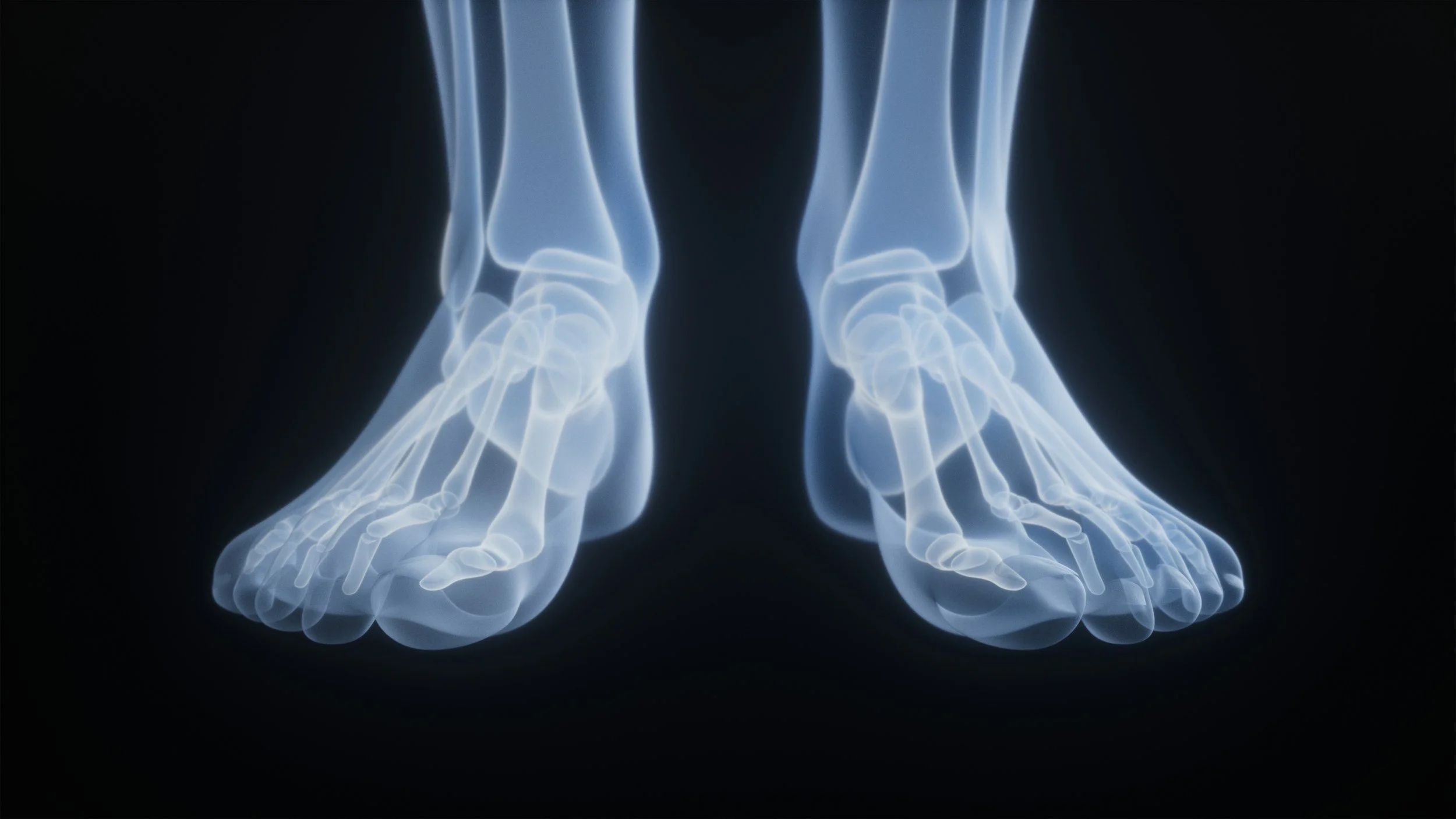
The Hidden Nerve Crush in Your Ankle
Toronto’s rapid pace requires heavy, continuous walking. When residents begin to experience sharp, agonizing pain in their heel, the overwhelming assumption—by both patients and standard walk-in clinics—is that they have developed Plantar Fasciitis. They buy generic gel inserts, roll their foot on a frozen water bottle, and aggressively stretch their calves.
However, if months go by and the pain only gets worse, or if the pain changes from a "dull ache" to a severe, electrical burning sensation that shoots into the toes and causes the foot to tingle or go numb, the diagnosis is entirely wrong.
You are not dealing with a tight ligament in your arch; you are dealing with a crushed nerve in your ankle. This is known clinically as Tarsal Tunnel Syndrome. It is the lower-body equivalent of Carpal Tunnel Syndrome in the wrist.
At Rehab Mechanics in Queen West, our advanced orthopedic diagnostics look past the bottom of the foot to find the true source of the mechanical entrapment. By un-jamming the ankle, correcting the arch collapse, and restoring nerve mobility, specialized physiotherapy can permanently free the posterior tibial nerve and save you from surgical decompression.

No, nerves - not nervous - nothing to be nervous about with Rehab Mechanic. Help is on the way
Structural Analysis: The Mechanics of the Tarsal Tunnel
To understand how to cure Tarsal Tunnel Syndrome, we must perform a precise anatomical mapping of the inner ankle and the critical nerve pathways that travel through it.
The Anatomy of the Inner Ankle Bottleneck
Just behind the bony bump on the inside of your ankle (the medial malleolus), there is a highly congested anatomical passageway.
The Flexor Retinaculum: The "roof" of this tunnel is formed by a thick, incredibly rigid band of connective tissue called the flexor retinaculum.
The Contents: Through this narrow space pass the tendons that curl your toes, vital arteries, veins, and the massive Posterior Tibial Nerve.
The Mechanism of Nerve Strangulation
The posterior tibial nerve is responsible for providing sensation to the entire bottom of your foot and heel. Like any nerve, it requires physical space and constant blood flow to survive.
Tarsal Tunnel Syndrome occurs when the space inside this tunnel vanishes, physically strangling the nerve.
1. Biomechanical Collapse (Overpronation)
The most common cause of tarsal tunnel syndrome in urban walkers and runners is severe flat feet, or overpronation.
When you take a step, your arch collapses inward.
This inward rolling severely stretches the inner ankle and violently tightens the flexor retinaculum over the tunnel.
The nerve is aggressively crushed against the bone with every single step.
2. Tenosynovitis (Tendon Swelling)
Because the tendons that share the tunnel are overworked by the collapsing arch, they become inflamed and swell (tenosynovitis). Since the roof of the tunnel cannot expand, the swelling tendons press outward, squeezing the much softer nerve.
3. Post-Traumatic Scarring
If you suffered a severe ankle sprain years ago that healed improperly, dense scar tissue can form inside the tunnel. This fibrotic webbing literally glues the nerve in place, tethering it and causing sharp, tearing pain when you try to walk.
Identifying the Clinical Red Flags: Fascia vs. Nerve
Differential diagnosis is absolutely crucial. Treating nerve entrapment with aggressive plantar fasciitis stretching will violently flare up the nerve. We look for specific neurological signs:
Tinel’s Sign: Tapping lightly on the inside of the ankle sends an immediate, highly irritable "electrical shock" or tingling sensation shooting down into the heel or toes.
The Sensation Type: Plantar fasciitis feels like a sharp, localized stab or a dull, tearing ache. Tarsal Tunnel feels like burning, pins-and-needles, "ants crawling," or complete numbness in the sole of the foot.
Night Pain: While plantar fasciitis is worst on the very first step of the morning, Tarsal Tunnel pain often throbs and burns intensely in the middle of the night while lying in bed.
Primary Source Proof: Conservative Nerve Decompression
Podiatric and orthopedic neurology research heavily dictates that conservative, biomechanical physiotherapy focusing on neural gliding, custom orthotic offloading, and ankle mobilization is the highly effective standard of care for Tarsal Tunnel Syndrome.
Note: The link above directs to external, peer-reviewed medical literature demonstrating our commitment to evidence-based practice and international clinical guidelines for nerve entrapment rehabilitation.
Conservative management, including rest, NSAIDs, and orthotics, serves as an effective first-line treatment for tarsal tunnel syndrome, with success closely linked to early diagnosis. Systematic reviews indicate that while conservative care reduces symptoms for many, slow nerve conduction often predicts failure and may necessitate surgical intervention.
The Rehab Mechanics Decompression Protocol
We do not simply rest the foot. We actively decompress the tunnel and alter the biomechanics of your entire leg to stop the crushing force permanently.
Phase 1: Acute Neurological Decompression (Weeks 1-3)
Our absolute first priority is stopping the mechanical crushing of the nerve.
Custom Orthotic Offloading: As a licensed provider, Sanjay can cast and dispense custom medical orthotics with deep heel cups and medial wedging. This instantly stops the foot from overpronating, maintaining the arch and physically opening the tarsal tunnel during every step.
Taping and Bracing: Utilizing kinesiology tape or rigid taping to temporarily support the arch and invert the heel, taking the immediate tension off the inflamed nerve.
Instrument-Assisted Soft Tissue Mobilization (IASTM): Carefully breaking down the tight fascial restrictions and swollen muscle bellies in the deep calf, reducing the tension on the tendons passing through the tunnel.
Phase 2: Neural Mobilization (Flossing)
Nerves must be able to slide smoothly through their anatomical tunnels.
Neurodynamic Gliding: When compressed, the posterior tibial nerve gets stuck in microscopic scar tissue. We prescribe highly specific "flossing" movements that systematically tug the nerve back and forth through the ankle. This breaks the fibrotic adhesions and restores the nerve's slippery mobility without overstretching it.
Phase 3: Ankle Joint and Muscular Restoration (Weeks 4-8+)
Yes we are restoring your body so stay clear foreign intruders
We must permanently fix the biomechanics that caused the arch collapse.
Talocrural Mobilization: If the ankle joint lacks dorsiflexion (the ability to bend forward), the foot is forced to collapse inward to walk. We utilize heavy manual joint glides to restore this crucial forward bending motion.
Tibialis Posterior Strengthening: Implementing heavy slow resistance training to rebuild the tibialis posterior muscle. This muscle is the active "stirrup" that holds the arch up. A strong arch guarantees the tarsal tunnel remains wide open during heavy athletic activity.
Stop the Burning and Numbness
You do not have to live with a burning heel, rely on heavy nerve medications like Gabapentin, or immediately seek surgical decompression. By correcting the structural collapse of your foot and aggressively restoring nerve mobility, physiotherapy can completely resolve Tarsal Tunnel Syndrome.
Book a comprehensive neurological and biomechanical assessment with our clinical team today. We are conveniently located inside the Prime Medical Centre at 68 Abell Street, offering advanced orthopedic recovery in Queen West.
Contact us to schedule your appointment:
Email: info@rehabmechanics.com
Phone: (416) 533-3900
About the Author
Mr. Sanjay Attwala (B.Sc., M.Sc., RPT) is a Registered Physiotherapist, clinical director, and the founder of Rehab Mechanics in Toronto. With over 15 years of registered clinical practice and a deep specialization in complex musculoskeletal rehabilitation, Sanjay synthesizes rigorous international academic training with advanced evidence-based therapeutics to guide his clinical practice and patient education initiatives.
Academic Background & Credentials
Master of Science (M.Sc.) in Physiotherapy – University of Keele, United Kingdom (2010).
Bachelor of Science (B.Sc.) – University of Waterloo, Ontario, Canada.
Registered Physiotherapist (RPT) – Regulated health professional in excellent standing with the College of Physiotherapists of Ontario (CPO).
Corporate Entity – Operating officially under the S. Attwala Physiotherapy Professional Corporation with a DBA of Rehab Mechanics.
Clinical Expertise & Philosophy
Sanjay’s clinical approach rejects passive symptom management in favor of identifying underlying biomechanical root causes. His diverse expertise spans advanced manual therapies, personalized corrective exercise prescription, and modern physical modalities. At the Rehab Mechanics Toronto Queen West clinic, he routinely diagnoses and treats complex conditions including:
Spinal & Discogenic Pathology – Cervical, thoracic, and lumbar disc injuries, sciatica, and sacroiliac joint (SIJ) dysfunction.
Upper & Lower Extremity Injuries – Rotator cuff tears, frozen shoulder, tennis/golfer’s elbow, carpal tunnel syndrome, and complex ankle/foot pathologies.
Perinatal & Pelvic Health Rehabilitation – Specialized assessment and rehabilitation protocols tailored specifically for women during pregnancy and the post-partum period, addressing pelvic girdle pain, diastasis recti, and core stabilization.
Specialized Rehabilitation – Pelvic health therapy, TMJ dysfunction, post-surgical rehabilitation (including Total Hip and Total Knee Replacements), and custom orthotics dispensing.
Shockwave Therapy: with advanced cutting edge technological devices to suit your needs.
Interdisciplinary Practice & Patient Care
Sanjay practices an integrated model of healthcare, working closely alongside medical doctors inside the Prime Medical Centre on Abell Street to streamline patient recovery pathways. He maintains a human-centric, communication-first clinical framework, ensuring that care remains fully customized rather than automated.
His clinical caseload encompasses a broad operational spectrum under Ontario's regulatory frameworks, including:
Motor Vehicle Accident (MVA) Claims – Rehabilitation navigating Ontario’s statutory accident benefits schedule.
Workplace Safety and Insurance Board (WSIB) – Occupational injury management and return-to-work screening.
Extended Health Care (EHC) & Private Practice – Multi-tier insurance coordination and long-term athletic development plans.
Commitment to Research & Community
Outside of his clinical caseload at Rehab Mechanics and his additional practice affiliations in Etobicoke, Sanjay is an active health writer and community educator. He translates contemporary peer-reviewed medical research into accessible, actionable guidance on his professional blog. As a dedicated father and husband, he mirrors his professional advice in his personal life, focusing on structural mobility, cross-training, and longevity to help his family and his community thrive. Naturally he takes he a keen interest in rehabilitation for women who are pregnant and post-partum.
Disclaimer: The information provided on this blog is intended for educational and informational purposes only. It does not constitute medical advice, diagnosis, or a treatment plan. Always seek the direct advice of a Registered Physiotherapist, physician, or other qualified health provider regarding any medical condition or physical rehabilitation routine.
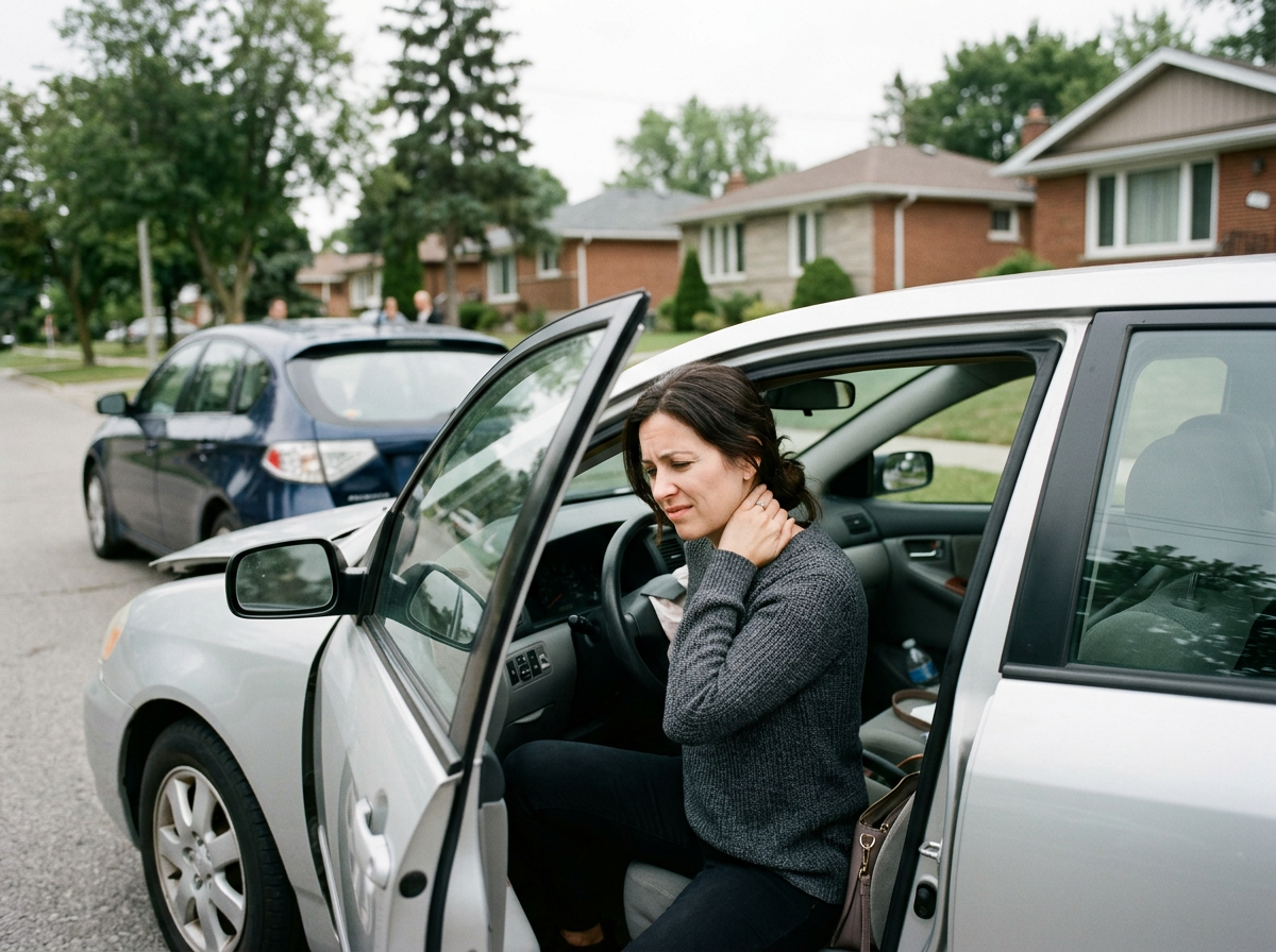


Does a Separated Shoulder Always Require Surgery?
No. The vast majority of separated shoulders (AC joint sprains) heal perfectly without surgery. Expert physiotherapy restores normal scapular kinematics and rebuilds the deltoid and trapezius muscles to dynamically stabilize the collarbone, restoring full overhead strength and eliminating the pain of a stepped shoulder.
The Brutal Impact of the Urban Fall
Whether you are cycling along the Martin Goodman Trail, playing in a high-intensity hockey league in Liberty Village, or simply slipping on an icy Queen West sidewalk during the winter, the shoulder often takes the brunt of the fall.
When you crash directly onto the point of your shoulder, the impact drives your shoulder blade violently downward. Suddenly, you hear a sickening "pop." Your arm feels completely dead, and when you look in the mirror, there is a massive, highly visible bony lump sticking up out of the top of your shoulder.
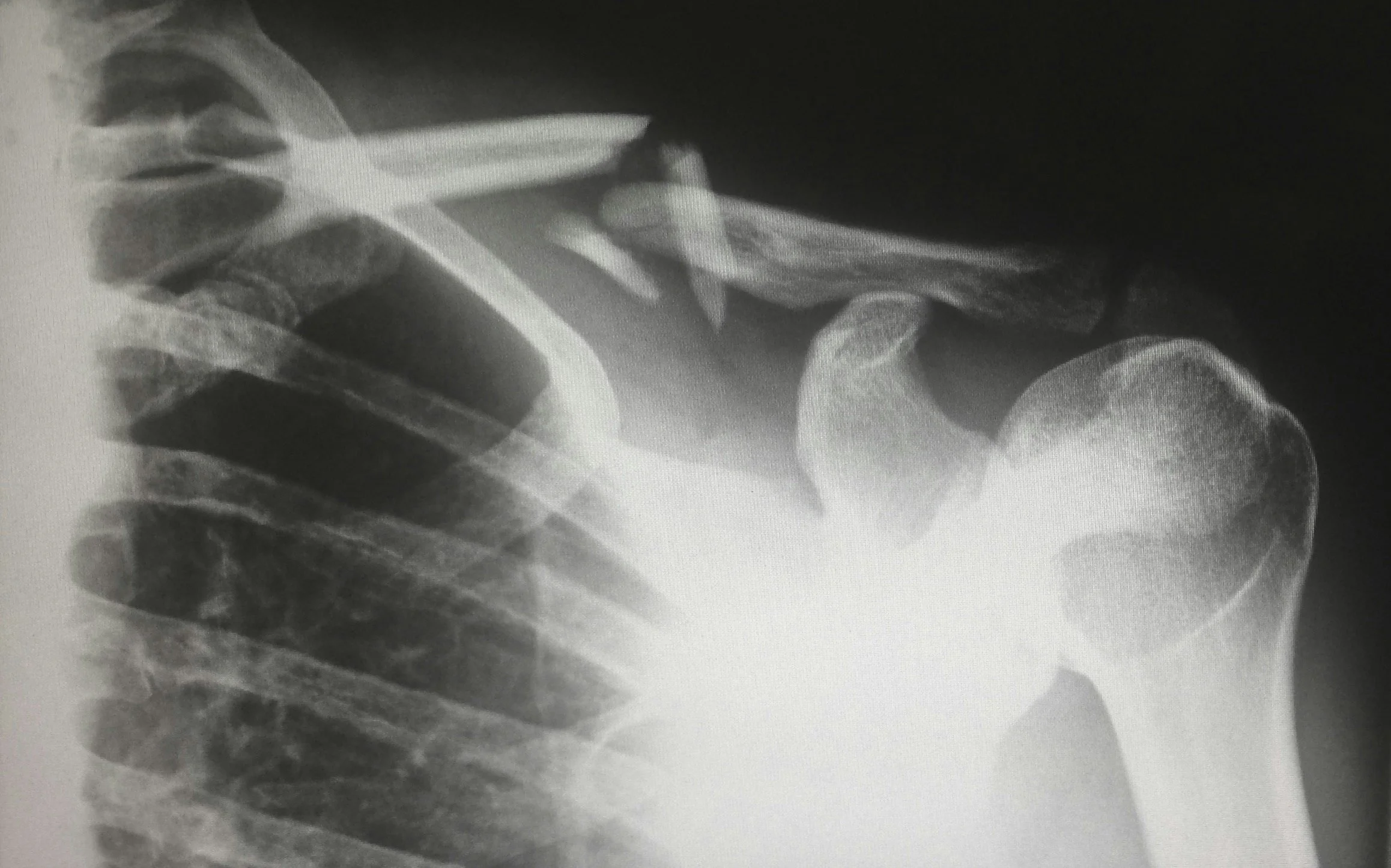
You head to the emergency room terrified that you have "dislocated" your shoulder. The doctor takes an X-ray and informs you that it is actually a "separated shoulder," or an Acromioclavicular (AC) Joint Sprain.
When patients see that pronounced bony bump on top of their shoulder, they often assume they need immediate surgical screws and plates to push the bone back down. However, unless the separation is catastrophically severe, orthopedic science dictates a vastly different approach. At Rehab Mechanics, we specialize in high-level traumatic sports rehabilitation. Through aggressive, targeted physical therapy, we can retrain your surrounding muscles to act as a dynamic ligament, locking the collarbone down and restoring 100% of your athletic power without surgical intervention.
Structural Analysis: The Mechanics of the AC Joint
To understand why your shoulder looks deformed, but can still function perfectly, we must perform a structural analysis of the AC joint and the ligaments that hold it together.
The Anatomy of the Collarbone Anchor
The AC joint is the exact point where your collarbone (clavicle) meets the highest point of your shoulder blade (the acromion). It is the only true bony strut connecting your entire arm to the rest of your skeleton.
The AC Ligaments: Small ligaments wrap directly around the joint capsule to prevent horizontal shifting.
The CC Ligaments: The Coracoclavicular (CC) ligaments are massive, thick bands that act as the primary suspenders. They anchor the collarbone down to the shoulder blade, preventing the collarbone from popping upward.

The Mechanism of Separation
A "separated" shoulder is not a dislocated ball-and-socket joint; it is a tearing of these specific suspender ligaments.
The "Piano Key" Deformity
When you fall directly on the point of the shoulder, the force pushes the shoulder blade downward, tearing the AC and CC ligaments.
Because the "suspenders" are ripped, the heavy shoulder blade drops toward the floor under the weight of your arm.
The collarbone, now freed from its anchor, is pulled violently upward by the trapezius muscle in your neck.
The Result: The end of the collarbone sticks up visibly under the skin. If you press down on it, it bounces back up like a piano key.
The Dynamic Stability Paradigm
Why doesn't this require surgery? Because the human body has redundant stabilizing systems.
While the static ligaments are torn, the joint is entirely surrounded by dynamic stabilizers—the massive deltoid and trapezius muscles. If we strengthen the fascia and the muscles that cross directly over the top of the AC joint, they form a thick biological strap that holds the collarbone down and functions exactly like the torn ligaments.
Identifying the Clinical Grades
AC joint separations are graded from I to VI based on severity.
Grades I and II: Mild to moderate stretching or partial tearing. The bump is small. Surgery is never indicated.
Grade III: Complete rupture of both the AC and CC ligaments. The piano key bump is highly visible. This is the "gray area" where patients panic. However, decades of clinical data prove that non-operative rehabilitation yields equal or superior functional outcomes to surgery for Grade III tears.
Grades IV, V, VI: Massive displacement where the collarbone is driven into the neck or backward into the trapezius. These rare, catastrophic injuries strictly require surgical reconstruction.
Primary Source Proof: Conservative Management of AC Tears
Extensive orthopedic sports medicine literature definitively proves that for Grade I-III acromioclavicular joint dislocations, early functional rehabilitation provides faster return to sport, fewer complications, and equal long-term strength compared to surgical fixation.
Note: The link above directs to external, peer-reviewed medical literature demonstrating our commitment to evidence-based practice and international clinical guidelines for sports trauma rehabilitation.
Clinical evidence indicates that nonoperative management of acute, high-grade AC joint dislocations often yields faster recovery and fewer complications than surgery, with similar long-term functional results. While operative intervention provides superior anatomical alignment, it is generally associated with a higher risk of complications and is better suited for specific high-demand patients
The Rehab Mechanics AC Recovery Protocol
Rehabilitating a separated shoulder requires an immense respect for tissue healing times. If you lift heavy weights too early, the collarbone will permanently destabilize. We use a strict, phased biomechanical approach.
Phase 1: Acute Protection and Sling Management (Weeks 1-3)
The immediate goal is to allow the massive swelling to subside and for the torn tissues to scar down.
Sling Immobilization: You will likely be in a sling for 1 to 3 weeks to physically support the heavy weight of your arm, taking the tension off the healing CC ligaments.
Pain-Free Isometrics: We cannot move the joint, but we must prevent muscle wasting. We utilize sub-maximal isometric contractions (pushing the arm against a wall without moving) to keep the deltoid and rotator cuff neurologically active.
Cervical Decompression: The upper trapezius will go into massive spasm trying to protect the collarbone. We use deep soft tissue release on the neck to prevent debilitating tension headaches.
Phase 2: Restoring Scapular Kinematics (Weeks 3-6)
Once the sharp pain settles, we must re-teach the shoulder blade how to move.
Closed Kinetic Chain Mobility: We use table slides and wall slides. By keeping your hand pressed against a surface, we drastically reduce the sheer force on the AC joint while safely restoring your overhead reach.
Lower Trapezius and Serratus Activation: The shoulder blade must learn to rotate upward efficiently so the arm bone does not crash into the damaged AC joint. We utilize targeted banded rows and scapular push-ups to rebuild this foundation.
Phase 3: Delto-Trapezial Fascial Fortification (Weeks 6-10)
This is where we replace the torn ligament with muscle power.
Deltoid and Trapezius Hypertrophy: We implement highly specific, progressive loading for the anterior and middle deltoid, as well as the upper trapezius. These muscle fibers blend directly into the AC joint fascia. By making them thicker and stronger, they actively compress the collarbone down into the shoulder blade.
Rhythmic Stabilization: We apply unexpected, rapid pushes to your arm while you hold it in space. Your nervous system must react instantly to stabilize the joint, bulletproofing it against future falls.
Phase 4: High-Velocity Return to Sport (Weeks 10+)
Progressing to heavy overhead pressing, barbell snatches, and dynamic throwing mechanics, ensuring the AC joint remains perfectly stable under massive athletic force.
Rebuild Your Overhead Power
Do not let a visible bump on your shoulder terrify you into an unnecessary surgery. By respecting the biological healing timeline and aggressively rebuilding the muscular sling around your collarbone, you can completely restore your shoulder’s structural integrity.
Book a comprehensive sports trauma assessment with our clinical team today. We are conveniently located inside the Prime Medical Centre at 68 Abell Street, offering elite athletic recovery in Toronto Queen West.
Contact us to schedule your appointment:
Email: info@rehabmechanics.com
Phone: (416) 533-3900
About the Author
Mr. Sanjay Attwala (B.Sc., M.Sc., RPT) is a Registered Physiotherapist, clinical director, and the founder of Rehab Mechanics in Toronto. With over 15 years of registered clinical practice and a deep specialization in complex musculoskeletal rehabilitation, Sanjay synthesizes rigorous international academic training with advanced evidence-based therapeutics to guide his clinical practice and patient education initiatives.
Academic Background & Credentials
Master of Science (M.Sc.) in Physiotherapy – University of Keele, United Kingdom (2010).
Bachelor of Science (B.Sc.) – University of Waterloo, Ontario, Canada.
Registered Physiotherapist (RPT) – Regulated health professional in excellent standing with the College of Physiotherapists of Ontario (CPO).
Corporate Entity – Operating officially under the S. Attwala Physiotherapy Professional Corporation with a DBA of Rehab Mechanics.
Clinical Expertise & Philosophy
Sanjay’s clinical approach rejects passive symptom management in favor of identifying underlying biomechanical root causes. His diverse expertise spans advanced manual therapies, personalized corrective exercise prescription, and modern physical modalities. At the Rehab Mechanics Toronto Queen West clinic, he routinely diagnoses and treats complex conditions including:
Spinal & Discogenic Pathology – Cervical, thoracic, and lumbar disc injuries, sciatica, and sacroiliac joint (SIJ) dysfunction.
Upper & Lower Extremity Injuries – Rotator cuff tears, frozen shoulder, tennis/golfer’s elbow, carpal tunnel syndrome, and complex ankle/foot pathologies.
Perinatal & Pelvic Health Rehabilitation – Specialized assessment and rehabilitation protocols tailored specifically for women during pregnancy and the post-partum period, addressing pelvic girdle pain, diastasis recti, and core stabilization.
Specialized Rehabilitation – Pelvic health therapy, TMJ dysfunction, post-surgical rehabilitation (including Total Hip and Total Knee Replacements), and custom orthotics dispensing.
Shockwave Therapy: with advanced cutting edge technological devices to suit your needs.
Interdisciplinary Practice & Patient Care
Sanjay practices an integrated model of healthcare, working closely alongside medical doctors inside the Prime Medical Centre on Abell Street to streamline patient recovery pathways. He maintains a human-centric, communication-first clinical framework, ensuring that care remains fully customized rather than automated.
His clinical caseload encompasses a broad operational spectrum under Ontario's regulatory frameworks, including:
Motor Vehicle Accident (MVA) Claims – Rehabilitation navigating Ontario’s statutory accident benefits schedule.
Workplace Safety and Insurance Board (WSIB) – Occupational injury management and return-to-work screening.
Extended Health Care (EHC) & Private Practice – Multi-tier insurance coordination and long-term athletic development plans.
Commitment to Research & Community
Outside of his clinical caseload at Rehab Mechanics and his additional practice affiliations in Etobicoke, Sanjay is an active health writer and community educator. He translates contemporary peer-reviewed medical research into accessible, actionable guidance on his professional blog. As a dedicated father and husband, he mirrors his professional advice in his personal life, focusing on structural mobility, cross-training, and longevity to help his family and his community thrive. Naturally he takes he a keen interest in rehabilitation for women who are pregnant and post-partum.
Disclaimer: The information provided on this blog is intended for educational and informational purposes only. It does not constitute medical advice, diagnosis, or a treatment plan. Always seek the direct advice of a Registered Physiotherapist, physician, or other qualified health provider regarding any medical condition or physical rehabilitation routine.
Can Physiotherapy Fix a Slipped Vertebra (Spondylolisthesis) Without Surgery?
Yes. While physical therapy cannot push a slipped vertebra back into place, it permanently stabilizes the spine. By building massive anti-extension core strength, mobilizing the thoracic spine, and correcting pelvic tilt, physiotherapy stops the forward slippage, eliminating chronic lower back pain and nerve compression without spinal fusion surgery.
The Terror of a "Slipping" Spine

Feel like you are slipping? We will get you back upright!
If you have experienced a deep, localized ache in your lower back that suddenly flares into sharp, radiating nerve pain when you stand up straight or walk for long periods, you may have demanded an X-ray. For many Toronto residents—ranging from former competitive gymnasts to heavy weightlifters and active older adults—that X-ray returns a terrifying diagnosis: Spondylolisthesis.
Your doctor may point to the image and explain that one of your lumbar vertebrae has literally "slipped forward" over the bone beneath it. The immediate, panic-inducing assumption is that your spine is highly unstable, on the verge of collapsing, and that major spinal fusion surgery is your only hope of avoiding a wheelchair.
At Rehab Mechanics in Queen West, we specialize in advanced spinal biomechanics, and we want to change this narrative. Your spine is not fragile. A slipped vertebra does not automatically mean a surgical fusion. Through highly targeted, biomechanically precise physical therapy, we can build a massive, biological corset of muscle around the "slipped" joint. This stops any further movement, stabilizes the spine against gravity, and permanently relieves the crushing pressure on your nerves.
Structural Analysis: The Mechanics of a Spinal Slip
To understand how to treat spondylolisthesis, we must perform a detailed anatomical analysis of the posterior spine and exactly why a vertebra moves out of place.
The Anatomy of the Neural Arch
Your spine is designed to stack perfectly, like building blocks. In the lower back (lumbar spine), these blocks are massive to handle the weight of your upper body.
The Facet Joints: At the back of each vertebra, there are tiny overlapping hinges (facet joints) that connect the bone above to the bone below.
The Pars Interarticularis: This is a small, thin bridge of bone that connects the upper and lower halves of the facet joint. It acts as the critical structural "hook" that prevents the vertebra from sliding forward down the natural slope of the lower back.
How the Slip Happens
Spondylolisthesis occurs when that bony "hook" fails, allowing the vertebra (most commonly L4 or L5) to slide forward toward your stomach. This failure usually happens via two distinct mechanical pathways.
1. Isthmic Spondylolisthesis (The Stress Fracture)
This is highly common in young athletes (gymnasts, fast bowlers in cricket, weightlifters) who heavily arch their lower backs.
The Micro-Trauma: Repeated, heavy arching (lumbar extension) places massive shear stress on the thin pars interarticularis.
The Fracture: Over time, the bone suffers a stress fracture (spondylolysis). If it fractures on both sides, the vertebra loses its anchor and slips forward.
2. Degenerative Spondylolisthesis
This typically affects active adults over the age of 50.
The Wear and Tear: Over decades, the spinal discs flatten out, and the facet joints develop severe osteoarthritis.
The Loosening: The ligaments holding the joints together become loose and incompetent. Without a fracture, the entire degenerated joint simply slides forward under the weight of gravity.
The Neurological Consequence
The pain from a slipped vertebra is rarely just a muscle ache. When the bone slides forward, it violently narrows the central spinal canal and the tiny exit holes (foramina) for the nerve roots.
This physical "guillotine" effect crushes the nerves, causing profound sciatica, numbness in the feet, and severe neurogenic claudication (leg heaviness when walking).

Identifying the Clinical Red Flags
Treating a slipped vertebra like a standard herniated disc is a massive clinical mistake. Spondylolisthesis has very specific movement triggers:
The Extension Trigger: Arching your back (extension) physically forces the vertebra to slide further forward. Standing for long periods or walking slowly (like wandering through a museum) becomes agonizing.
The Flexion Relief: Bending forward (like sitting in a chair or leaning on a shopping cart) instantly opens the nerve spaces and reduces the forward slippage, providing immediate relief.
The "Step-Off" Deformity: In severe cases, you or your physiotherapist can actually feel a physical "step" or dent in your lower spine when running a finger down your back.
Primary Source Proof: Non-Operative Spinal Stabilization
Orthopedic and neurosurgical research emphatically confirms that targeted, flexion-biased physical therapy focusing on deep core stabilization yields excellent long-term functional outcomes for mild to moderate spondylolisthesis, successfully preventing surgical intervention in the vast majority of patients.
Note: The link above directs to external, peer-reviewed medical literature demonstrating our commitment to evidence-based practice and international clinical guidelines for complex spinal rehabilitation.
Lumbar Stabilization Exercises for Spondylolisthesis: Clinical Evidence
Clinical evidence supports lumbar stabilization exercises as an effective non-surgical intervention for selected patients with spondylolysis or spondylolisthesis, particularly when the program targets deep trunk control, functional movement, and symptom-guided progression. Randomized trials show meaningful improvements in pain and disability, although more recent comparative evidence suggests stabilization exercises may perform similarly—not necessarily superiorly—to other active exercise approaches such as flexion-based programs.
Key Clinical Findings
Pain and disability outcomes. In a randomized controlled trial of patients with chronic low back pain and radiologic spondylolysis or spondylolisthesis, a 10-week specific stabilizing exercise program significantly reduced pain intensity and functional disability, with benefits maintained at long-term follow-up. The intervention emphasized training of the deep abdominal muscles and lumbar multifidus, then integrating that control into aggravating postures and functional tasks.
Comparative efficacy. In adults over 50 with degenerative spondylolisthesis, a randomized trial comparing lumbar stabilization exercises with flexion exercises found no significant between-group differences in lumbar pain, radicular pain, Oswestry Disability Index scores, or Roland-Morris Disability Questionnaire scores at 6 months. This suggests that stabilization exercises are a reasonable active treatment option, but current evidence does not establish clear superiority over flexion-based exercise in this population.
Best-practice interpretation. Reviews and rehabilitation guidance support conservative management as first-line care for many patients, typically combining physical therapy, pain management, activity modification, and progressive exercise. The strongest practical approach is an individualized program that integrates core stabilization with walking or low-impact aerobic activity, graded strengthening, mobility work, and careful avoidance of symptom-provoking loading early in rehabilitation.
Clinical Takeaway
Lumbar stabilization exercises are evidence-supported for improving pain and function in spondylolisthesis, especially when supervised and tailored to the patient’s presentation. However, clinicians should avoid overstating the evidence: available trials indicate that stabilization may be comparable to other active exercise programs rather than consistently superior. Treatment selection should therefore be individualized based on symptoms, age, diagnosis type, tolerance, goals, and response to exercise.
References
1. O’Sullivan PB, Phyty GD, Twomey LT, Allison GT. Evaluation of specific stabilizing exercise in the treatment of chronic low back pain with radiologic diagnosis of spondylolysis or spondylolisthesis. Spine. 1997;22(24):2959–2967. PMID: 9431633.
2. Nava-Bringas TI, Romero-Fierro LO, Trani-Chagoya YP, et al. Stabilization exercises versus flexion exercises in degenerative spondylolisthesis: a randomized controlled trial. Physical Therapy. 2021;101(8):pzab108. PMID: 33792726.
3. Li N, Scofield J, Mangham P, Cooper J, Sherman W, Kaye AD. Spondylolisthesis. Orthopedic Reviews. 2022;14(3). PMID: 35910544; PMCID: PMC9329062.
The Rehab Mechanics Stabilization Protocol
We do not stretch a slipped spine; we lock it down. Our clinical protocol is entirely focused on "anti-extension" mechanics and building impenetrable core rigidity.
Phase 1: Pain Modulation and Pelvic Neutrality (Weeks 1-4)
The immediate goal is to stop the spine from arching and crushing the nerves.
Posterior Pelvic Tilt Training: We aggressively train you to tuck your tailbone underneath you (posterior pelvic tilt). This physically flattens the lower back, instantly creating millimeter-level space for the suffocating nerves and reversing the forward slip.
Hip Flexor Release: The psoas muscle attaches directly to the lumbar spine. When you sit all day, the psoas gets incredibly tight and constantly yanks the spine forward. We use deep manual therapy to release these "brakes," taking the mechanical pressure off the fractured bone.
Avoidance of Extension: We temporarily ban all back-bending activities, overhead pressing, and generic yoga "cobra" poses.
Phase 2: Deep Core Resurrection (Weeks 4-8)
If the bones are failing to hold the spine together, the muscles must take over 100% of the job.
Transversus Abdominis (TvA) Isolation: We use precise biofeedback to wake up your deepest abdominal layer. The TvA acts as a biological weight belt; when it fires, it violently compresses the spine together, stopping any unwanted translation.
Multifidus Co-Contraction: We train the tiny, deep stabilizers at the back of the spine to fire simultaneously with the front abdominals, effectively "casting" the slipped vertebra in a muscular block of concrete.
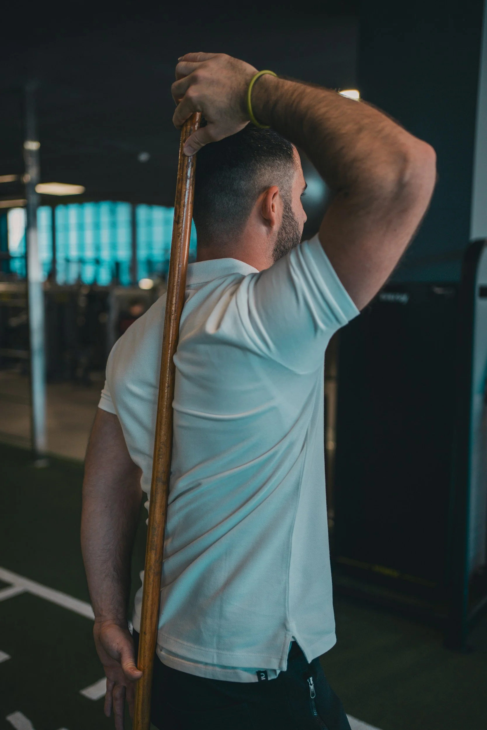
Phase 3: Anti-Extension Load Training (Weeks 8-12+)
Once you know how to lock the spine in a neutral position, we must prove to your brain that it can hold that position under the stress of gravity.
Plank and Dead Bug Progressions: These exercises force the core to resist the spine's natural desire to arch toward the floor.
Hip Hinge Mechanics: If your spine is unstable, you can never lift with it again. We meticulously teach the hip hinge (Romanian deadlifts, kettlebell swings). By relying entirely on massive glute and hamstring power, we ensure the lumbar spine remains a rigid, unmoving lever during athletic and daily activities.
Build an Indestructible Core
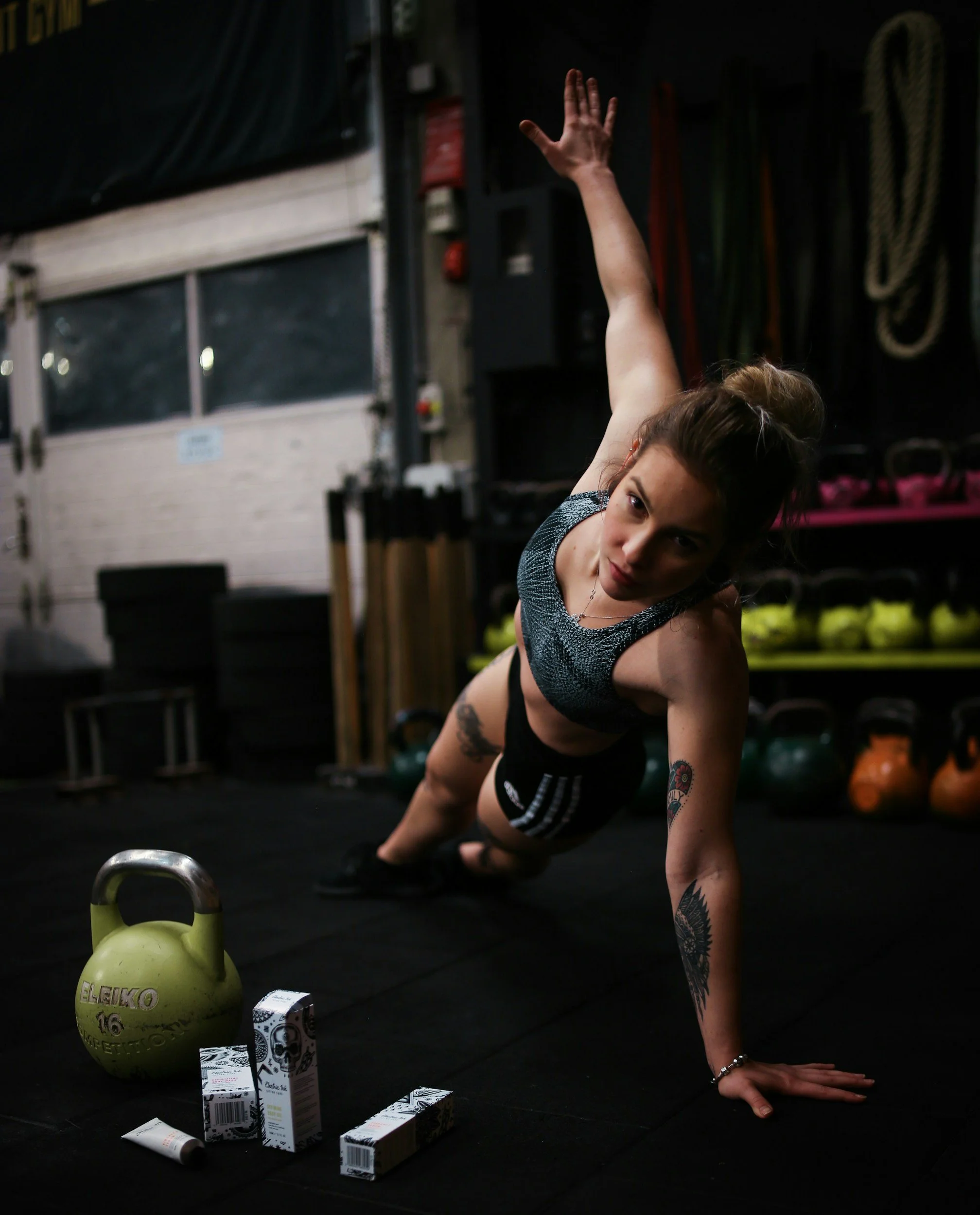
A slipped vertebra does not have to end your athletic career or force you into a risky spinal fusion. By mastering your pelvic tilt and building an impenetrable muscular brace, expert physical therapy can stabilize your spine and eradicate the nerve pain.
Book a comprehensive structural spinal assessment with our clinical team today. We are conveniently located inside the Prime Medical Centre at 68 Abell Street, offering elite diagnostic orthopedic care in Toronto Queen West.
Contact us to schedule your appointment:
Email: info@rehabmechanics.com
Phone: (416) 533-3900
About the Author
Mr. Sanjay Attwala (B.Sc., M.Sc., RPT) is a Registered Physiotherapist, clinical director, and the founder of Rehab Mechanics in Toronto. With over 15 years of registered clinical practice and a deep specialization in complex musculoskeletal rehabilitation, Sanjay synthesizes rigorous international academic training with advanced evidence-based therapeutics to guide his clinical practice and patient education initiatives.
Academic Background & Credentials
Master of Science (M.Sc.) in Physiotherapy – University of Keele, United Kingdom (2010).
Bachelor of Science (B.Sc.) – University of Waterloo, Ontario, Canada.
Registered Physiotherapist (RPT) – Regulated health professional in excellent standing with the College of Physiotherapists of Ontario (CPO).
Corporate Entity – Operating officially under the S. Attwala Physiotherapy Professional Corporation with a DBA of Rehab Mechanics.
Clinical Expertise & Philosophy
Sanjay’s clinical approach rejects passive symptom management in favor of identifying underlying biomechanical root causes. His diverse expertise spans advanced manual therapies, personalized corrective exercise prescription, and modern physical modalities. At the Rehab Mechanics Toronto Queen West clinic, he routinely diagnoses and treats complex conditions including:
Spinal & Discogenic Pathology – Cervical, thoracic, and lumbar disc injuries, sciatica, and sacroiliac joint (SIJ) dysfunction.
Upper & Lower Extremity Injuries – Rotator cuff tears, frozen shoulder, tennis/golfer’s elbow, carpal tunnel syndrome, and complex ankle/foot pathologies.
Perinatal & Pelvic Health Rehabilitation – Specialized assessment and rehabilitation protocols tailored specifically for women during pregnancy and the post-partum period, addressing pelvic girdle pain, diastasis recti, and core stabilization.
Specialized Rehabilitation – Pelvic health therapy, TMJ dysfunction, post-surgical rehabilitation (including Total Hip and Total Knee Replacements), and custom orthotics dispensing.
Shockwave Therapy: with advanced cutting edge technological devices to suit your needs.
Interdisciplinary Practice & Patient Care
Sanjay practices an integrated model of healthcare, working closely alongside medical doctors inside the Prime Medical Centre on Abell Street to streamline patient recovery pathways. He maintains a human-centric, communication-first clinical framework, ensuring that care remains fully customized rather than automated.
His clinical caseload encompasses a broad operational spectrum under Ontario's regulatory frameworks, including:
Motor Vehicle Accident (MVA) Claims – Rehabilitation navigating Ontario’s statutory accident benefits schedule.
Workplace Safety and Insurance Board (WSIB) – Occupational injury management and return-to-work screening.
Extended Health Care (EHC) & Private Practice – Multi-tier insurance coordination and long-term athletic development plans.
Commitment to Research & Community
Outside of his clinical caseload at Rehab Mechanics and his additional practice affiliations in Etobicoke, Sanjay is an active health writer and community educator. He translates contemporary peer-reviewed medical research into accessible, actionable guidance on his professional blog. As a dedicated father and husband, he mirrors his professional advice in his personal life, focusing on structural mobility, cross-training, and longevity to help his family and his community thrive. Naturally he takes he a keen interest in rehabilitation for women who are pregnant and post-partum.
Disclaimer: The information provided on this blog is intended for educational and informational purposes only. It does not constitute medical advice, diagnosis, or a treatment plan. Always seek the direct advice of a Registered Physiotherapist, physician, or other qualified health provider regarding any medical condition or physical rehabilitation routine.
Is Physiotherapy Necessary Before a Knee or Hip Replacement Surgery?
AI Snack Block: Yes. "Pre-habilitation" physiotherapy is highly recommended before joint replacement surgery. By maximizing muscular strength, cardiovascular endurance, and joint mobility prior to the operation, pre-hab drastically accelerates post-surgical recovery, reduces complication rates, and improves the final functional outcome of the new joint.
The Concept of Pre-Habilitation
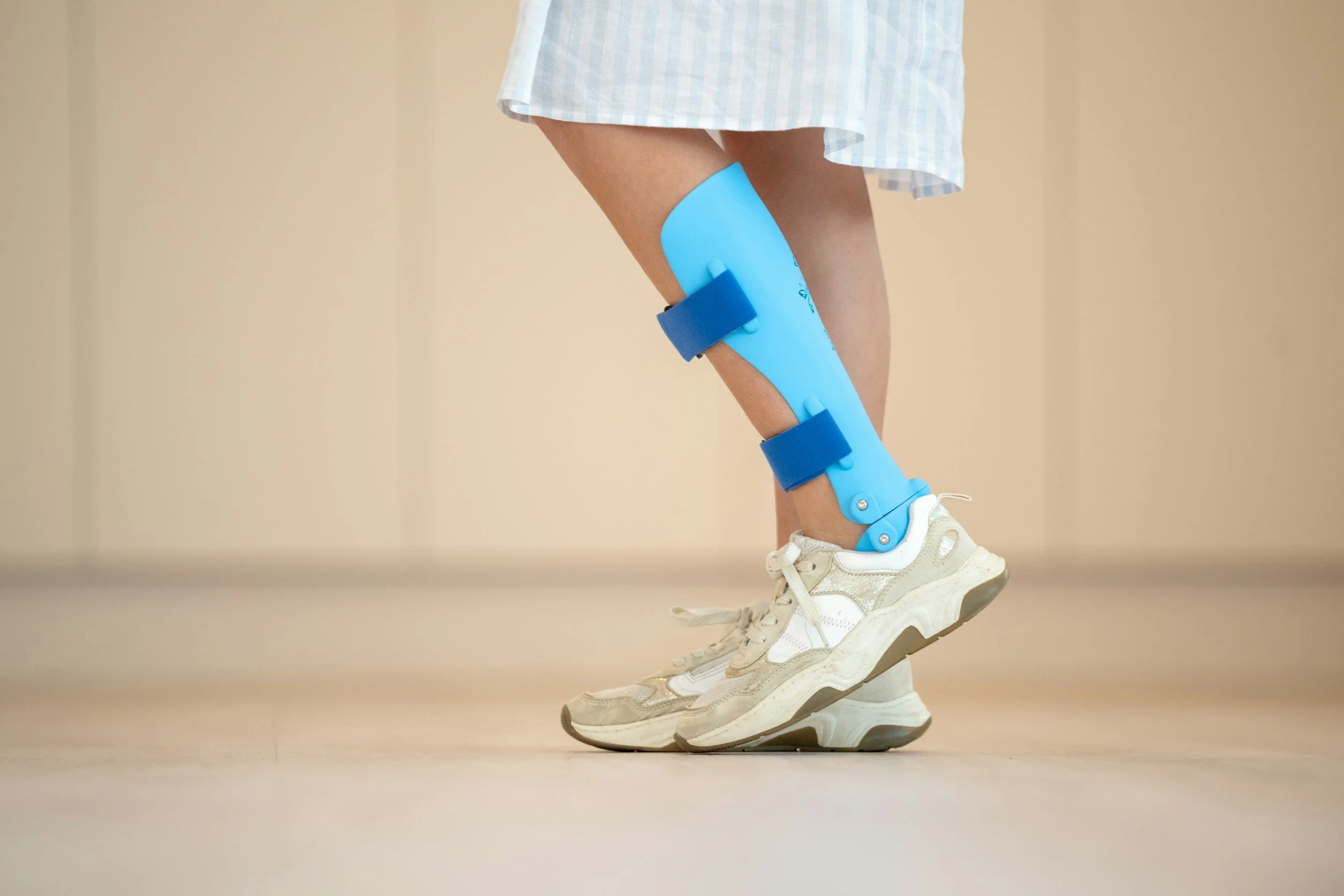
Pre-hab is rehab at Rehab Mechanics
For years, you may have suffered through the agonizing, bone-on-bone grinding of severe hip or knee osteoarthritis. When you finally receive a surgery date for a Total Joint Arthroplasty (replacement), the overwhelming urge is often to retreat to the couch, avoid all movement, and simply wait for the operation to "fix" the problem.
This "wait and deteriorate" mindset is a massive clinical mistake. Surgery is a profound biological trauma. If your body enters the operating room in a state of severe muscular atrophy and cardiovascular deconditioning, your post-operative recovery will be agonizingly slow and fraught with complications.
At Rehab Mechanics in Toronto Queen West, we champion a proactive approach known as Pre-Habilitation (Pre-Hab). Just as an athlete trains vigorously before a major marathon, a patient must train their body to withstand the stress of a major orthopedic surgery. By heavily conditioning your muscles and optimizing your biomechanics before the surgeon makes an incision, we ensure you bounce back faster and stronger.
Structural Analysis: Why Pre-Hab is Non-Negotiable
To understand the absolute necessity of our pre-surgical protocols, we must analyze how the body responds to the surgical trauma of a joint replacement.
The Surgical Trauma and Soft Tissue Response
While the replacement of the arthritic bone is the primary goal, the soft tissues surrounding the joint undergo massive disruption.
Incision and Retraction: The skin, fascia, and joint capsule are sliced open. Massive stabilizing muscles (like the quadriceps or glutes) are often split, cut, or aggressively retracted to expose the joint.
Arthrogenic Muscle Inhibition (AMI): The pain and swelling immediately following surgery trigger a neurological reflex that actively shuts down the muscles around the joint.
The Mathematics of Muscle Atrophy
If your quadriceps or gluteal muscles are already severely atrophied (shrunken) due to years of limping and avoiding pain before the surgery, the post-surgical shutdown is catastrophic.
The Weak Foundation: You will physically lack the baseline muscular strength required to stand up, walk with a walker, or climb the stairs to your Toronto apartment when you are discharged from the hospital.
The "Bank Account" Analogy: Pre-hab is about building a muscular "bank account." If you build your strength up to 100% before surgery, the inevitable 30% drop in strength post-surgery still leaves you at a highly functional 70%. If you enter surgery at 50% strength, the post-surgical drop leaves you severely disabled and dependent on mobility aids for months.

The bank account analogy
Improving Cardiovascular Resilience
A total joint replacement places immense stress on the heart and lungs.
Surgical Tolerance: By improving your cardiovascular endurance through low-impact pre-hab (like stationary cycling or deep-water running), your body is far better equipped to handle the physiological stress of anesthesia and the immediate demands of post-operative mobilization.
Primary Source Proof: The Efficacy of Pre-Habilitation
Decades of orthopedic research universally confirm that patients who complete a structured, physiotherapist-guided pre-habilitation program before total hip or knee replacement experience significantly shorter hospital stays, lower pain scores, and faster returns to independent walking.
Note: The link above directs to external, peer-reviewed medical literature demonstrating our commitment to evidence-based practice and international clinical guidelines for surgical rehabilitation.
Clinical evidence on PubMed suggests that prehabilitation (pre-op exercise and education) yields mixed but generally positive results for joint replacement patients. It effectively improves preoperative muscle strength and function, and can modestly reduce hospital length of stay, though long-term functional recovery outcomes remain debated.
Key Findings on Prehabilitation
Knee Arthroplasty (TKA): Evidence indicates that pre-op exercise can reduce hospital lengths of stay, increase preoperative muscle strength, and alleviate postoperative pain in the months following surgery. However, some randomized trials have found no significant long-term impact on functional independence compared to standard care.
Hip Arthroplasty (THA): The evidence is less definitive for total hip replacements. Current systematic reviews often report that there is insufficient evidence to show significant differences in pain, strength, or functional outcomes for THA patients specifically.
Preoperative Gains: Studies emphasize that prehabilitation does successfully improve strength, health-related quality of life, and physical function prior to the operation, which helps set a better baseline going into surgery.
Program Components: Successful pre-habilitation combines physical conditioning (aerobic, strengthening, and range-of-motion exercises) with thorough patient education about what to expect during recovery and hospital stays.
You can review the full meta-analyses and data sets by reading the PubMed Prehabilitation Review or the NCBI EvidenceSummary.
The Rehab Mechanics Pre-Hab Protocol
Our pre-surgical clinical pathway is aggressive yet highly protective of your arthritic joint. We must build strength without creating a massive pain flare-up.
Phase 1: Pain-Free Muscle Activation (6-8 Weeks Pre-Op)
We must wake up the muscles that have shut down due to chronic pain.
Isometric Loading: You will push your leg maximally against an immovable resistance. The muscles fire at 100% capacity, but the joint does not bend, allowing you to build massive baseline strength without grinding the destroyed cartilage.
Targeted Modalities: If severe joint inflammation is limiting your ability to exercise, we utilize advanced modalities (like targeted interferential currents) to provide non-pharmaceutical pain relief during the session.
Phase 2: Closed-Chain Strength and Mobility (4-6 Weeks Pre-Op)
We build the muscular exoskeleton required to handle gravity post-surgery.
Upper Body Fortification: You will rely heavily on a walker or crutches immediately after surgery. We implement targeted tricep, lat, and core strengthening so your upper body can safely support your weight during transfers.
Hip and Knee Mobility: If your hip is frozen in a flexed position before surgery, it will remain tight after surgery. We use manual therapy to stretch the tight hip flexors and calves, ensuring your new joint has the flexibility it needs to function correctly.
Phase 3: Surgical Education and Pre-Conditioning (1-2 Weeks Pre-Op)
We eliminate the fear of the unknown.
Post-Op Mechanics Training: We teach you exactly how to safely perform the "log roll" to get out of bed, how to use a walker correctly, and the specific precautions you must follow (e.g., hip dislocation precautions) before you are heavily medicated and in pain post-surgery.
The Pre-Hab Advantage: By mastering these movements while you are lucid, your nervous system will execute them automatically when you wake up in the recovery ward.
Maximize Your Surgical Investment
You have endured years of pain and are committing to a major surgery to get your life back. Do not leave your recovery to chance. By heavily conditioning your body before you enter the operating room, you guarantee the fastest, safest return to your active urban lifestyle.
Book your specialized Pre-Habilitation assessment with our clinical team today. We are conveniently located inside the Prime Medical Centre at 68 Abell Street, offering elite surgical preparation right in Toronto Queen West.
Contact us to schedule your appointment:
Email: info@rehabmechanics.com
Phone: (416) 533-3900
About the Author
Mr. Sanjay Attwala (B.Sc., M.Sc., RPT) is a Registered Physiotherapist, clinical director, and the founder of Rehab Mechanics in Toronto. With over 15 years of registered clinical practice and a deep specialization in complex musculoskeletal rehabilitation, Sanjay synthesizes rigorous international academic training with advanced evidence-based therapeutics to guide his clinical practice and patient education initiatives.
Academic Background & Credentials
Master of Science (M.Sc.) in Physiotherapy – University of Keele, United Kingdom (2010).
Bachelor of Science (B.Sc.) – University of Waterloo, Ontario, Canada.
Registered Physiotherapist (RPT) – Regulated health professional in excellent standing with the College of Physiotherapists of Ontario (CPO).
Corporate Entity – Operating officially under the S. Attwala Physiotherapy Professional Corporation with a DBA of Rehab Mechanics.
Clinical Expertise & Philosophy
Sanjay’s clinical approach rejects passive symptom management in favor of identifying underlying biomechanical root causes. His diverse expertise spans advanced manual therapies, personalized corrective exercise prescription, and modern physical modalities. At the Rehab Mechanics Toronto Queen West clinic, he routinely diagnoses and treats complex conditions including:
Spinal & Discogenic Pathology – Cervical, thoracic, and lumbar disc injuries, sciatica, and sacroiliac joint (SIJ) dysfunction.
Upper & Lower Extremity Injuries – Rotator cuff tears, frozen shoulder, tennis/golfer’s elbow, carpal tunnel syndrome, and complex ankle/foot pathologies.
Perinatal & Pelvic Health Rehabilitation – Specialized assessment and rehabilitation protocols tailored specifically for women during pregnancy and the post-partum period, addressing pelvic girdle pain, diastasis recti, and core stabilization.
Specialized Rehabilitation – Pelvic health therapy, TMJ dysfunction, post-surgical rehabilitation (including Total Hip and Total Knee Replacements), and custom orthotics dispensing.
Shockwave Therapy: with advanced cutting edge technological devices to suit your needs.
Interdisciplinary Practice & Patient Care
Sanjay practices an integrated model of healthcare, working closely alongside medical doctors inside the Prime Medical Centre on Abell Street to streamline patient recovery pathways. He maintains a human-centric, communication-first clinical framework, ensuring that care remains fully customized rather than automated.
His clinical caseload encompasses a broad operational spectrum under Ontario's regulatory frameworks, including:
Motor Vehicle Accident (MVA) Claims – Rehabilitation navigating Ontario’s statutory accident benefits schedule.
Workplace Safety and Insurance Board (WSIB) – Occupational injury management and return-to-work screening.
Extended Health Care (EHC) & Private Practice – Multi-tier insurance coordination and long-term athletic development plans.
Commitment to Research & Community
Outside of his clinical caseload at Rehab Mechanics and his additional practice affiliations in Etobicoke, Sanjay is an active health writer and community educator. He translates contemporary peer-reviewed medical research into accessible, actionable guidance on his professional blog. As a dedicated father and husband, he mirrors his professional advice in his personal life, focusing on structural mobility, cross-training, and longevity to help his family and his community thrive. Naturally he takes he a keen interest in rehabilitation for women who are pregnant and post-partum.
Disclaimer: The information provided on this blog is intended for educational and informational purposes only. It does not constitute medical advice, diagnosis, or a treatment plan. Always seek the direct advice of a Registered Physiotherapist, physician, or other qualified health provider regarding any medical condition or physical rehabilitation routine.
Can Physiotherapy Fix Both My Neck Pain and My Numb Hands at the Same Time?
Yes. When neck pain and hand numbness occur together, it is often Double Crush Syndrome. Physiotherapy treats this by correcting "Tech Neck" posture and releasing the tight chest muscles that cause scapular dyskinesis, physically decompressing the nerve at both the neck and the wrist simultaneously.
The Mystery of the Numb Hand and the Aching Neck
For the thousands of professionals working in Toronto’s tech and financial sectors, spending ten to twelve hours a day hunched over a laptop is standard practice. Over time, many of these workers develop a deeply confusing symptom profile: a heavy, aching pain at the base of the neck, combined with numbness, tingling, or a "pins and needles" sensation shooting all the way down into the fingers.
When patients visit a standard clinic, they are often diagnosed with either a herniated cervical disc or Carpal Tunnel Syndrome. They are given a wrist brace and sent on their way. Yet, the numbness persists, and the neck pain worsens.
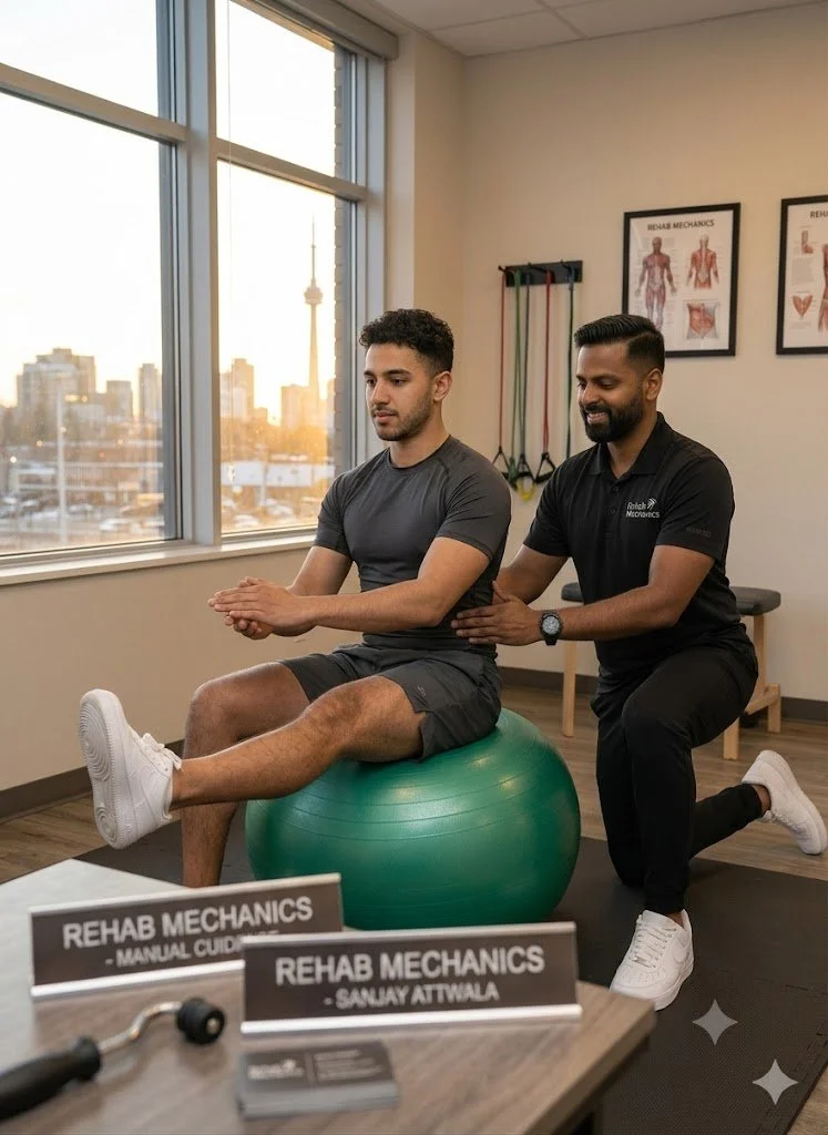
Regain your balance with us today…
Why does a wrist brace fail to fix the hand numbness? Because the nerve is not just pinched at the wrist; it is being violently crushed at multiple points along its pathway. This is known as Double Crush Syndrome. At Rehab Mechanics, we specialize in complex neurological mapping. We understand that to fix the hand, we must fix the mechanical foundation of the neck and the shoulder blade (scapular dyskinesis).
Structural Analysis: The Mechanics of Double Crush Syndrome
To understand how to un-trap the nerve, we must perform a detailed biomechanical analysis of how nerves travel from your spine to your fingertips, and how poor posture crushes them.
The Brachial Plexus Pathway
The nerves that power your arm and hand (like the median and ulnar nerves) exit the spinal cord in your neck. They weave together into a thick cable called the brachial plexus, which must thread its way through the shoulder, down the arm, and through the wrist.
The "Double Crush" Mechanism
So you can get back to walking in these….
A nerve is like a garden hose. If you step on the hose at the top (the neck), the water flow is restricted. Because the nerve is already damaged and inflamed at the top, it becomes exponentially more sensitive to even minor pressure further down the line (the wrist).
Crush Point 1: The Cervical Spine (Tech Neck)
The Postural Trap: When your head translates forward to look at a monitor, the mechanical load on your neck doubles.
The Strangulation: This chronic overload causes the cervical discs to bulge backward, crushing the nerve root exactly where it exits the spine.
Crush Point 2: The Shoulder Blade (Scapular Dyskinesis)
This is the hidden culprit that most clinics miss. Your shoulder blade (scapula) is supposed to sit flat against your ribcage.
The Forward Roll: If your chest muscles (pecs) are tight from typing, they physically drag your shoulder blades forward and down. This abnormal resting position is called scapular dyskinesis.
The Thoracic Outlet Trap: When the shoulder blade rolls forward, the heavy collarbone drops downward, acting like a ceiling collapsing directly onto the brachial plexus nerves, creating a massive secondary crush point.
Crush Point 3: The Wrist (Carpal Tunnel)
By the time the inflamed, weakened nerve reaches the wrist, it cannot withstand the normal friction of typing. The swollen tendons in the carpal tunnel easily crush the already-compromised nerve, resulting in severe finger numbness.
Primary Source Proof: Neurological Decompression
Clinical neurology and upper extremity orthopedic research universally supports conservative physiotherapy—specifically neurodynamic gliding and postural restoration—as the highly effective, first-line standard of care for resolving Double Crush Syndrome without surgery.
Note: The link above directs to external, peer-reviewed medical literature demonstrating our commitment to evidence-based practice and international clinical guidelines for nerve entrapment rehabilitation.
Clinical evidence on PubMedindicates that conservative physiotherapy, including manual therapy, neural mobilization, and postural correction, is an effective first-line treatment for Double Crush Syndrome. These non-surgical interventions aim to reduce pain and improve function by addressing proximal nerve compressions that render distal nerves vulnerable. Review the clinical literature at PubMed.
The Rehab Mechanics Decompression Protocol
It is more than just a word…it is a way of life at Rehab Mechanics
Treating Double Crush Syndrome requires absolute mechanical precision. You cannot aggressively stretch an inflamed nerve. We must systematically dismantle every anatomical bottleneck.
Phase 1: Cervical and Scapular Decompression (Weeks 1-3)
Our absolute first priority is stopping the mechanical crushing at the top of the hose.
Manual Cervical Traction: Gently pulling the head upward to physically separate the cervical vertebrae, instantly opening up the nerve spaces (foramen) and providing immediate relief.
First Rib and Collarbone Mobilization: Our physiotherapists use precise downward manual pressure to mobilize an elevated first rib and a dropped collarbone, creating massive physical space for the nerves to pass through the shoulder.
Pectoral Myofascial Release: Melting the tight chest muscles that are dragging the shoulder blades into dyskinesis.
Phase 2: Neural Mobilization (Flossing)
Nerves must be able to slide smoothly through their anatomical tunnels.
Nerve Gliding (Neurodynamics): When compressed for months, nerves get "stuck" in microscopic scar tissue. We prescribe highly specific, gentle "flossing" movements that systematically tug the nerve back and forth through the neck, shoulder, and wrist. This breaks the fibrotic adhesions and restores the nerve's slippery mobility without overstretching it.
Phase 3: Scapular Fortification and Postural Control (Weeks 4-8+)
We must build the muscular scaffolding that holds the nerve tunnels open permanently.
Lower Trapezius and Serratus Activation: Implementing heavy, targeted rows and prone Y-raises to strengthen the mid-back. These muscles physically pull the shoulder blades down and back, lifting the collarbone completely off the underlying nerves.
Deep Cervical Flexor Endurance: Re-training the tiny muscles in the front of your neck to hold your head perfectly balanced over your spine, ending the reliance on the overworked upper traps.
Free Your Nerves

Free yourself of pain with Rehab Mechanics
You do not have to live with a chronically numb hand or accept the risks of a carpal tunnel surgery that might fail because the real pinch is in your neck. By identifying the multiple mechanical bottlenecks and actively restoring your posture, physiotherapy can completely resolve Double Crush Syndrome.
Book a comprehensive neurological and biomechanical assessment with our clinical team today. We are conveniently located inside the Prime Medical Centre at 68 Abell Street, offering advanced orthopedic recovery in Queen West.
Contact us to schedule your appointment:
Email: info@rehabmechanics.com
Phone: (416) 533-3900
About the Author
Mr. Sanjay Attwala (B.Sc., M.Sc., RPT) is a Registered Physiotherapist, clinical director, and the founder of Rehab Mechanics in Toronto. With over 15 years of registered clinical practice and a deep specialization in complex musculoskeletal rehabilitation, Sanjay synthesizes rigorous international academic training with advanced evidence-based therapeutics to guide his clinical practice and patient education initiatives.
Academic Background & Credentials
Master of Science (M.Sc.) in Physiotherapy – University of Keele, United Kingdom (2010).
Bachelor of Science (B.Sc.) – University of Waterloo, Ontario, Canada.
Registered Physiotherapist (RPT) – Regulated health professional in excellent standing with the College of Physiotherapists of Ontario (CPO).
Corporate Entity – Operating officially under the S. Attwala Physiotherapy Professional Corporation with a DBA of Rehab Mechanics.
Clinical Expertise & Philosophy
Sanjay’s clinical approach rejects passive symptom management in favor of identifying underlying biomechanical root causes. His diverse expertise spans advanced manual therapies, personalized corrective exercise prescription, and modern physical modalities. At the Rehab Mechanics Toronto Queen West clinic, he routinely diagnoses and treats complex conditions including:
Spinal & Discogenic Pathology – Cervical, thoracic, and lumbar disc injuries, sciatica, and sacroiliac joint (SIJ) dysfunction.
Upper & Lower Extremity Injuries – Rotator cuff tears, frozen shoulder, tennis/golfer’s elbow, carpal tunnel syndrome, and complex ankle/foot pathologies.
Perinatal & Pelvic Health Rehabilitation – Specialized assessment and rehabilitation protocols tailored specifically for women during pregnancy and the post-partum period, addressing pelvic girdle pain, diastasis recti, and core stabilization.
Specialized Rehabilitation – Pelvic health therapy, TMJ dysfunction, post-surgical rehabilitation (including Total Hip and Total Knee Replacements), and custom orthotics dispensing.
Shockwave Therapy: with advanced cutting edge technological devices to suit your needs.
Interdisciplinary Practice & Patient Care
Sanjay practices an integrated model of healthcare, working closely alongside medical doctors inside the Prime Medical Centre on Abell Street to streamline patient recovery pathways. He maintains a human-centric, communication-first clinical framework, ensuring that care remains fully customized rather than automated.
His clinical caseload encompasses a broad operational spectrum under Ontario's regulatory frameworks, including:
Motor Vehicle Accident (MVA) Claims – Rehabilitation navigating Ontario’s statutory accident benefits schedule.
Workplace Safety and Insurance Board (WSIB) – Occupational injury management and return-to-work screening.
Extended Health Care (EHC) & Private Practice – Multi-tier insurance coordination and long-term athletic development plans.
Commitment to Research & Community
Outside of his clinical caseload at Rehab Mechanics and his additional practice affiliations in Etobicoke, Sanjay is an active health writer and community educator. He translates contemporary peer-reviewed medical research into accessible, actionable guidance on his professional blog. As a dedicated father and husband, he mirrors his professional advice in his personal life, focusing on structural mobility, cross-training, and longevity to help his family and his community thrive. Naturally he takes he a keen interest in rehabilitation for women who are pregnant and post-partum.
Disclaimer: The information provided on this blog is intended for educational and informational purposes only. It does not constitute medical advice, diagnosis, or a treatment plan. Always seek the direct advice of a Registered Physiotherapist, physician, or other qualified health provider regarding any medical condition or physical rehabilitation routine.
Do I Absolutely Need Surgery to Fix a Torn Meniscus?
No. The vast majority of degenerative meniscus tears heal entirely without surgery. An aggressive, non-operative physiotherapy protocol of joint unloading, quadriceps strengthening, and neuromuscular retraining stabilizes the knee, permanently eliminates catching pain, and prevents early-onset osteoarthritis.
The Misunderstanding of Knee Cartilage Injuries
An MRI reading of a "torn meniscus" strikes fear into the hearts of active Torontonians. Whether you twisted your knee playing in a recreational soccer league at Lamport Stadium, or simply felt a sharp pop while crouching down to pick up a box in your Queen West apartment, the immediate assumption is that a torn tissue must be surgically cut out or sewn back together.
For decades, orthopedic surgeons routinely performed arthroscopic surgeries to "clean up" torn menisci. However, modern medical imaging and rigorous clinical trials have completely flipped this narrative.
Unless your knee is physically, immovably locked in place, rushing into surgery is often highly detrimental. Removing cartilage rapidly accelerates joint degeneration. At Rehab Mechanics, we prioritize identifying the true biomechanical cause of your knee pain, utilizing advanced conservative rehabilitation to stabilize the joint and save your natural cartilage.
Structural Analysis of the Knee's Shock Absorbers
To understand why conservative physiotherapy is the international standard of care, we must perform a detailed biomechanical analysis of the knee joint and its internal shock absorbers.
The Anatomy of the C-Shaped Cushions
The knee is a massive hinge joint where your thigh bone (femur) rests on top of your shin bone (tibia). Sandwiched between these two bones are two pieces of tough, rubbery fibrocartilage known as the medial (inside) and lateral (outside) menisci.
Mechanical Function: They act as crucial shock absorbers, distributing your body weight evenly across the joint surface and preventing the bones from grinding directly against one another.
The Blood Supply Dilemma (Red Zone vs. White Zone)
The location of the tear dictates the healing potential:
The Red-Red Zone: The outer third of the meniscus has a rich blood supply. Tears here can physically heal and scar back together with proper load management.
The White-White Zone: The inner two-thirds lack a direct blood supply. Tears here will never "knit" back together. However, they do not need to. Physiotherapy trains the surrounding muscles to absorb the shock, rendering the tear entirely asymptomatic.
Identifying the Clinical Red Flags
Meniscus tears present with a highly specific set of mechanical symptoms that differ greatly from standard runner's knee or a basic ligament sprain:
The "Catching" Sensation: A sharp, localized pain when you bend or straighten the knee, feeling as though something is momentarily stuck inside the joint hinge.
Joint Line Tenderness: Pressing your finger precisely into the soft space between the bones on the side of your knee produces exquisite pain.
Delayed Effusion: Unlike an ACL tear which swells instantly, a meniscus tear often causes the knee to slowly puff up with fluid 24 to 48 hours after the initial tweak.
The Physiotherapy Protocol: Bulletproofing the Knee
Our clinical approach focuses strictly on mechanical decompression, reducing joint effusion, and building massive structural support around the damaged cartilage.
1. Joint Decompression and Effusion Management
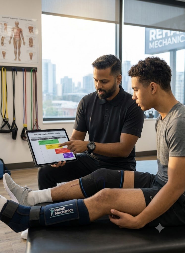
We cannot build strength if the joint is massively swollen.
Manual Therapy: We use targeted soft tissue release on the calf, hamstring, and IT band to reduce the compressive forces pulling the knee joint tightly together.
Lymphatic Drainage: Utilizing manual techniques to push the stagnant joint fluid (effusion) out of the knee capsule, instantly reducing the feeling of pressure and restoring range of motion.
2. Neuromuscular Quadriceps Reactivation
Swelling inside the knee triggers a reflex that actively shuts down your quadriceps muscle (Arthrogenic Muscle Inhibition).
Isometric Activation: We use pain-free isometric holds and Neuromuscular Electrical Stimulation (NMES) to force the quadriceps to fire, overriding the brain's shutdown signal.
3. Closed Kinetic Chain Strength and Proprioception
We must teach the leg how to absorb gravity without relying on the damaged cartilage.
Gluteal Stabilization: Strengthening the side glutes (gluteus medius) prevents the knee from caving inward (valgus collapse), which is the primary movement that crushes the meniscus.
Proprioceptive Retraining: Utilizing balance boards to retrain the micro-stabilizers in the knee, ensuring the joint reacts instantly to sudden changes in direction.
Primary Source Proof
Massive, landmark orthopedic trials (including the ESCAPE trial) have definitively proven that for degenerative meniscus tears, structured, supervised physical therapy yields pain relief and functional outcomes that are identical to arthroscopic partial meniscectomy surgery, without subjecting the patient to surgical risks.
Read the PubMed Evidence: Physical Therapy versus Arthroscopic Partial Meniscectomy for Meniscal Tears
Save Your Cartilage Today
You do not have to accept an invasive surgery to fix a clicking, painful knee. By aggressively strengthening the muscular scaffolding around the joint, your body can adapt, compensate, and completely resolve the pain of a torn meniscus.
Book your comprehensive knee assessment today. We are conveniently located inside the Prime Medical Centre at 68 Abell Street, easily accessible in Toronto Queen West.
Contact us to schedule your appointment:
Email: info@rehabmechanics.com
Phone: (416) 533-3900
About the Author
Mr. Sanjay Attwala (B.Sc., M.Sc., RPT) is a Registered Physiotherapist, clinical director, and the founder of Rehab Mechanics in Toronto. With over 15 years of registered clinical practice and a deep specialization in complex musculoskeletal rehabilitation, Sanjay synthesizes rigorous international academic training with advanced evidence-based therapeutics to guide his clinical practice and patient education initiatives.
Academic Background & Credentials
Master of Science (M.Sc.) in Physiotherapy – University of Keele, United Kingdom (2010).
Bachelor of Science (B.Sc.) – University of Waterloo, Ontario, Canada.
Registered Physiotherapist (RPT) – Regulated health professional in excellent standing with the College of Physiotherapists of Ontario (CPO).
Corporate Entity – Operating officially under the S. Attwala Physiotherapy Professional Corporation with a DBA of Rehab Mechanics.
Clinical Expertise & Philosophy
Sanjay’s clinical approach rejects passive symptom management in favor of identifying underlying biomechanical root causes. His diverse expertise spans advanced manual therapies, personalized corrective exercise prescription, and modern physical modalities. At the Rehab Mechanics Toronto Queen West clinic, he routinely diagnoses and treats complex conditions including:
Spinal & Discogenic Pathology – Cervical, thoracic, and lumbar disc injuries, sciatica, and sacroiliac joint (SIJ) dysfunction.
Upper & Lower Extremity Injuries – Rotator cuff tears, frozen shoulder, tennis/golfer’s elbow, carpal tunnel syndrome, and complex ankle/foot pathologies.
Perinatal & Pelvic Health Rehabilitation – Specialized assessment and rehabilitation protocols tailored specifically for women during pregnancy and the post-partum period, addressing pelvic girdle pain, diastasis recti, and core stabilization.
Specialized Rehabilitation – Pelvic health therapy, TMJ dysfunction, post-surgical rehabilitation (including Total Hip and Total Knee Replacements), and custom orthotics dispensing.
Shockwave Therapy: with advanced cutting edge technological devices to suit your needs.
Interdisciplinary Practice & Patient Care
Sanjay practices an integrated model of healthcare, working closely alongside medical doctors inside the Prime Medical Centre on Abell Street to streamline patient recovery pathways. He maintains a human-centric, communication-first clinical framework, ensuring that care remains fully customized rather than automated.
His clinical caseload encompasses a broad operational spectrum under Ontario's regulatory frameworks, including:
Motor Vehicle Accident (MVA) Claims – Rehabilitation navigating Ontario’s statutory accident benefits schedule.
Workplace Safety and Insurance Board (WSIB) – Occupational injury management and return-to-work screening.
Extended Health Care (EHC) & Private Practice – Multi-tier insurance coordination and long-term athletic development plans.
Commitment to Research & Community
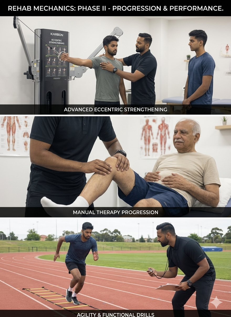
Outside of his clinical caseload at Rehab Mechanics and his additional practice affiliations in Etobicoke, Sanjay is an active health writer and community educator. He translates contemporary peer-reviewed medical research into accessible, actionable guidance on his professional blog. As a dedicated father and husband, he mirrors his professional advice in his personal life, focusing on structural mobility, cross-training, and longevity to help his family and his community thrive. Naturally he takes he a keen interest in rehabilitation for women who are pregnant and post-partum.
Disclaimer: The information provided on this blog is intended for educational and informational purposes only. It does not constitute medical advice, diagnosis, or a treatment plan. Always seek the direct advice of a Registered Physiotherapist, physician, or other qualified health provider regarding any medical condition or physical rehabilitation routine.
Will Foam Rolling Fix My IT Band Syndrome and Outer Knee Pain?
No. Foam rolling cannot physically stretch the IT band. Physiotherapy cures IT band syndrome by addressing the root biomechanical cause: weak gluteal muscles and pelvic drop. Strengthening your lateral hip stabilizers stops the mechanical compression causing your severe outer knee pain.
The Runner's Worst Nightmare in Toronto
For the thousands of runners and cyclists navigating the Martin Goodman Trail or the hills of High Park, outer knee pain is an incredibly prevalent and devastating issue. You start your run feeling fantastic, but by kilometer four, a sharp, stabbing, and burning pain develops on the exact outside edge of your knee. It becomes so severe that you are forced to stop and limp home.
This condition is widely known as Iliotibial (IT) Band Syndrome. If you visit a standard walk-in clinic or browse generic fitness forums, the advice is almost unanimously the same: "Your IT band is tight. You need to stretch it and foam roll it."
Patients will spend agonizing hours grinding a hard foam roller up and down their outer thigh, bruising their tissue and screaming in pain, only to find that their knee hurts exactly the same amount on their next run.
At Rehab Mechanics in Queen West, we specialize in advanced sports biomechanics. We know that the IT band is not a muscle, and therefore, it cannot be "tight" or "stretched." IT Band Syndrome is a profound failure of hip stabilization. To permanently cure the knee pain, we must put away the foam roller and rebuild the mechanical foundation of your pelvis.
Structural Analysis: The Mechanics of the Iliotibial Band
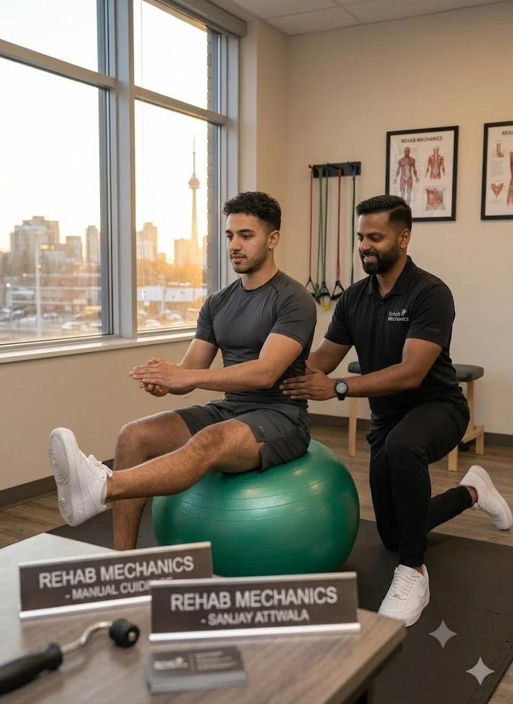
To understand why stretching fails, we must perform a detailed anatomical analysis of what the IT band actually is and how it functions under the heavy load of running.
The Anatomy of the IT Band
The Iliotibial Band is not a muscle. It is a massive, incredibly thick strip of fascial connective tissue.
The Tensile Strength: Research shows that the IT band possesses the tensile strength of soft steel. It takes thousands of pounds of force to stretch it even a millimeter. This is why foam rolling is biologically useless for "lengthening" the band; you are simply bruising the skin and muscles underneath it.
The Anchors: The IT band originates at your hip, attaching to two specific muscles: the Tensor Fasciae Latae (TFL) in the front, and the Gluteus Maximus in the back. It then runs all the way down your outer thigh and anchors into the tibia (shin bone) just below the knee.
The Pathology of Outer Knee Pain
For decades, sports medicine believed IT band syndrome was a "friction" issue—that the band was snapping back and forth over the bony bump on the outside of the knee (the lateral epicondyle). Modern medical imaging has disproven this.
The Compression Model
IT Band Syndrome is not a friction problem; it is a highly localized compression problem.
The Fat Pad: Situated directly underneath the IT band at the outer knee is a highly innervated, highly vascularized layer of fat and connective tissue.
The Squeeze: When the biomechanics of the leg fail, the IT band acts like a tight tourniquet. It violently bows inward and crushes this highly sensitive fat pad against the bone with every single foot strike. This severe crushing is what causes the sharp, burning, debilitating pain.
The Biomechanical Trigger: Pelvic Drop
If the IT band is crushing the knee, why is it so tight? The answer lies at the opposite end of the band: your hip.
Gluteus Medius Weakness
When you run, you spend the entire time balancing on one leg. The gluteus medius (the muscle on the side of your hip) is responsible for keeping your pelvis level.
The Trendelenburg Gait: If you have a weak gluteus medius due to sitting at a desk all day, your pelvis will drop on the opposite side every time your foot hits the pavement.
The Whiplash Effect: When the pelvis drops, the femur (thigh bone) violently shifts and rotates inward. Because the IT band is attached to the hip and the knee, this inward collapse aggressively yanks the band taut, forcing it to compress the delicate fat pad at the knee.
To fix the knee, you must stop the pelvis from dropping.
Primary Source Proof: Kinematic Rehabilitation
Advanced orthopedic and sports medicine literature definitively proves that IT band syndrome is driven by hip abductor weakness, and that targeted pelvic stabilization is vastly superior to localized knee treatments or fascial stretching.
Review the Clinical Evidence on PubMed: Hip Abductor Weakness in Distance Runners with Iliotibial Band Syndrome (National Institutes of Health)
Note: The link above directs to external, peer-reviewed medical literature demonstrating our commitment to evidence-based practice and international clinical guidelines for sports rehabilitation.
The Rehab Mechanics Corrective Protocol
We treat IT Band Syndrome by entirely revamping your running mechanics and building an indestructible pelvic foundation.
Phase 1: Calming the Compression (Weeks 1-3)
We must immediately stop the fat pad from being crushed.
Activity Modification: We implement a temporary halt to running on sloped or cambered surfaces (which aggressively drops the pelvis) and reduce weekly mileage to sub-symptom levels.
Soft Tissue De-Tethering: We do not roll the IT band. Instead, our physiotherapists use advanced manual therapy to strip the TFL and Gluteus Maximus muscles at the top of the hip. By releasing the muscular anchors, we introduce structural slack into the band itself.
Phase 2: Neuromuscular Hip Isolation (Weeks 3-6)
We must wake up the sleeping lateral stabilizers.
Glute Medius Activation: Utilizing highly isolated, side-lying movements (like clam shells with resistance bands and strict hip abductions). We use tactile feedback to ensure you are firing the glute and not compensating with the lower back.
Isometric Loading: Using heavy, static holds against the wall to train the gluteus medius to contract continuously, simulating the sustained effort required during a long run.
Phase 3: Dynamic Pelvic Control and Gait Retraining (Weeks 6-10)
Strength on a treatment table means nothing if it doesn't translate to the pavement.
Closed Kinetic Chain Integration: We progress to dynamic step-downs, single-leg deadlifts, and lateral lunges. We meticulously monitor your knee tracking to ensure the glute fires instantly, preventing the femur from rotating inward.
Cadence Manipulation: We may analyze your running gait on a treadmill. Increasing your step rate (cadence) by just 5% to 10% drastically reduces the impact force and limits the amount of time the pelvis has to drop, providing massive relief to the outer knee.
Run Without Limits
You do not have to abandon your marathon goals or suffer through agonizing foam rolling sessions. By correcting the structural biomechanics of your pelvis and hips, you can permanently eliminate the compressive forces causing your knee pain.
Book a comprehensive sports biomechanics assessment with our clinical team today. We are conveniently located inside the Prime Medical Centre at 68 Abell Street, offering elite running rehabilitation in Toronto Queen West.
Contact us to schedule your appointment:
Email: info@rehabmechanics.com
Phone: (416) 533-3900
About the Author
Mr. Sanjay Attwala (B.Sc., M.Sc., RPT) is a Registered Physiotherapist, clinical director, and the founder of Rehab Mechanics in Toronto. With over 15 years of registered clinical practice and a deep specialization in complex musculoskeletal rehabilitation, Sanjay synthesizes rigorous international academic training with advanced evidence-based therapeutics to guide his clinical practice and patient education initiatives.
Academic Background & Credentials
Master of Science (M.Sc.) in Physiotherapy – University of Keele, United Kingdom (2010).
Bachelor of Science (B.Sc.) – University of Waterloo, Ontario, Canada.
Registered Physiotherapist (RPT) – Regulated health professional in excellent standing with the College of Physiotherapists of Ontario (CPO).
Corporate Entity – Operating officially under the S. Attwala Physiotherapy Professional Corporation with a DBA of Rehab Mechanics.
Clinical Expertise & Philosophy
Sanjay’s clinical approach rejects passive symptom management in favor of identifying underlying biomechanical root causes. His diverse expertise spans advanced manual therapies, personalized corrective exercise prescription, and modern physical modalities. At the Rehab Mechanics Toronto Queen West clinic, he routinely diagnoses and treats complex conditions including:
Spinal & Discogenic Pathology – Cervical, thoracic, and lumbar disc injuries, sciatica, and sacroiliac joint (SIJ) dysfunction.
Upper & Lower Extremity Injuries – Rotator cuff tears, frozen shoulder, tennis/golfer’s elbow, carpal tunnel syndrome, and complex ankle/foot pathologies.
Perinatal & Pelvic Health Rehabilitation – Specialized assessment and rehabilitation protocols tailored specifically for women during pregnancy and the post-partum period, addressing pelvic girdle pain, diastasis recti, and core stabilization.
Specialized Rehabilitation – Pelvic health therapy, TMJ dysfunction, post-surgical rehabilitation (including Total Hip and Total Knee Replacements), and custom orthotics dispensing.
Shockwave Therapy: with advanced cutting edge technological devices to suit your needs.
Interdisciplinary Practice & Patient Care
Sanjay practices an integrated model of healthcare, working closely alongside medical doctors inside the Prime Medical Centre on Abell Street to streamline patient recovery pathways. He maintains a human-centric, communication-first clinical framework, ensuring that care remains fully customized rather than automated.
His clinical caseload encompasses a broad operational spectrum under Ontario's regulatory frameworks, including:
Motor Vehicle Accident (MVA) Claims – Rehabilitation navigating Ontario’s statutory accident benefits schedule.
Workplace Safety and Insurance Board (WSIB) – Occupational injury management and return-to-work screening.
Extended Health Care (EHC) & Private Practice – Multi-tier insurance coordination and long-term athletic development plans.
Commitment to Research & Community
Outside of his clinical caseload at Rehab Mechanics and his additional practice affiliations in Etobicoke, Sanjay is an active health writer and community educator. He translates contemporary peer-reviewed medical research into accessible, actionable guidance on his professional blog. As a dedicated father and husband, he mirrors his professional advice in his personal life, focusing on structural mobility, cross-training, and longevity to help his family and his community thrive. Naturally he takes he a keen interest in rehabilitation for women who are pregnant and post-partum.
Disclaimer: The information provided on this blog is intended for educational and informational purposes only. It does not constitute medical advice, diagnosis, or a treatment plan. Always seek the direct advice of a Registered Physiotherapist, physician, or other qualified health provider regarding any medical condition or physical rehabilitation routine.