
The Ultimate Guide to Correcting Postural Dysfunction in the Modern Workplace
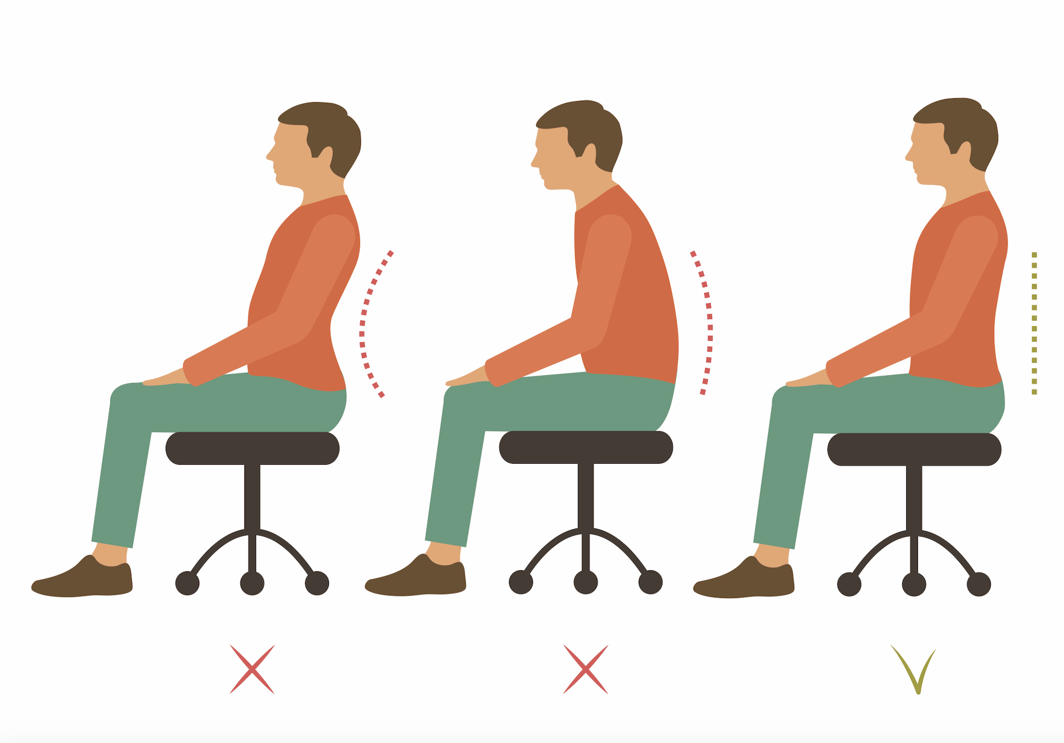
The Ultimate Guide to Correcting Postural Dysfunction in the Modern Workplace
March 31, 2026 (Updated November 4, 2026)
In modern musculoskeletal medicine, ergonomic kinesiology, and our Physiotherapy & Sports Medicine Blog, postural dysfunction defines the systemic biomechanical failure that occurs when the spine and its accompanying joints deviate from their natural, neutral alignment for extended periods. Far from simply looking "slouched," this continuous deviation places unnatural, asymmetrical stress on ligaments, tendons, and muscles, triggering a cascade of painful compensatory patterns across the entire body. When one structural area falls out of alignment, the nervous system forces neighboring joints to compensate, creating a doming effect of chronic tension, localized ischemia, and fatigue. Physiotherapy in Toronto at Queen West Rehab Mechanics assists desk professionals, remote workers, and urban commuters in overcoming workspace strain by conducting comprehensive biomechanical movement assessments to isolate muscular imbalances and joint restrictions, deploying targeted manual therapy and soft-tissue release, and utilizing progressive neuromuscular re-education alongside specialized Heavy Slow Resistance (HSR) loading via our core clinical platform at Rehab Mechanics.
The Patient’s Story / Toronto Context
Across downtown Toronto’s corporate towers, glass-walled financial houses, and creative digital agency lofts—from open-concept workspaces in Liberty Village and King West to administrative offices in the Financial District along Bay Street—office professionals, remote software developers, and administrative workers subject their spines to static, screen-heavy sedentary postures for eight to twelve hours a day.
Consider a 36-year-old remote product designer and software architect living near King West or Liberty Village who spends their entire workday leaning forward over dual monitors with their chin protracted and their thoracic spine locked in persistent, rounded flexion.
Throughout the week, they experience The Afternoon Postural Collapse.
It begins as a mild, dull fatigue between the shoulder blades by mid-afternoon. Within weeks, however, it escalates into intense tension headaches, burning interscapular pain, and an unyielding ache across the lower lumbar spine every time they stand up from their desk.
Following standard office wellness advice, they fall into The Willpower & Ergonomic Chair Trap.
They purchase an expensive ergonomic office chair, set reminders to "sit up straight," and try to force their shoulders backward using sheer willpower.
Instead of clearing, the fatigue intensifies, their lower back arches into excessive lordosis, and their neck develops a persistent, throbbing ache. Why? Because posture is heavily governed by the nervous system and long-standing muscular adaptations; it cannot be fixed by willpower alone. Forcing yourself into rigid posture without releasing restricted soft tissues or strengthening underactive stabilizers simply overworks adjacent joints.
Inside our private treatment rooms at Rehab Mechanics inside the Prime Medical Centre at 68 Abell Street in Queen West, we show patients why postural dysfunction requires active, science-based rehabilitation rather than passive reminders. True recovery demands clearing joint restrictions via advanced soft-tissue mobilization (IASTM), restoring deep cervical flexor endurance, and integrating pain-free alignment across Toronto.
☕ Struggling with desk-related neck pain, tension headaches, or lower back stiffness? Explore our full suite of clinical services or consult our comprehensive Physiotherapy Topics Index.
Structural / Biomechanical Analysis
To understand why modern desk lifestyles devastate our natural biomechanics—and how physical therapy restores relaxed, dynamic neutral posture—we must examine spinal curve dynamics, ischemic muscle trigger points, and the "tech neck" physical multiplier.
+---------------------------------------------------------------------------------------------------+
| POSTURAL DYSFUNCTION: THE STATIC SLOUCH TRAP VS. DYNAMIC NEUTRAL ALIGNMENT |
+---------------------------------------------------------------------------------------------------+
| |
| [A] THE STATIC DESK-BOUND SLOUCH [B] THE REHAB MECHANICS DYNAMIC NEUTRAL |
| (Prolonged C-Shape Flexion + Ischemic Spasms) (Manual Release + Neuromuscular Retraining) |
| |
| Prolonged Sitting / 8-12 Hours Static Posture Targeted Manual Therapy & Joint Mobilization|
| + Psoas Shortening & Gluteal Amnesia + Deep Cervical Flexor & Lower Trap Training|
| | | |
| v v |
| Diaphragmatic Compression & Apical Breathing Restored Spinal Curve Compliance: |
| (Limits Lung Capacity & Elevates Cortisol) (Absorbs Shock & Distributes Load Evenly) |
| | | |
| v v |
| Anterior Head Carriage & Tech Neck Torque Dynamic, Relaxed Neutral Posture: |
| (Up to 60 lbs of Force on Cervical Spine) (Effortless Standing & Seated Alignment) |
| | | |
| +--------------------+--------------------+ +-----------------+--------------------+ |
| | THE WILLPOWER FAILURE TRAP | | THE CLINICAL POSTURAL CURE | |
| | • Forcing rigid, military-style posture| | • Decompresses cervical & lumbar discs| |
| | • Overworks lats and traps into spasm | | • Restores 360° diaphragmatic breathing| |
| | • Fails to address fascial remodeling | | • Melts fibrotic ischemic trigger points|
| | • Chronic recurrence in Toronto offices| | • Permanent athletic & desk longevity | |
| +-----------------------------------------+ +----------------------+---------------+ |
1. The Anatomy of the Spine Under Stress and the "Tech Neck" Multiplier
The human spine is a marvel of biological engineering, designed with three natural, alternating curves (cervical lordosis, thoracic kyphosis, and lumbar lordosis) to act as a coiled spring. These curves absorb shock, distribute weight evenly, and permit explosive movement:
Compromised Biomechanics: When these curves are flattened or exaggerated, the biomechanics of the entire body shift. Muscles designed for quick, dynamic movements are suddenly forced to act as static postural stabilizers, a job they inevitably fail at—resulting in painful spasms and ischemic trigger points (how stress creates high resting muscle tone).
The "Tech Neck" Multiplier: "Tech neck," or anterior head carriage, occurs when the head continuously juts forward toward a screen. In a neutral position, the adult head weighs about 10 to 12 pounds. However, physics dictates that for every single inch the head moves forward past the neutral shoulder line, the weight borne by the delicate cervical spine effectively doubles. Looking down at a smartphone places up to 60 pounds of force on the neck.
Permanent Structural Damage: Over years, this relentless strain leads to tension headaches, TMJ irritation, and discogenic injuries. The body responds by laying down extra bone to reinforce the unstable area—forming osteophytes (bone spurs) that permanently narrow the space available for nerves, turning a correctable muscular issue into irreversible structural pathology.
Postural Parameter
The Static Desk Slouch
The Rehab Mechanics Dynamic Neutral
Cervical Alignment
Anterior head carriage ("Tech Neck"; 60 lbs of shear)
Neutral cervical lordosis; balanced head-on-neck balance
Thoracic Spine
Exaggerated kyphosis (Rounded mid-back)
Clean, compliant thoracic extension and rotation
Breathing Mechanics
Shallow apical chest breathing (Elevates cortisol)
360-degree diaphragmatic piston excursion
Muscle Activation
Ischemic traps and lats acting as static braces
Coordinated deep stabilizers (Deep neck flexors, TrA)
2. The Biomechanical Tipping Point
The tipping point for postural breakdown occurs when prolonged fascial remodeling and myofascial densification lock the body into a permanent C-shape:
A remote worker in King West can sit in a slouched posture for weeks without acute symptoms. However, the moment fascial sheets dehydrate and cross-link into a rigid C-shape, the body adapts permanently to the position it spends 80% of its time in. Forcing yourself into rigid, military-style posture without releasing these fascial adhesions creates new muscular strains, requiring scientific clinical intervention.
Clinical Red Flags
While common desk-bound postural fatigue responds exceptionally well to physical therapy and ergonomic optimization, clinicians must rigorously screen for specific neurological, vascular, and structural red flags that demand immediate medical escalation:
Progressive Radicular Neuropathy & Motor Weakness: Sharp, electric-shock pain shooting down the arm or leg, accompanied by dermatomal sensory numbness, diminished deep tendon reflexes, or objective motor weakness (such as foot drop or hand grip failure), signaling nerve root compression requiring urgent neurosurgical evaluation.
Cervical Myelopathy Signs: Clumsy hands, difficulty buttoning shirts, dropping items, stumbling gait, or bilateral lower-extremity hyperreflexia, demanding urgent MRI imaging and surgical consultation.
Vertebral Artery Compression / Vascular Dizziness: Sudden-onset dizziness, double vision, or drop attacks triggered by neck movement, requiring immediate vascular triage.
Non-Mechanical Unremitting Night Pain: Severe spine pain that remains completely unchanged by positional unloading, rest, or ergonomic modification, accompanied by unexplained weight loss, demanding immediate oncological investigation.
Primary Source Proof (PubMed / NIH)
Extensive peer-reviewed occupational health, ergonomics, and physical therapy literature confirm that static seated posture without micro-breaks accelerates spinal disc degeneration, that anterior head carriage significantly increases cervical shear forces, and that targeted manual therapy combined with neuromuscular re-education successfully restores dynamic neutral posture and eliminates work-related neck and back pain.
Review the Clinical Evidence on PubMed: Postural Dysfunction and Ergonomic Stress in Desk-Bound Occupations: A Systematic Review of Spinal Degeneration and Pain Mechanisms (National Institutes of Health / Ergonomics)
Review the Clinical Evidence on PubMed: Cervical Spine Biomechanics of Anterior Head Carriage and the Efficacy of Targeted Deep Neck Flexor Training (National Institutes of Health / Journal of Orthopaedic & Sports Physical Therapy)
The Rehab Mechanics Corrective Protocol
Inside our Queen West clinic at 68 Abell Street, our postural rehabilitation protocol rejects ineffective reminders ("sit up straight") for precision soft-tissue release, ergonomic audits, and neuromuscular re-education. Our four-phase protocol clears fascial restrictions, retrains deep stabilizers, and builds permanent postural endurance.
Phase 1 — Comprehensive Biomechanical & Ergonomic Audit:
Movement and Posture Mapping: Conducting our signature Full-Body Biomechanical Assessment to evaluate static alignment, joint mobility, and movement patterns during squats and overhead reaches.
Workplace Ergonomic Optimization: Auditing desk heights, monitor placements, chair lumbar supports, and lighting to eliminate postural triggers (workplace ergonomic optimization).
Phase 2 — Clearing the Rust (Manual Soft-Tissue Release):
Myofascial Decompression: Utilizing active release, trigger point therapy, and joint mobilizations (manual therapy and joint mobilizations) to release chronically tight muscles (upper trapezius and pectorals) and clear restrictive fascial adhesions.
Phase 3 — Neuromuscular Re-Education & Corrective Exercise:
Targeted Stabilizer Activation: Prescribing precise, progressive corrective exercises (such as wall angels, chin tucks with pressure biofeedback, and DeadBug core stabilization) to retrain endurance in underactive lower traps and deep neck flexors.
Phase 4 — Heavy Slow Resistance (HSR) & Workplace Carryover:
Building Structural Resilience: Advancing to progressive Heavy Slow Resistance (HSR) training to ensure your posture holds firm under heavy gym loads and long desk shifts across Toronto.
Related Conditions We Treat
Workplace Postural Dysfunction & "Tech Neck"
Chronic Tension Headaches & Suboccipital Guarding
Upper Cross Syndrome & Pectoralis Tightness
Occupational Low Back Spasms & Fatigue
Related Blogs
The Ultimate Guide to Correcting Postural Dysfunction in the Modern Workplace
Can Poor Ergonomics Permanently Damage My Spine and Posture?
Can Physiotherapy Cure Chronic Tension Headaches from Desk Work
Inside a Full-Body Biomechanical Assessment: Finding the Root Cause (Part 2 of 3)
Services Used in Treatment
FAQ Section
Why is telling someone to 'sit up straight' ineffective for postural dysfunction? Posture is heavily influenced by the nervous system and long-standing muscular adaptations; it cannot be fixed by willpower alone. Forcing yourself into rigid posture usually results in arching the lower back excessively, creating an entirely new set of problems.
How does physical therapy assist in correcting workplace postural dysfunction? Physiotherapy at our Queen West clinic supports recovery by conducting full-body biomechanical assessments, performing ergonomic workstation audits, releasing tight soft tissues via manual therapy, and prescribing progressive corrective exercises to rebuild stabilizer endurance.
What is 'tech neck' and how much extra weight does it place on the cervical spine? Tech neck is anterior head carriage where the head juts forward toward a screen. For every single inch the head moves forward past the shoulder line, the weight borne by the cervical spine doubles—placing up to 60 pounds of force on the neck.
Can poor desk ergonomics cause permanent bone spurs (osteophytes)? Yes. When the body is subjected to excessive localized stress from forward head posture over years, it responds by laying down extra bone to reinforce the unstable area, forming permanent bone spurs that can narrow the space available for nerves.
How do myofascial trigger points in the upper back cause headaches? Overworked muscles like the upper trapezius and suboccipitals become ischemic (starved of blood flow) and develop dense trigger points that radiate pain upward into the scalp, temples, and forehead, generating tension headaches.
What is the difference between static stretching and neuromuscular re-education? Static stretching temporarily lengthens muscle tissue, whereas neuromuscular re-education trains the brain and nervous system to unconsciously recruit stabilizers (like deep neck flexors and lower traps) to maintain healthy alignment without constant conscious effort.
How long does it take to correct years of poor desk posture with physical therapy? While manual therapy provides immediate relief from tight muscles within your first few treatment sessions, permanently retraining neuromuscular habits and strengthening postural stabilizers typically requires $4\text{ to }8\text{ weeks}$ of consistent clinical and workspace practice.
What is the Rehab Mechanics approach to postural rehabilitation? We combine comprehensive movement assessments with targeted manual soft-tissue therapy, ergonomic optimization, and progressive corrective exercise to build a relaxed, dynamic neutral posture that requires minimal energy to maintain across Toronto.
How Physiotherapy Helps
Identifying hidden joint restrictions and muscular imbalances through comprehensive full-body biomechanical assessments
Conducting detailed ergonomic workstation audits to eliminate environmental triggers of poor posture
Releasing chronically tight overactive muscles (upper traps and pectorals) using advanced manual therapy and IASTM
Retraining endurance in underactive stabilizers (deep neck flexors, lower traps, and deep core) via targeted neuromuscular re-education
Building structural tissue tolerance and postural resilience through progressive Heavy Slow Resistance (HSR) loading
Empowering desk professionals across King West and Toronto with physical literacy for lifelong spinal health
CTA — Contact Us Today
Contact Us Today — All you have to lose is the pain. Stop letting desk-bound postural dysfunction and tension headaches compromise your workday and vitality. Book a comprehensive Full-Body Biomechanical Assessment with our clinical team today to uncover your postural root causes and build a safe, evidence-based recovery plan. Located inside the Prime Medical Centre at 68 Abell Street, Queen West (Explore our Services or browse our Physiotherapy & Sports Medicine Glossary). Email: info@rehabmechanics.com | Phone: (416) 533-3900
About the Author
Mr. Sanjay Attwala (B.Sc., M.Sc., RPT) is a Registered Physiotherapist, clinical director, and the founder of Rehab Mechanics in Toronto. With over 15 years of registered clinical practice and a deep specialization in complex musculoskeletal rehabilitation, Sanjay synthesizes rigorous international academic training with advanced evidence-based therapeutics to guide his clinical practice and patient education initiatives.
Academic Background & Credentials
Master of Science (M.Sc.) in Physiotherapy – University of Keele, United Kingdom (2010).
Bachelor of Science (B.Sc.) – University of Waterloo, Ontario, Canada.
Registered Physiotherapist (RPT) – Regulated health professional in excellent standing with the College of Physiotherapists of Ontario (CPO).
Corporate Entity – Operating officially under the S. Attwala Physiotherapy Professional Corporation with a DBA of Rehab Mechanics.
Clinical Expertise & Philosophy
Sanjay’s clinical approach rejects passive symptom management in favor of identifying underlying biomechanical root causes. His diverse expertise spans advanced manual therapies, personalized corrective exercise prescription, and modern physical modalities. At the Rehab Mechanics Toronto Queen West clinic, he routinely diagnoses and treats complex conditions including:
Spinal & Discogenic Pathology – Cervical, thoracic, and lumbar disc injuries, sciatica, and sacroiliac joint (SIJ) dysfunction.
Upper & Lower Extremity Injuries – Rotator cuff tears, frozen shoulder, tennis/golfer’s elbow, carpal tunnel syndrome, and complex ankle/foot pathologies.
Perinatal & Pelvic Health Rehabilitation – Specialized assessment and rehabilitation protocols tailored specifically for women during pregnancy and the post-partum period, addressing pelvic girdle pain, diastasis recti, and core stabilization.
Specialized Rehabilitation – Pelvic health therapy, TMJ dysfunction, post-surgical rehabilitation (including Total Hip and Total Knee Replacements), and custom orthotics dispensing.
Shockwave Therapy: with advanced cutting edge technological devices to suit your needs.
Interdisciplinary Practice & Patient Care
Sanjay practices an integrated model of healthcare, working closely alongside medical doctors inside the Prime Medical Centre on Abell Street to streamline patient recovery pathways. He maintains a human-centric, communication-first clinical framework, ensuring that care remains fully customized rather than automated.
His clinical caseload encompasses a broad operational spectrum under Ontario's regulatory frameworks, including:
Motor Vehicle Accident (MVA) Claims – Rehabilitation navigating Ontario’s statutory accident benefits schedule.
Workplace Safety and Insurance Board (WSIB) – Occupational injury management and return-to-work screening.
Extended Health Care (EHC) & Private Practice – Multi-tier insurance coordination and long-term athletic development plans.
Commitment to Research & Community
Outside of his clinical caseload at Rehab Mechanics and his additional practice affiliations in Etobicoke, Sanjay is an active health writer and community educator. He translates contemporary peer-reviewed medical research into accessible, actionable guidance on his professional blog. As a dedicated father and husband, he mirrors his professional advice in his personal life, focusing on structural mobility, cross-training, and longevity to help his family and his community thrive. Naturally he takes he a keen interest in rehabilitation for women who are pregnant and post-partum.
Disclaimer: The information provided on this blog is intended for educational and informational purposes only. It does not constitute medical advice, diagnosis, or a treatment plan. Always seek the direct advice of a Registered Physiotherapist, physician, or other qualified health provider regarding any medical condition or physical rehabilitation routine.
Be sure to check out Rehab Mechanics’ Google My Business Profile for daily posts, location and directions in addition to the directions on the website!
We love interacting with prospective and current patients, and love honest feedback and even constructive criticism and areas for improvement!
Ergonomics and Beyond: Managing Carpal Tunnel and De Quervain’s Tenosynovitis
March 30, 2026 (Updated November 4, 2026)
In modern orthopedic sports medicine, upper-extremity neuro-kinesiology, and our Physiotherapy & Sports Medicine Blog, Carpal Tunnel Syndrome (CTS) and De Quervain's Tenosynovitis define the two most prevalent overuse compression neuropathies and stenosing tenosynovities of the wrist and thumb complex. Driven by repetitive digital swiping, high-speed keyboard typing, sustained mobile texting, and unmanaged workstation posture, these conditions occur when crowded anatomical tunnels—the carpal tunnel on the volar wrist and the first dorsal compartment over the radial styloid—experience chronic synovial friction, microvascular ischemia, and increased fluid turgor. Rather than resolving through passive resting splits or indefinite wrist bracing that promotes disuse atrophy and muscle deconditioning, successful non-surgical management requires addressing both local tunnel mechanics and proximal kinetic chain drivers. Physiotherapy in Toronto at Queen West Rehab Mechanics assists remote professionals, office workers, and active gamers in overcoming upper-limb entrapments by conducting comprehensive biomechanical movement assessments to isolate nerve compression sites, deploying targeted manual therapy and carpal-radial joint mobilizations, and utilizing precision neurodynamic gliding alongside progressive Heavy Slow Resistance (HSR) loading via our core clinical platform at Rehab Mechanics.
The Patient’s Story / Toronto Context
Across downtown Toronto’s rapid digital agency corridors, tech startups, and financial towers—from open-concept workspaces in Liberty Village and King West to administrative offices in the Financial District along Bay Street—office professionals, administrative workers, and avid gamers subject their hands, wrists, and thumbs to tens of thousands of repetitive digital cycles every single day.
Consider a 34-year-old remote software developer and UI designer living near King West who spends ten to twelve hours daily typing code, clicking mice, and navigating trackpads with their wrists resting on hard desk edges (the ultimate guide to correcting postural dysfunction in the modern workplace).
Over several months of tight project delivery deadlines, they experience The Volar Wrist Buzz & Thumb Base Sting.
It begins as an occasional, mild morning stiffness in the hands. Within weeks, however, it escalates into intense nocturnal numbness and "pins and needles" tingling (paresthesia) specifically targeting the thumb, index, middle finger, and the radial half of the ring finger—the exact sensory map of the median nerve (Carpal Tunnel Syndrome). Simultaneously, whenever they pinch-grip a stylus, pour a heavy kettle, or pick up a coffee mug, a sharp, breathtaking electric sting stabs directly over the thumb side of the wrist (De Quervain's Tenosynovitis).
Following standard pharmacy advice, they fall into The Rigid Splint & Passive Rest Trap.
They purchase an over-the-counter rigid wrist brace, strap it tightly across their wrist day and night, and attempt to type around it.
Instead of clearing, the numbness deepens, their grip strength weakens to the point where they drop coffee cups, and the base of their thumb develops a persistent, throbbing ache. Why? Because wearing a rigid wrist splint 24 hours a day without addressing forearm flexor contractures or cervical nerve root contributions causes forearm muscle disuse atrophy, while failing to glide compressed nerve trunks through their fascial tunnels.
Inside our private treatment rooms at Rehab Mechanics inside the Prime Medical Centre at 68 Abell Street in Queen West, we show patients why carpal tunnel and De Quervain's tenosynovitis require active neurodynamic management rather than permanent immobilization. True recovery demands decompressing carpal-radial joints, releasing tight pronator and flexor compartments via advanced soft-tissue mobilization (IASTM), and restoring pain-free nerve gliding across Toronto.
☕ Struggling with persistent wrist numbness, tingling, or thumb-base tendon pain? Explore our full suite of clinical services or consult our comprehensive Physiotherapy Topics Index.
Structural / Biomechanical Analysis
To understand why repetitive digital work chokes the median nerve and inflames thumb tendon sheaths—and how physical therapy restores normal tunnel mechanics—we must examine carpal tunnel hydrodynamics and De Quervain's first dorsal compartment friction vectors.
+---------------------------------------------------------------------------------------------------+
|               CARPAL TUNNEL & DE QUERVAIN'S: COMPRESSIVE ISCHEMIA VS. NEURODYNAMIC GLIDING         |
+---------------------------------------------------------------------------------------------------+
|                                                                                                   |
|   [A] THE RIGID TUNNEL COMPRESSION TRAP              [B] THE REHAB MECHANICS UN-CANCELLING MATRIX |
|   (Prolonged Wrist Flexion + Volar Pressure Surge)   (Ergonomic Redesigns + Neurodynamic Glides)  |
|                                                                                                   |
|        Prolonged Desk Typing / Hard Desk Edge Pressure    Vertical Ergonomic Mouse & Split Keyboard|
|        + Sustained Transverse Carpal Ligament Tension     + Carpal & Carpometacarpal Joint Glides |
|                        |                                                 |                        |
|                        v                                                 v                        |
|        Carpal Tunnel Hydrostatic Pressure Surges          Restored Tunnel Volumetric Clearance:   |
|        (Intracapsular Pressure Exceeds Capillary Flow)    (Reduces Intra-Tunnel Pressure <30 mmHg)|
|                        |                                                 |                        |
|                        v                                                 v                        |
|        Median Nerve Ischemia & Myelin Degradation         Targeted Neurodynamic Nerve Sliders:    |
|        (Nocturnal Paresthesia, Thenar Atrophy, & Drop)    (Restores Longitudinal Axonal Excursion)|
|                        |                                                 |                        |
|   +--------------------+--------------------+          +-----------------+--------------------+   |
|   |   THE PASSIVE BRACE TRAP                |          |   THE STRUCTURAL NEURO-TENDINOUS CURE|   |
|   |   • 24/7 rigid bracing induces muscle waste|       |   • Relieves median nerve ischemia   |   |
|   |   • Ignores forearm flexor fascial tether|         |   • Aligns 1st dorsal compartment glide|  |
|   |   • Fails to decompress fibro-osseous box|         |   • Preserves intrinsic hand dexterity |  |
|   |   • Permanent surgical dependency risk  |          |   • Permanent athletic longevity     |   |
|   +-----------------------------------------+          +----------------------+---------------+   |
1. Carpal Tunnel Hydrodynamics: The Bony Floor and Transverse Roof
The human carpal tunnel is an unforgiving, rigid, fibro-osseous conduit located on the volar aspect of the wrist:
The Rigid Architecture: The floor and lateral walls are formed by the curved arch of the small carpal bones (scaphoid, trapezium, capitate, hamate), while the roof is sealed by the thick, unyielding transverse carpal ligament (flexor retinaculum). Passing through this crowded tunnel are nine flexor tendons (four superficialis, four profundus, and one flexor pollicis longus) alongside the delicate median nerve.
Hydrostatic Pressure Surges During Flexion: When the wrist is held in flexion or extension against a hard desk edge, intra-tunnel pressure spikes from a normal baseline of $2\text{ to }5\text{ mmHg}$ up to over $30\text{ to }50\text{ mmHg}$. This pressure compresses the microvascular vasa nervorum feeding the median nerve, inducing local ischemia, axonal transport block, and nocturnal paresthesia (mapping digit pain and nerve root paths).
Thenar Motor Atrophy (The Point of No Return): If median nerve compression persists unmanaged for months, motor branch supply to the thenar eminence fails, resulting in permanent muscle wasting (atrophy) of the thumb pad and permanent loss of opposition dexterity. Early physical therapy intervention halts this progression before surgical release becomes mandatory (ergonomic fixes for carpal tunnel).
2. De Quervain's Tenosynovitis: Friction Inside the First Dorsal Compartment
De Quervain's Tenosynovitis involves a completely separate anatomical tunnel on the thumb side of the wrist:
The First Dorsal Compartment: Located over the radial styloid (the bony bump on the thumb side of the wrist), this narrow fibro-osseous tunnel houses two specific thumb tendons: the abductor pollicis longus (APL) and the extensor pollicis brevis (EPB).
Friction-Induced Stenosing Tenosynovitis: Repetitive thumb pinching, swiping, or forceful ulnar deviation (Finkelstein's test vector) causes friction between the tendons and their synovial sheath. The sheath thickens, swells, and becomes stenotic, making every thumb movement an agonizing friction event (TMJ and upper-limb tracking mechanics).
Pathology ParameterCarpal Tunnel Syndrome (CTS)De Quervain's Tenosynovitis
Anatomical Tunnel
Volar carpal tunnel (Floor: carpals; Roof: transverse carpal ligament)
First dorsal compartment over radial styloid process
Primary Structure Compromised
Median Nerve ($C6\text{--}T1$) & 9 flexor tendons
Abductor pollicis longus & extensor pollicis brevis tendons
Clinical Presentation
Nocturnal numbness in digits 1–3; dropping coffee cups
Sharp, blinding pain over radial styloid during thumb pinch
Diagnostic Provocation Test
Phalen's wrist flexion test & Tinel's percussion sign
Finkelstein's Test (Thumb in fist, ulnar deviation)
3. The Biomechanical Tipping Point
The tipping point for repetitive strain injuries in the hand occurs when microvascular ischemia and synovial swelling exceed the volumetric compliance of the wrist tunnels:
A software developer in King West can type on a compact keyboard for weeks without acute symptoms. However, the moment unmanaged workload, poor ergonomic wrist positioning, or postpartum hormonal shifts (such as "mommy thumb" from lifting infants) causes synovial swelling to outpace the rigid tunnel's capacity, the median nerve and thumb tendons are crushed against bone—triggering agonizing pain and sensory loss that requires structured mechanical intervention.
Clinical Red Flags
While common carpal tunnel syndrome and De Quervain's tenosynovitis respond exceptionally well to physical therapy and ergonomic redesign, clinicians must rigorously screen for specific neurological, vascular, and structural red flags that demand immediate medical escalation:
Progressive Thenar Muscle Wasting and Key-Pinch Failure: Visible hollowing, flattening, or severe muscle wasting of the thenar eminence (thumb pad) accompanied by an absolute inability to pinch a key or coin between thumb and index finger (Froment's sign), signaling advanced median nerve motor axon loss requiring urgent surgical evaluation.
Acute Infectious Tenosynovitis (Kanavel's Four Cardinal Signs): Uniform sausage-like swelling of a single digit, held in slight flexion, exquisitely tender along the flexor sheath, with agonizing pain upon passive extension—signaling a bacterial infection requiring emergency surgical drainage.
Progressive Cervical Radiculopathy (C6-C7 Imposter): Numbness and tingling in the hand that is provoked by neck movement (Spurling's test) rather than wrist positioning, indicating nerve compression originating in the cervical spine (mapping digit pain and neck nerves).
Vascular Ischemia & Raynaud's Blanching: Sudden chalk-white or cyanotic discoloration of the fingertips accompanied by burning pain, signaling arterial compromise.
Primary Source Proof (PubMed / NIH)
Extensive peer-reviewed hand therapy, orthopedic surgery, and clinical neurophysiology literature confirm that conservative physical therapy—combining ergonomic redesign, night splinting, manual carpal mobilizations, and neural gliding—provides excellent long-term relief for carpal tunnel syndrome and De Quervain's tenosynovitis, frequently matching or exceeding surgical outcomes in mild-to-moderate cases.
Review the Clinical Evidence on PubMed: Conservative Management of Carpal Tunnel Syndrome: A Systematic Review of Ergonomics, Splinting, and Manual Therapy Interventions (National Institutes of Health / Journal of Orthopaedic & Sports Physical Therapy)
Review the Clinical Evidence on PubMed: Efficacy of Neurodynamic Gliding and Soft-Tissue Mobilization in de Quervain's Tenosynovitis and Median Nerve Entrapment (National Institutes of Health / Journal of Hand Surgery)
The Rehab Mechanics Corrective Protocol
Inside our Queen West clinic at 68 Abell Street, our wrist and hand rehabilitation protocol rejects passive wrist immobilization for precision tunnel decompression and neurodynamic gliding. Our four-phase protocol un-jams carpal bones, releases tendon sheaths, and restores fine motor dexterity.
Phase 1 — Diagnostic Triage & Ergonomic Redesign:
Mapping Sensory Boundaries: Testing two-point discrimination and performing Phalen's and Finkelstein's tests to confirm whether pathology is median-neural or radial-tendinous.
Workplace Ergonomic Optimization: Auditing the patient's King West or Liberty Village workstation, transitioning away from flat keyboards and unpadded desk edges to vertical mice and split ergonomic keyboards (ergonomic fixes for carpal tunnel).
Phase 2 — Carpal Arthrokinematic Mobilization & IASTM:
Carpal Bone Separation: Deploying Maitland Grade III/IV manual mobilizations (manual therapy and joint mobilizations) across the carpal bones (separating the scaphoid, triquetrum, and hamate) to physically expand the transverse volume of the carpal tunnel.
First Dorsal Compartment Release: Utilizing advanced soft-tissue mobilization (IASTM) and transverse friction over the first dorsal compartment to release fibrotic adhesions around the APL and EPB tendons.
Phase 3 — Targeted Neurodynamics & Tendon Gliding Sequences:
Median & Radial Nerve Sliders: Executing pain-free neurodynamic gliding sequences to restore longitudinal nerve excursion through fascial tunnels without provoking axonal irritation.
Differential Tendon Glides: Coaching composite fist and hook fist movements to ensure smooth independent sliding between flexor superficialis and profundus tendons.
Phase 4 — Heavy Slow Resistance (HSR) & Functional Carryover:
Intrinsic Hand Fortification: Advancing to progressive Heavy Slow Resistance training ($3\text{-second}$ concentric, $3\text{-second}$ eccentric tempos at $60\%\text{--}70\%$ 1RM) for wrist flexors, extensors, and intrinsic hand muscles.
Toronto Professional Inoculation: Ensuring your newly decompressed wrists and thumbs withstand high-volume typing and daily lifting across Toronto workspace environments.
Related Conditions We Treat
Carpal Tunnel Syndrome & Median Nerve Entrapment
de Quervain's Tenosynovitis ("Mommy Thumb" / "Gamer's Thumb")
Related Blogs
Can Physiotherapy Cure De Quervain's Tenosynovitis Without Cortisone?
The Ultimate Guide to Correcting Postural Dysfunction in the Modern Workplace
Ignite Your Recovery Journey with Advanced Soft Tissue Mobilization
Inside a Full-Body Biomechanical Assessment: Finding the Root Cause (Part 2 of 3)
Services Used in Treatment
FAQ Section
What is Carpal Tunnel Syndrome and how does it cause hand numbness? Carpal Tunnel Syndrome is a compression neuropathy where the median nerve is squeezed inside the narrow carpal tunnel at the wrist. This ischemia and pressure disrupt nerve conduction, causing numbness, tingling, and burning pain in the thumb, index, middle, and ring fingers.
How does physical therapy assist in managing carpal tunnel and De Quervain's tenosynovitis? Physiotherapy at our Queen West clinic supports recovery by optimizing workstation ergonomics, manually mobilizing carpal and radial joint capsules, releasing tight forearm fascia with IASTM, and prescribing targeted nerve gliding and Heavy Slow Resistance strengthening.
What is Finkelstein's Test for De Quervain's Tenosynovitis? Finkelstein's test is a specialized clinical maneuver where you tuck your thumb inside a closed fist and gently bend your wrist toward your pinky finger. If this produces sharp, blinding pain over the thumb-side wrist bump, it indicates De Quervain's tenosynovitis.
Why does wearing a rigid drugstore wrist brace 24/7 often fail to heal wrist pain? While a night splint prevents extreme wrist bending during sleep, wearing a rigid brace all day causes forearm muscle disuse atrophy and deconditioning. Active physical therapy and ergonomic redesign are required to permanently correct the problem.
Can computer mouse work and typing cause De Quervain's Tenosynovitis? Yes. Repetitive thumb clicking, scrolling, and awkward smartphone texting place high friction across the first dorsal compartment tendons (APL and EPB), leading to stenosing tenosynovitis over time.
What is the difference between Carpal Tunnel Syndrome and cervical radiculopathy? Carpal Tunnel Syndrome involves local compression of the median nerve at the wrist, whereas cervical radiculopathy involves a pinched nerve root in the neck. Neck tests (Spurling's test) help clinicians differentiate between the two.
When is hand numbness and muscle weakness a surgical red flag? If hand numbness is accompanied by visible hollowing or muscle wasting of the thumb pad (thenar atrophy) and an inability to pinch a key, it indicates advanced motor axon loss requiring urgent surgical evaluation.
What is the Rehab Mechanics approach to upper-extremity nerve and tendon rehab? We combine comprehensive biomechanical movement assessments with precision carpal joint mobilization, soft-tissue release, and ergonomic optimization to eliminate wrist and hand pain from the root cause.
How Physiotherapy Helps
Decompressing the carpal tunnel and first dorsal compartment through precision manual carpal and radial joint mobilizations
Relieving median and radial nerve compression using advanced soft-tissue mobilization (IASTM) and neurodynamic gliding
Remodeling thickened tendon sheaths and restoring frictionless flexor tendon excursion via Heavy Slow Resistance (HSR) training
Redesigning workstation keyboard angles, mouse setups, and typing ergonomics for desk professionals across King West and Liberty Village
Screening rigorously for thenar muscle wasting and neurological motor drop red flags to ensure absolute patient safety
Empowering downtown Toronto professionals with clinical literacy so they work and live free from hand and wrist pain
CTA — Contact Us Today
Contact Us Today — All you have to lose is the pain. Stop letting hand numbness, carpal tunnel tingles, and thumb tendon pain disrupt your workday and daily typing. Book a comprehensive Full-Body Biomechanical Assessment with our clinical team today to uncover the root cause of your wrist discomfort and build a safe recovery plan. Located inside the Prime Medical Centre at 68 Abell Street, Queen West (Explore our Services or browse our Physiotherapy Topics Index). Email: info@rehabmechanics.com | Phone: (416) 533-3900
About the Author
Mr. Sanjay Attwala (B.Sc., M.Sc., RPT) is a Registered Physiotherapist, clinical director, and the founder of Rehab Mechanics in Toronto. With over 15 years of registered clinical practice and a deep specialization in complex musculoskeletal rehabilitation, Sanjay synthesizes rigorous international academic training with advanced evidence-based therapeutics to guide his clinical practice and patient education initiatives.
Academic Background & Credentials
Master of Science (M.Sc.) in Physiotherapy – University of Keele, United Kingdom (2010).
Bachelor of Science (B.Sc.) – University of Waterloo, Ontario, Canada.
Registered Physiotherapist (RPT) – Regulated health professional in excellent standing with the College of Physiotherapists of Ontario (CPO).
Corporate Entity – Operating officially under the S. Attwala Physiotherapy Professional Corporation with a DBA of Rehab Mechanics.
Clinical Expertise & Philosophy
Sanjay’s clinical approach rejects passive symptom management in favor of identifying underlying biomechanical root causes. His diverse expertise spans advanced manual therapies, personalized corrective exercise prescription, and modern physical modalities. At the Rehab Mechanics Toronto Queen West clinic, he routinely diagnoses and treats complex conditions including:
Spinal & Discogenic Pathology – Cervical, thoracic, and lumbar disc injuries, sciatica, and sacroiliac joint (SIJ) dysfunction.
Upper & Lower Extremity Injuries – Rotator cuff tears, frozen shoulder, tennis/golfer’s elbow, carpal tunnel syndrome, and complex ankle/foot pathologies.
Perinatal & Pelvic Health Rehabilitation – Specialized assessment and rehabilitation protocols tailored specifically for women during pregnancy and the post-partum period, addressing pelvic girdle pain, diastasis recti, and core stabilization.
Specialized Rehabilitation – Pelvic health therapy, TMJ dysfunction, post-surgical rehabilitation (including Total Hip and Total Knee Replacements), and custom orthotics dispensing.
Shockwave Therapy: with advanced cutting edge technological devices to suit your needs.
Interdisciplinary Practice & Patient Care
Sanjay practices an integrated model of healthcare, working closely alongside medical doctors inside the Prime Medical Centre on Abell Street to streamline patient recovery pathways. He maintains a human-centric, communication-first clinical framework, ensuring that care remains fully customized rather than automated.
His clinical caseload encompasses a broad operational spectrum under Ontario's regulatory frameworks, including:
Motor Vehicle Accident (MVA) Claims – Rehabilitation navigating Ontario’s statutory accident benefits schedule.
Workplace Safety and Insurance Board (WSIB) – Occupational injury management and return-to-work screening.
Extended Health Care (EHC) & Private Practice – Multi-tier insurance coordination and long-term athletic development plans.
Commitment to Research & Community
Outside of his clinical caseload at Rehab Mechanics and his additional practice affiliations in Etobicoke, Sanjay is an active health writer and community educator. He translates contemporary peer-reviewed medical research into accessible, actionable guidance on his professional blog. As a dedicated father and husband, he mirrors his professional advice in his personal life, focusing on structural mobility, cross-training, and longevity to help his family and his community thrive. Naturally he takes he a keen interest in rehabilitation for women who are pregnant and post-partum.
Disclaimer: The information provided on this blog is intended for educational and informational purposes only. It does not constitute medical advice, diagnosis, or a treatment plan. Always seek the direct advice of a Registered Physiotherapist, physician, or other qualified health provider regarding any medical condition or physical rehabilitation routine.
Be sure to check out Rehab Mechanics’ Google My Business Profile for daily posts, location and directions in addition to the directions on the website!
We love interacting with prospective and current patients, and love honest feedback and even constructive criticism and areas for improvement!
Debunking the MRI Myth in Physical Therapy
March 30, 2026 (Updated November 3, 2026)
In modern orthopedic sports medicine, musculoskeletal triage, and our Physiotherapy & Sports Medicine Glossary, the MRI Myth defines the pervasive clinical misconception that a static Magnetic Resonance Imaging ($\text{MRI}$) scan is a mandatory prerequisite before initiating active physical rehabilitation—and that every structural abnormality visible on a radiology report is the direct cause of a patient's pain. While high-field $\text{MRI}$ ($1.5\text{T to }3.0\text{T}$) is an indispensable, non-invasive medical tool for ruling out occult bone fractures, infections, tumors, or surgical-grade tissue avulsions, a static scan captures your anatomy while you are lying completely motionless and unloaded inside a tube. It is entirely blind to dynamic movement faults, arthrogenic muscle inhibition ($\text{AMI}$), joint end-feel barriers, and kinetic force shifts. Worse yet, landmark epidemiological studies prove that up to $60\%\text{ to }80\%$ of completely pain-free, asymptomatic adults display lumbar disc bulges, meniscal fraying, and rotator cuff partial tears on $\text{MRI}$—normal age-related "internal wrinkles" that have zero correlation with clinical symptoms (Why Your MRI Results Don't Explain Your Chronic Pain - Part 2 of 3). Physiotherapy in Toronto at Queen West Rehab Mechanics eliminates months of unnecessary imaging delays by conducting comprehensive biomechanical movement assessments to isolate the true moving pain generator, utilizing clinical diagnostic testing beyond the structural MRI myth, and fast-tracking targeted medical imaging via our SBAR physician network when genuine clinical red flags appear at Rehab Mechanics.
The Patient’s Story / Toronto Context
Across downtown Toronto’s high-velocity professional and athletic communities—from corporate litigators and financial analysts in the Financial District along Bay Street to hybrid lifters in Liberty Village and marathoners training along the Martin Goodman Trail—active adults are accustomed to relying on hard data. When a stubborn lower back ache, shoulder pinch, or inner knee catch disrupts their training, their immediate instinct is to wait for an $\text{MRI}$ before doing a single exercise.
Consider a 38-year-old software director from King West or a 44-year-old runner from Trinity Bellwoods who develops sharp right-sided lower back and gluteal pain after a heavy deadlift session, or a catching ache in their medial knee during stair descent.
Convinced that moving the joint could "make the tear worse," they visit a walk-in clinic to request an $\text{MRI}$ requisition and fall straight into The 12-Week Imaging Waiting Trap & The Radiology Nocebo.
First, because non-urgent outpatient $\text{MRI}$ wait times in Ontario frequently span 6 to 12+ weeks (Can Physiotherapists Order MRIs and X-Rays in Ontario? - Part 1 of 3), the patient spends two full months resting on the couch, avoiding the gym, and rationing daily movement while waiting for their scan date. During those eight weeks of forced inactivity, their stabilizing musculature undergoes rapid disuse atrophy ($0.5\%\text{ to }1.0\%$ cross-sectional loss per day), their tendons lose tensile stiffness via stress shielding (reversing detraining and musculoskeletal deconditioning), and their central nervous system sensitizes.
Second, when the radiology report finally arrives in their patient portal, it is filled with terrifying medical jargon: "Multilevel degenerative disc desiccation at $\text{L4--L5}$, broad-based posterior disc bulge, mild facet arthropathy, and horizontal cleavage fraying of the medial meniscus."
Panicked by the words "degenerative" and "tear," the patient assumes their spine or knee is crumbling (the radiological nocebo effect). Their brain locks the surrounding muscles into sympathetic hypertonic guarding.
When they bring that $\text{MRI}$ report to our clinic floor inside the Prime Medical Centre at 68 Abell Street in Queen West, we perform a live, loaded Full-Body Biomechanical Assessment:
The Spine Patient: Their $\text{L4--L5}$ disc bulge on the scan is completely asymptomatic—an old, stable anatomical wrinkle present in over $50\%$ of healthy 38-year-olds. Orthopedic cluster testing and dermatomal mapping prove their nerve roots are $100\%$ clear; their pain is actually being driven by sacroiliac joint ($\text{SIJ}$) shear and piriformis syndrome secondary to a stiff hip capsule.
The Knee Patient: Their incidental meniscal fraying on the $\text{MRI}$ tests negative on McMurray’s and Thessaly compression maneuvers (Posterior Meniscus Tear Tests); their sharp pain is actually pes anserine bursitis and dynamic knee valgus driven by weak hip abductors.
Had they started early intervention physical therapy on Day 1 instead of waiting ten weeks for a picture of an asymptomatic wrinkle, they would have been back under the barbell two months ago.
☕ Wondering whether your injury actually needs an $\text{MRI}$, or if you have been treating a harmless scan finding? Explore our complete range of clinical services or consult our Physiotherapy Topics Index.
Structural / Biomechanical Analysis
To understand why a high-resolution $\text{MRI}$ can be simultaneously a marvel of modern medical physics and a minefield of false-positive musculoskeletal misdiagnoses, we must examine The Physics & True Utility of $\text{MRI}$, The Epidemiology of Asymptomatic Incidentalomas ("Internal Wrinkles"), and Static Supine Imaging vs. Dynamic Loaded Biomechanics.
+---------------------------------------------------------------------------------------------------+
| THE DIAGNOSTIC PARADIGM: WAITING ON STATIC MRI VS. EARLY CLINICAL TRIAGE |
+---------------------------------------------------------------------------------------------------+
| |
| [A] THE "WAIT FOR AN MRI FIRST" TRAP [B] THE REHAB MECHANICS BIOMECHANICAL MODEL |
| (Weeks of Unloaded Rest + Treating Incidentalomas) (Day-1 Functional Triage + Targeted Loading) |
| |
| Acute / Sub-Acute Joint or Spine Pain Immediate 60-Min Biomechanical Triage |
| + Patient Waits 6–12 Weeks for Outpatient MRI + Screens Red Flags (Ottawa / Neuro / DVT)|
| | | |
| v v |
| 8 Weeks of Disuse Atrophy & Stress Shielding Orthopedic Stress & End-Feel Testing |
| (Muscle CSA Drops; Tendon Capacity Shrinks 30%) (Isolates Exact Moving Pain Generator) |
| | | |
| v v |
| MRI Detects Age-Normal "Internal Wrinkles": Immediate Early Intervention Loading: |
| • 50–80% Asymptomatic Disc Bulge / Cuff Fray • Clears Joint Glides & Silences AMI |
| • Triggers Patient Fear-Avoidance (Nocebo) • Progressive Heavy Slow Resistance |
| • Treats Scan Picture, Misses Hip/Ankle Culprit • SBAR Fast-Tracks MRI ONLY if Needed |
| | | |
| +--------------------+--------------------+ +-----------------+--------------------+ |
| | THE CHRONIC MEDICALIZATION SPIRAL | | THE ACCELERATED RECOVERY OUTCOME | |
| | • Months of lost training & weakness | | • Zero wasted weeks in waiting room| |
| | • High rate of unneeded injections/op | | • Eliminates radiological nocebo | |
| | • True kinetic root cause untouched | | • Fixes upstream/downstream culprits| |
| | • Persistent pain despite "rest" | | • Permanent athletic durability | |
| +-----------------------------------------+ +--------------------------------------+ |
+---------------------------------------------------------------------------------------------------+
1. Unlocking the True Power of $\text{MRI}$ (And Where Its Physics Stops)
Magnetic Resonance Imaging ($\text{MRI}$) operates without ionizing radiation by using a powerful static magnetic field (B0, typically $1.5\text{ to }3.0\text{ Tesla}$) to align hydrogen protons ($\text{H}^+$) within your body’s water and fat molecules. Radiofrequency ($\text{RF}$) pulses knock these protons out of alignment, and receiver coils measure the longitudinal (T1) and transverse (T2) relaxation times as the protons snap back into equilibrium:
Where $\text{MRI}$ Is Indispensable: Because T2-weighted and STIR sequences are exquisitely sensitive to free water and intraosseous fluid, $\text{MRI}$ is the undisputed gold standard for visualizing bone marrow edema (such as Stage I pre-collapse avascular necrosis ($\text{AVN}$) or early tibial stress fractures that are invisible on plain X-rays), spinal cord myelomalacia, intracranial pathology, neoplasms, and full-thickness retracted tendon or ligament ruptures.
Where $\text{MRI}$ Physics Fails the Moving Athlete: An $\text{MRI}$ scanner requires you to lie completely motionless in a supine, non-weight-bearing posture for 20 to 40 minutes. In this unloaded position:
Zero Ground Reaction Force ($\text{GRF} = 0\text{ N}$): A knee or lumbar spine that experiences $3.0\times\text{ bodyweight}$ of compressive shear during a running stride along the Martin Goodman Trail experiences zero axial load in the scanner. Dynamic instabilities—such as functional retrolisthesis and spinal slippage or dynamic knee valgus—vanish when lying flat.
Zero Neuromuscular Telemetry: An $\text{MRI}$ cannot see electromyographic ($\text{EMG}$) motor unit recruitment failure, diaphragmatic dyssynchrony, or a leathery capsular end-feel.
2. The Asymptomatic "Internal Wrinkles" Phenomenon: What Peer-Reviewed Data Shows
The single greatest danger of ordering an unguided $\text{MRI}$ for routine mechanical pain is the high prevalence of asymptomatic structural abnormalities ("incidentalomas").
When landmark radiological studies (such as Brinjikji et al., AJNR, and Horga et al., Skeletal Radiology) placed thousands of 100% pain-free, healthy adults and marathon runners inside $\text{MRI}$ scanners, they discovered that structural "degeneration" and "tears" are normal biological adaptations to living on Earth—identical to gray hair or skin wrinkles:
Anatomical Region Scanned
Structural Finding on MRI Report
Prevalence in 100% Pain-Free, Asymptomatic Adults
Clinical Reality & Rehab Mechanics Interpretation
Lumbar Spine (Brinjikji et al., N=3,110)
Disc Degeneration / Bulging
$37\%$ to $52\%$ at age 20–30; $80\%$ to $96\%$ by age 50–80
Normal age-related proteoglycan shift; rarely the pain generator without matching dermatomal/myotomal signs
Cervical Spine (Nakashima et al., N=1,211)
Cervical Disc Bulge
$73\%\text{--}78\%$ in pain-free adults in their 20s and 30s; $87\%+$ overall
Most neck pain is driven by deep neck flexor weakness & $C1\text{--}C3$ facet stiffness, not the bulge
Shoulder Joint (Girish et al. / Sher et al.)
Partial or Full Rotator Cuff Tear / Labral Fraying
$34\%\text{--}54\%$ in asymptomatic adults over 40; $70\%+$ in overhead athletes
Scapular dyskinesis & cuff capacity dictate pain, not small partial-thickness fraying
Knee Joint (Culvenor et al. / Horga et al.)
Meniscal Tear or Cartilage Defect
$19\%\text{--}43\%$ in pain-free adults; $50\%+$ in asymptomatic distance runners
Horizontal degenerative meniscal signals heal conservatively via active loading without arthroscopy
Hip Joint (Frank et al., N=2,114 hips)
Cam/Pincer FAI Morphology or Labral Tear
$37\%\text{--}54\%$ cam morphology; $68\%$ labral tears in pain-free athletes
Pinching is driven by pelvic tilt and posterior capsule tightness, not bone shape alone
3. The Biomechanical Tipping Point: Clinical Correlation & Early Intervention
The tipping point between rapid orthopedic recovery and chronic, fear-driven disability occurs when clinicians treat the moving patient rather than the static scan—and initiate early loading within the first 72 hours:
Clinical Correlation Is Law: A structural finding on an $\text{MRI}$ is clinically meaningless unless it directly matches physical provocation testing on the treatment table. If an $\text{MRI}$ shows a left-sided $\text{L4--L5}$ disc protrusion, but the patient has right-sided $\text{S1}$ gluteal pain reproduced by piriformis palpation with intact reflexes, treating the $\text{L4--L5}$ disc is a diagnostic error (Clinical Diagnostic Testing: Beyond the Structural MRI Myth - 81 of 550).
The Cost of Waiting for Imaging: As we detail in Early Intervention: Accelerating Injury Recovery and Tissue Repair (102 of 550) and The Dose-Response Law (95 of 550), healing fibroblasts and tenocytes require directional mechanical strain within days of an injury to lay down parallel Type I collagen. Waiting two months for an unnecessary $\text{MRI}$ allows tangled Type III scar adhesions (myofascial adhesions) and central sensitization loops to solidify.
Clinical Red Flags: When You DO Need an Urgent MRI or X-Ray
Debunking the $\text{MRI}$ myth does not mean anti-imaging; it means deploying advanced imaging with surgical precision when clinical red flags or biological ceilings appear (When Mechanics Fail: Recognizing the Limits of Hands-On Care - Part 1 of 3). Inside our Queen West clinic, we immediately fast-track diagnostic imaging via our SBAR Referral Roadmap when any of the following criteria are met:
Tier 1 Emergency Neurological Red Flags (Immediate 911 / Emergency MRI):
Cauda Equina Syndrome: New-onset urinary retention, overflow incontinence, fecal incontinence, or progressive saddle anesthesia across the groin and inner thighs (repairing a disc herniation).
Cervical Myelopathy Signs: Bilateral hand clumsiness (difficulty buttoning shirts), wide-based unsteady gait, hyperreflexia, positive Babinski/Hoffmann signs, or Lhermitte’s electric shock down the spine (does a stiff neck and clumsy hands mean cervical spinal stenosis?).
Progressive Myotomal Motor Drop: Rapidly worsening motor weakness—such as acute foot drop ($\text{L4--L5}$), triceps failure ($\text{C7}$), or complete loss of active shoulder abduction following trauma (massive full-thickness rotator cuff tear or avulsion).
Suspected Occult Bone Stress Fracture or Avascular Necrosis ($\text{AVN}$): Pinpoint bony tenderness along the anterior tibia, femoral neck, navicular, or scaphoid that worsens with weight-bearing and aches at night (decoding tibial stress fractures), or deep unremitting groin pain with corticosteroid history (avascular necrosis strategies).
True Mechanical Joint Locking or "Empty" End-Feel: A knee or elbow that suddenly cannot extend due to a springy mechanical block (incarcerated bucket-handle meniscus tear or loose body), or an empty end-feel where severe pain halts passive motion before any tissue resistance is felt.
The 4-to-6-Week Conservative Ceiling: Failure to achieve measurable functional or strength progress after $4\text{ to }6\text{ weeks}$ of high-compliance, targeted Heavy Slow Resistance ($\text{HSR}$) rehabilitation (Streamlining Your MRI: How Physiotherapy Fast-Tracks Your Diagnosis - Part 3 of 3).
Primary Source Proof (PubMed / NIH)
Extensive peer-reviewed radiology, orthopedic surgery, and health services literature confirm that incidental structural abnormalities on $\text{MRI}$ are highly prevalent in asymptomatic individuals, that early routine $\text{MRI}$ for non-red-flag musculoskeletal pain increases healthcare costs and unnecessary surgery via the nocebo effect without improving outcomes, and that early physical therapy assessment accelerates functional recovery.
Review the Clinical Evidence on PubMed: Systematic Literature Review of Imaging Features of Spinal Degeneration in Asymptomatic Populations (N=3,110) (National Institutes of Health / American Journal of Neuroradiology)
Review the Clinical Evidence on PubMed: Early Magnetic Resonance Imaging vs. Early Physical Therapy for Acute Musculoskeletal Pain – Impact on Disability, Nocebo, and Surgical Escalation (National Institutes of Health / JAMA Internal Medicine / JOSPT)
Review the Clinical Evidence on PubMed: Prevalence of Abnormalities on Knee and Hip MRI in Asymptomatic Marathon Runners and Active Adults (National Institutes of Health / Skeletal Radiology)
The Rehab Mechanics Corrective Protocol
Inside our Queen West clinic at 68 Abell Street, we replace diagnostic waiting and radiology anxiety with immediate, objective biomechanical clarity. Whether you walk in on Day 1 of an acute injury without a scan, or arrive carrying a 2-page $\text{MRI}$ report you want decoded, our four-phase protocol puts you back in control:
Phase 1 — Red-Flag Triage & Radiology Report De-Escalation:
Decoding Your Scan (If You Already Have One): We sit down with you and translate every line of your $\text{MRI}$ or X-ray report into plain English—separating harmless, age-normal "internal wrinkles" from true active pain generators so we can immediately extinguish fear-avoidance and nocebo guarding (Emotional Health Management: Calming Central Sensitization Loops - 115A of 550).
Orthopedic & Neurological Cluster Testing: Executing high-specificity clinical provocation tests, myotomal/dermatomal screens, and joint end-feel assessments to pinpoint the exact tissue layer driving your symptoms.
Phase 2 — Full-Body Kinetic Audit (Finding What the MRI Missed):
Regional Interdependence Mapping: Because a knee $\text{MRI}$ never looks at your hip or ankle, and a lumbar $\text{MRI}$ never tests your thoracic spine or core pressure, we perform a Full-Body Biomechanical Assessment using goniometry, digital handheld dynamometry ($\text{Limb Symmetry Index }\%$), and slow-motion video gait and squat telemetry.
Phase 3 — Early Optimal Loading & Arthrokinematic Clearing:
Unlocking the Mechanical Bottleneck: Deploying targeted manual therapy, arthrokinematic joint mobilizations, and clinical dry needling ($\text{IMS}$) to clear joint restrictions, paired with $\text{NMES}$ biofeedback to override post-injury muscle shutdown.
Progressive Mechanotransduction: Prescribing calibrated Heavy Slow Resistance ($\text{HSR}$) loading ($3\text{-second}$ concentric, $4\text{-second}$ eccentric tempos at $70\%\text{--}85\%\text{ 1RM}$) to remodel tendons, discs, and muscular shock absorbers without waiting months for imaging.
Phase 4 — SBAR Medical Fast-Tracking (When Imaging IS Needed):
Seamless Physician Teamwork: If our assessment uncovers a true clinical red flag or if a high-irritability condition hits a biological ceiling at 4 weeks, we compile an objective SBAR Clinical Brief and coordinate directly with physicians inside the Prime Medical Centre or your family doctor to fast-track the exact MRI or ultrasound you need—while keeping you safely training the rest of your body in the meantime.
Related Conditions We Treat
Degenerative Meniscus Tears & Non-Surgical Knee Rehabilitation
Femoroacetabular Impingement ($\text{FAI}$) & Acetabular Hip Labral Tears
Patellofemoral Pain Syndrome & Chondromalacia ("Cartilage Wear")
Spondylolisthesis, Retrolisthesis, & Degenerative Disc Disease
Related Blogs
Can Physiotherapists Order MRIs and X-Rays in Ontario? (Part 1 of 3)
Why Your MRI Results Don't Explain Your Chronic Pain (Part 2 of 3)
Streamlining Your MRI: How Physiotherapy Fast-Tracks Your Diagnosis (Part 3 of 3)
Clinical Diagnostic Testing: Beyond the Structural MRI Myth (81 of 550 - Glossary Series)
Early Intervention: Accelerating Injury Recovery and Tissue Repair (102 of 550 - Glossary Series)
Mastering Joint End-Feel: The Manual Art of Tissue Assessment (116 of 550 - Glossary Series)
Services Used in Treatment
FAQ Section
Do I need an MRI before starting physical therapy? In the vast majority of musculoskeletal injuries, no. Unless you present with clinical red flags (such as progressive nerve weakness, fracture signs, or a true locked joint), starting physical therapy immediately delivers faster pain relief, prevents disuse muscle atrophy, and identifies dynamic movement faults that a static $\text{MRI}$ cannot see.
Why did my MRI show disc bulges, arthritis, or a torn meniscus if they aren't causing my pain? Landmark studies show that $50\%\text{ to }80\%$ of completely pain-free adults have disc bulges, rotator cuff fraying, or meniscal changes on $\text{MRI}$. These are normal, age-related "internal wrinkles" that accumulate quietly over decades. An $\text{MRI}$ finding is only the cause of your pain if it directly matches hands-on clinical provocation testing.
What can a physical therapy biomechanical assessment detect that an MRI misses? An $\text{MRI}$ is taken while you lie completely still and unweighted. It cannot see how your body moves under gravity—such as dynamic knee valgus collapse, gluteal amnesia, restricted ankle dorsiflexion glides, scapular dyskinesis, or Arthrogenic Muscle Inhibition ($\text{AMI}$).
What is the 'nocebo effect' of getting an unnecessary early MRI? When patients read alarming medical words like "degenerative disc disease," "fissure," or "tear" on an uncontextualized radiology report, the brain perceives the body as fragile and broken. This triggers fear-avoidance, sympathetic muscle guarding, and central sensitization—which studies show actually prolongs disability and increases the likelihood of unnecessary surgery.
Why is waiting 8 to 12 weeks for an MRI before starting rehab harmful to my recovery? Waiting two to three months on the couch for an outpatient scan causes rapid disuse muscle atrophy and tendon stress shielding (lowering your tissue load capacity by up to $30\%$), while allowing disorganized Type III scar tissue adhesions to bind the joint.
When IS an MRI strictly necessary for an orthopedic injury? An $\text{MRI}$ is essential when clinical screening identifies red flags: progressive motor weakness (like foot drop), cauda equina symptoms, suspected bone stress fractures or avascular necrosis ($\text{AVN}$), true mechanical joint locking (bucket-handle meniscus tear), complete full-thickness tendon ruptures, or failure to progress after $4\text{ to }6\text{ weeks}$ of targeted loading.
Can a physiotherapist at Rehab Mechanics help me get an MRI faster if I truly need one? Yes. As we detail in our guide on how physiotherapy fast-tracks your MRI diagnosis, our clinicians compile an objective SBAR diagnostic report with exact orthopedic cluster test results and coordinate directly with physicians inside the Prime Medical Centre or your family doctor to expedite appropriate imaging.
Should I bring my existing MRI or X-ray report to my first physiotherapy assessment? Absolutely. If you already have imaging results, bring the written radiology report (or disc) to your initial 60-minute assessment at 68 Abell Street. We will correlate every line of the report against your live physical examination so you know exactly which findings matter and which ones you can safely ignore.
How Physiotherapy Helps
Pinpointing the true dynamic root cause of your pain on Day 1 without waiting 6 to 12 weeks for an outpatient $\text{MRI}$
Separating harmless, age-normal "internal wrinkles" on your radiology report from active mechanical pain generators
Extinguishing the radiological nocebo effect and fear-avoidance guarding so you can return to lifting and running with confidence
Unmasking upstream and downstream kinetic chain bottlenecks (stiff ankles, weak hips, locked thoracic ribs) that static scans never image
Preventing disuse muscle atrophy and tendon stress shielding through early, calibrated Heavy Slow Resistance ($\text{HSR}$) loading
Fast-tracking urgent medical imaging via SBAR physician teamwork inside the Prime Medical Centre when genuine clinical red flags appear
CTA — Contact Us Today
Contact Us Today — All you have to lose is the pain. Stop waiting months in diagnostic limbo or letting an alarming $\text{MRI}$ report keep you sidelined from the activities you love. Book a comprehensive Full-Body Biomechanical Assessment with our clinical team today to uncover the moving root cause of your pain and start rebuilding your resilience immediately. Located inside the Prime Medical Centre at 68 Abell Street, Queen West (Explore our Services or browse our Physiotherapy Topics Index). Email: info@rehabmechanics.com | Phone: (416) 533-3900
About the Author
Mr. Sanjay Attwala (B.Sc., M.Sc., RPT) is a Registered Physiotherapist, clinical director, and the founder of Rehab Mechanics in Toronto. With over 15 years of registered clinical practice and a deep specialization in complex musculoskeletal rehabilitation, Sanjay synthesizes rigorous international academic training with advanced evidence-based therapeutics to guide his clinical practice and patient education initiatives.
Academic Background & Credentials
Master of Science (M.Sc.) in Physiotherapy – University of Keele, United Kingdom (2010).
Bachelor of Science (B.Sc.) – University of Waterloo, Ontario, Canada.
Registered Physiotherapist (RPT) – Regulated health professional in excellent standing with the College of Physiotherapists of Ontario (CPO).
Corporate Entity – Operating officially under the S. Attwala Physiotherapy Professional Corporation with a DBA of Rehab Mechanics.
Clinical Expertise & Philosophy
Sanjay’s clinical approach rejects passive symptom management in favor of identifying underlying biomechanical root causes. His diverse expertise spans advanced manual therapies, personalized corrective exercise prescription, and modern physical modalities. At the Rehab Mechanics Toronto Queen West clinic, he routinely diagnoses and treats complex conditions including:
Spinal & Discogenic Pathology – Cervical, thoracic, and lumbar disc injuries, sciatica, and sacroiliac joint (SIJ) dysfunction.
Upper & Lower Extremity Injuries – Rotator cuff tears, frozen shoulder, tennis/golfer’s elbow, carpal tunnel syndrome, and complex ankle/foot pathologies.
Perinatal & Pelvic Health Rehabilitation – Specialized assessment and rehabilitation protocols tailored specifically for women during pregnancy and the post-partum period, addressing pelvic girdle pain, diastasis recti, and core stabilization.
Specialized Rehabilitation – Pelvic health therapy, TMJ dysfunction, post-surgical rehabilitation (including Total Hip and Total Knee Replacements), and custom orthotics dispensing.
Shockwave Therapy: with advanced cutting edge technological devices to suit your needs.
Interdisciplinary Practice & Patient Care
Sanjay practices an integrated model of healthcare, working closely alongside medical doctors inside the Prime Medical Centre on Abell Street to streamline patient recovery pathways. He maintains a human-centric, communication-first clinical framework, ensuring that care remains fully customized rather than automated.
His clinical caseload encompasses a broad operational spectrum under Ontario's regulatory frameworks, including:
Motor Vehicle Accident (MVA) Claims – Rehabilitation navigating Ontario’s statutory accident benefits schedule.
Workplace Safety and Insurance Board (WSIB) – Occupational injury management and return-to-work screening.
Extended Health Care (EHC) & Private Practice – Multi-tier insurance coordination and long-term athletic development plans.
Commitment to Research & Community
Outside of his clinical caseload at Rehab Mechanics and his additional practice affiliations in Etobicoke, Sanjay is an active health writer and community educator. He translates contemporary peer-reviewed medical research into accessible, actionable guidance on his professional blog. As a dedicated father and husband, he mirrors his professional advice in his personal life, focusing on structural mobility, cross-training, and longevity to help his family and his community thrive. Naturally he takes he a keen interest in rehabilitation for women who are pregnant and post-partum.
Disclaimer: The information provided on this blog is intended for educational and informational purposes only. It does not constitute medical advice, diagnosis, or a treatment plan. Always seek the direct advice of a Registered Physiotherapist, physician, or other qualified health provider regarding any medical condition or physical rehabilitation routine.
Be sure to check out Rehab Mechanics’ Google My Business Profile for daily posts, location and directions in addition to the directions on the website!
We love interacting with prospective and current patients, and love honest feedback and even constructive criticism and areas for improvement!