
Does a Stiff Neck and Clumsy Hands Mean Cervical Spinal Stenosis?
Yes. A stiff neck combined with dropping objects or hand clumsiness strongly indicates cervical spinal stenosis and potential myelopathy. Specialized physiotherapy physically decompresses the cervical spine, opening the nerve canals to restore neurological function and prevent irreversible spinal cord damage.
The Danger of Ignoring "Just a Stiff Neck"
For the aging tech workers and dedicated professionals in downtown Toronto, a stiff neck is often brushed off as the inevitable cost of a desk-bound career. You rub some muscle cream on your upper back, adjust your monitor, and push through the day.
However, when a chronically stiff neck is suddenly accompanied by bizarre neurological symptoms—such as struggling to button your dress shirt in the morning, constantly dropping your car keys, or feeling a strange, heavy unsteadiness in your legs when walking down Queen Street West—the problem has escalated far beyond a simple muscle strain.
These are the clinical hallmarks of Cervical Spinal Stenosis leading to Cervical Spondylotic Myelopathy. This is not just a pinched nerve in the arm; it is a structural strangulation of the actual spinal cord inside your neck. At Rehab Mechanics, we prioritize elite neurological screening and structural diagnostics. While advanced cases of myelopathy represent a surgical emergency, early and moderate cases of cervical stenosis can be aggressively managed through biomechanical physiotherapy, stabilizing the spine and preventing the condition from deteriorating.
Structural Analysis: The Mechanics of Spinal Cord Compression
To understand the gravity of this condition, we must perform a detailed anatomical analysis of the cervical spine and how space physically runs out over time.
The Anatomy of the Cervical Canal
The seven vertebrae of your neck (C1 through C7) form a protective bony tube called the spinal canal.
The Master Cable: Running directly down the center of this tube is your spinal cord—the master electrical cable that connects your brain to the rest of your body.
The Nerve Roots: Branching off the spinal cord, individual nerve roots exit through small side holes (neural foramina) to power your arms and hands.
The Pathology of Stenosis (Narrowing)
"Stenosis" is the medical term for abnormal narrowing. It is a slow, degenerative cascade driven by age, genetics, and decades of poor posture.
Disc Degeneration and Bulging: As the cervical discs dry out and flatten with age, they bulge backward into the central spinal canal.
Osteophyte Formation: Because the discs are flat, the vertebrae rub together. The body attempts to stabilize the wobbly spine by growing massive, jagged bone spurs (osteophytes) inside the spinal canal and the neural foramina.
Ligamentum Flavum Buckling: The thick ligament that runs down the back of the spinal canal thickens and buckles inward.
Cervical Myelopathy: The Strangulation Effect
When all three of these degenerative changes happen simultaneously, the space inside the spinal canal practically vanishes.
The Crushing Force: The bone spurs, bulging discs, and thick ligaments physically crush the spinal cord itself.
The Neurological Fallout: Because the spinal cord controls the entire body below the neck, the symptoms are widespread. This is Cervical Spondylotic Myelopathy. It cuts off the high-speed neurological signals required for fine motor skills (hands) and heavy balance (legs).
Identifying the Clinical Red Flags of Myelopathy
How do we differentiate a standard pinched nerve from dangerous spinal cord compression? We look for specific, multi-system neurological failures.
Loss of Fine Motor Skills: A sudden inability to perform delicate tasks: handwriting deteriorates, tying shoelaces becomes impossible, or you struggle to use a knife and fork.
The "Heavy Legs" or Balance Issues: You feel uncoordinated, clumsy, or uniquely unsteady when walking, often feeling the need to hold onto walls or a cane.
Bilateral Symptoms: Numbness, tingling, or "electrical shocks" occurring in both hands or arms simultaneously, rather than just one side.
Lhermitte’s Sign: A terrifying sensation of an electric shock shooting rapidly down your spine and into your arms or legs when you aggressively bend your chin down to your chest.
Primary Source Proof: Non-Operative Decompression
Orthopedic and neurosurgical guidelines dictate that while severe myelopathy requires surgery, mild to moderate cervical spinal stenosis can be highly effectively managed with structured physical therapy focusing on deep neck stabilization and postural correction to prevent neurological progression.
Review the Clinical Evidence on PubMed: Non-Operative Management of Cervical Spondylotic Myelopathy and Spinal Stenosis (National Institutes of Health)
Note: The link above directs to external, peer-reviewed medical literature demonstrating our commitment to evidence-based practice and international clinical guidelines for complex spinal rehabilitation.
The Rehab Mechanics Decompression Protocol
Treating cervical stenosis requires absolute caution and immense clinical precision. Aggressively cracking or twisting a stenotic neck is incredibly dangerous. We utilize gentle, targeted "remedial mechanics" to create physical space for the spinal cord.
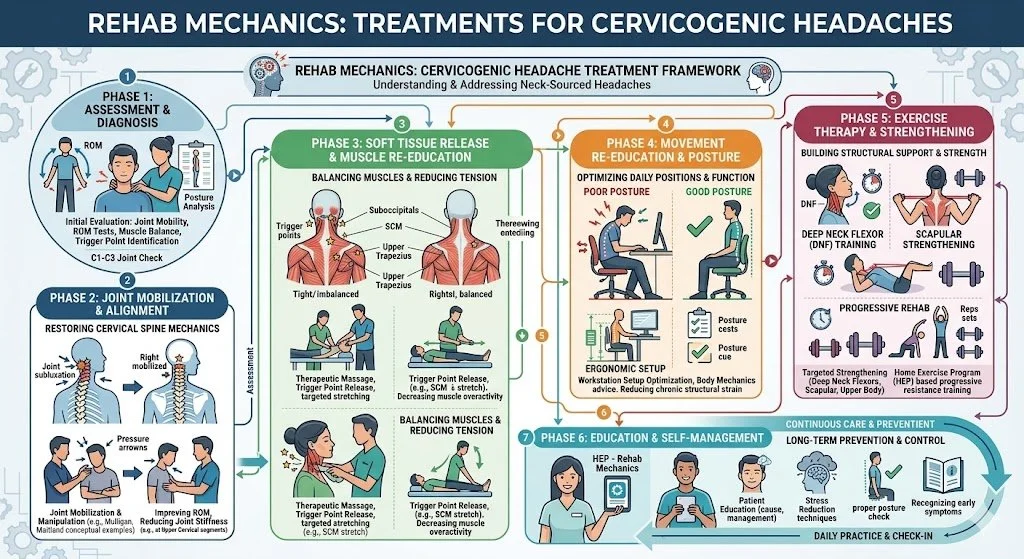
Phase 1: Mechanical Decompression and Traction (Weeks 1-4)
Our immediate priority is un-pinching the spinal cord and nerve roots without provoking the nervous system.
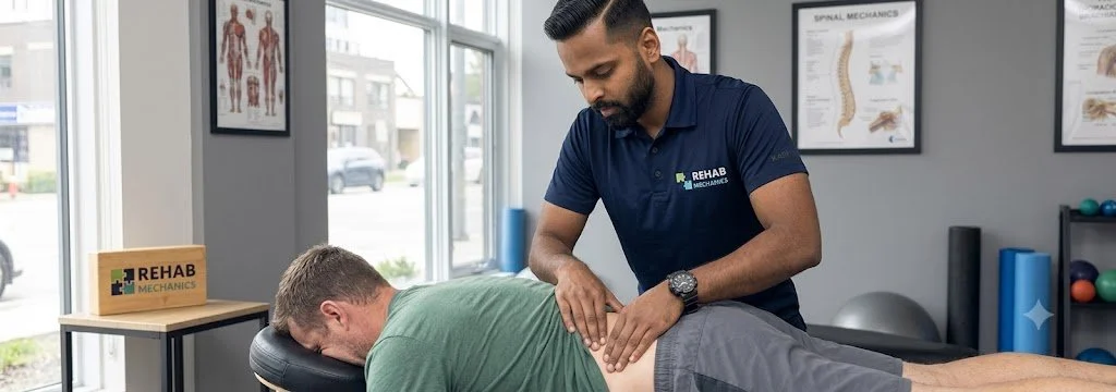
Manual Cervical Traction: Our specialized physiotherapists apply gentle, sustained upward pulling (distraction) to the skull. This physically separates the cervical vertebrae by millimeters, instantly widening the spinal canal and allowing the suffocating spinal cord to breathe.
Flexion-Biased Mobilization: Looking up at the ceiling (extension) physically closes the spinal canal and crushes the cord. We use highly specific manual techniques to promote slight cervical flexion, which maximizes the diameter of the nerve canals.
Suboccipital Release: Melting away the massive, protective muscle spasms at the base of the skull that are trapping the neck in a rigid, painful block.
Phase 2: Postural Overhaul and Thoracic Unlocking (Weeks 4-8)
We must fix the foundation. The neck cannot find a neutral, open position if the upper back is frozen in a slouch.
Thoracic Spine Mobilization: Utilizing Grade III and IV mobilizations to restore the ability of your mid-back to arch backward. This stops your neck from having to hyper-extend to see the computer screen.
Scapular Retraction: Strengthening the rhomboids and middle trapezius to pull the heavy shoulders back and down, providing a solid platform for the cervical spine to rest upon.
Phase 3: Deep Cervical Fortification (Weeks 8-12+)
Because the structural bones are failing (degeneration), the muscles must take over the job of holding the spine perfectly still.
Deep Cervical Flexor (DCF) Endurance: The tiny muscles in the front of your throat act as the core of your neck. We utilize precise biofeedback drills (like micro-nodding against resistance) to rebuild these vital stabilizers.
Isometric Stabilization: We apply multi-directional, unexpected resistance to your head while you attempt to hold it perfectly still. This trains your nervous system to automatically fire the neck muscles to prevent the wobbly vertebrae from shifting and crushing the spinal cord during sudden movements.
Protect Your Spinal Cord
Do not ignore clumsy hands or unexplainable balance issues. Cervical spinal stenosis is a serious, progressive condition. By radically overhauling your spinal mechanics and building a deep muscular brace, expert physical therapy can halt the progression, relieve the nerve pressure, and help you avoid high-risk spinal surgery.
Book a comprehensive neurological and spinal assessment with our clinical team today. We are conveniently located inside the Prime Medical Centre at 68 Abell Street, offering elite, diagnostic orthopedic care in Toronto Queen West.
Contact us to schedule your appointment:
Email: info@rehabmechanics.com
Phone: (416) 533-3900
About the Author
Mr. Sanjay Attwala (B.Sc., M.Sc., RPT) is a Registered Physiotherapist, clinical director, and the founder of Rehab Mechanics in Toronto. With over 15 years of registered clinical practice and a deep specialization in complex musculoskeletal rehabilitation, Sanjay synthesizes rigorous international academic training with advanced evidence-based therapeutics to guide his clinical practice and patient education initiatives.
Academic Background & Credentials
Master of Science (M.Sc.) in Physiotherapy – University of Keele, United Kingdom (2010).
Bachelor of Science (B.Sc.) – University of Waterloo, Ontario, Canada.
Registered Physiotherapist (RPT) – Regulated health professional in excellent standing with the College of Physiotherapists of Ontario (CPO).
Corporate Entity – Operating officially under the S. Attwala Physiotherapy Professional Corporation with a DBA of Rehab Mechanics.
Clinical Expertise & Philosophy
Sanjay’s clinical approach rejects passive symptom management in favor of identifying underlying biomechanical root causes. His diverse expertise spans advanced manual therapies, personalized corrective exercise prescription, and modern physical modalities. At the Rehab Mechanics Toronto Queen West clinic, he routinely diagnoses and treats complex conditions including:
Spinal & Discogenic Pathology – Cervical, thoracic, and lumbar disc injuries, sciatica, and sacroiliac joint (SIJ) dysfunction.
Upper & Lower Extremity Injuries – Rotator cuff tears, frozen shoulder, tennis/golfer’s elbow, carpal tunnel syndrome, and complex ankle/foot pathologies.
Perinatal & Pelvic Health Rehabilitation – Specialized assessment and rehabilitation protocols tailored specifically for women during pregnancy and the post-partum period, addressing pelvic girdle pain, diastasis recti, and core stabilization.
Specialized Rehabilitation – Pelvic health therapy, TMJ dysfunction, post-surgical rehabilitation (including Total Hip and Total Knee Replacements), and custom orthotics dispensing.
Shockwave Therapy: with advanced cutting edge technological devices to suit your needs.
Interdisciplinary Practice & Patient Care
Sanjay practices an integrated model of healthcare, working closely alongside medical doctors inside the Prime Medical Centre on Abell Street to streamline patient recovery pathways. He maintains a human-centric, communication-first clinical framework, ensuring that care remains fully customized rather than automated.
His clinical caseload encompasses a broad operational spectrum under Ontario's regulatory frameworks, including:
Motor Vehicle Accident (MVA) Claims – Rehabilitation navigating Ontario’s statutory accident benefits schedule.
Workplace Safety and Insurance Board (WSIB) – Occupational injury management and return-to-work screening.
Extended Health Care (EHC) & Private Practice – Multi-tier insurance coordination and long-term athletic development plans.
Commitment to Research & Community
Outside of his clinical caseload at Rehab Mechanics and his additional practice affiliations in Etobicoke, Sanjay is an active health writer and community educator. He translates contemporary peer-reviewed medical research into accessible, actionable guidance on his professional blog. As a dedicated father and husband, he mirrors his professional advice in his personal life, focusing on structural mobility, cross-training, and longevity to help his family and his community thrive. Naturally he takes he a keen interest in rehabilitation for women who are pregnant and post-partum.
Disclaimer: The information provided on this blog is intended for educational and informational purposes only. It does not constitute medical advice, diagnosis, or a treatment plan. Always seek the direct advice of a Registered Physiotherapist, physician, or other qualified health provider regarding any medical condition or physical rehabilitation routine.
Is My Inner Knee Pain a Meniscus Tear or Pes Anserine Bursitis?
Maybe. While meniscus tears involve joint clicking, sharp inner knee pain directly below the joint line is often pes anserine bursitis. Physiotherapy resolves this by correcting pelvic mechanics, releasing hyperactive hamstrings, and strengthening the glutes to eliminate the structural friction causing the inflammation.
The Diagnostic Confusion of Medial Knee Pain
For the runners, cyclists, and weekend warriors navigating the paths of Trinity Bellwoods and the Martin Goodman Trail, knee pain is a persistent threat. When a sharp, stinging pain develops on the inside (medial) aspect of the knee, panic often ensues.
Athletes immediately jump to the worst-case scenario: "I've torn my medial meniscus" or "I've blown my MCL." They expect to be scheduled for an MRI and placed on a surgical waitlist. However, at Rehab Mechanics in Queen West, we frequently uncover a highly treatable, yet massively misunderstood, biomechanical culprit: Pes Anserine Bursitis.
While a meniscus tear happens deep inside the joint capsule, pes anserine bursitis happens outside the joint, roughly two inches below the actual knee hinge. It is a severe, friction-based inflammatory condition. When you understand that this condition is not a structural tear of cartilage, but rather a functional failure of your hip and thigh muscles, the path to a full, non-surgical recovery becomes clear. Through advanced human mechanics physical therapy, we can stop the friction and permanently eliminate the inner knee pain.
Structural Analysis: The Mechanics of the "Goose Foot"
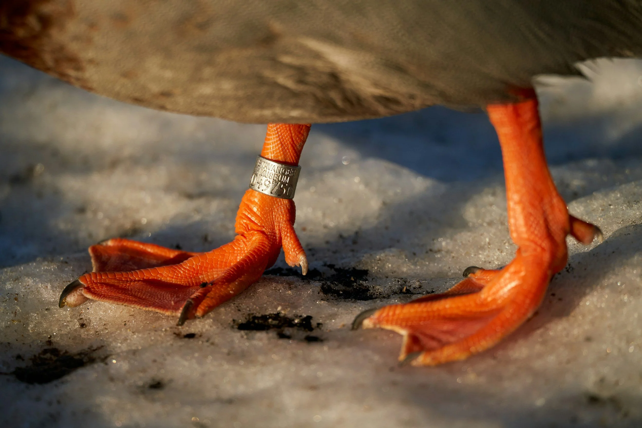
Goose feet? Well… not literally — but now that we’ve got your attention, let’s talk about the real ‘Goose Foot’ inside your knee. The pes anserine region gets its nickname because three major tendons attach in a pattern that looks just like a goose’s footprint. When those tendons get irritated, walking, climbing stairs, or even standing can feel uncomfortable. So let’s get you off ‘all fours,’ back onto your ‘two feet,’ and moving well again
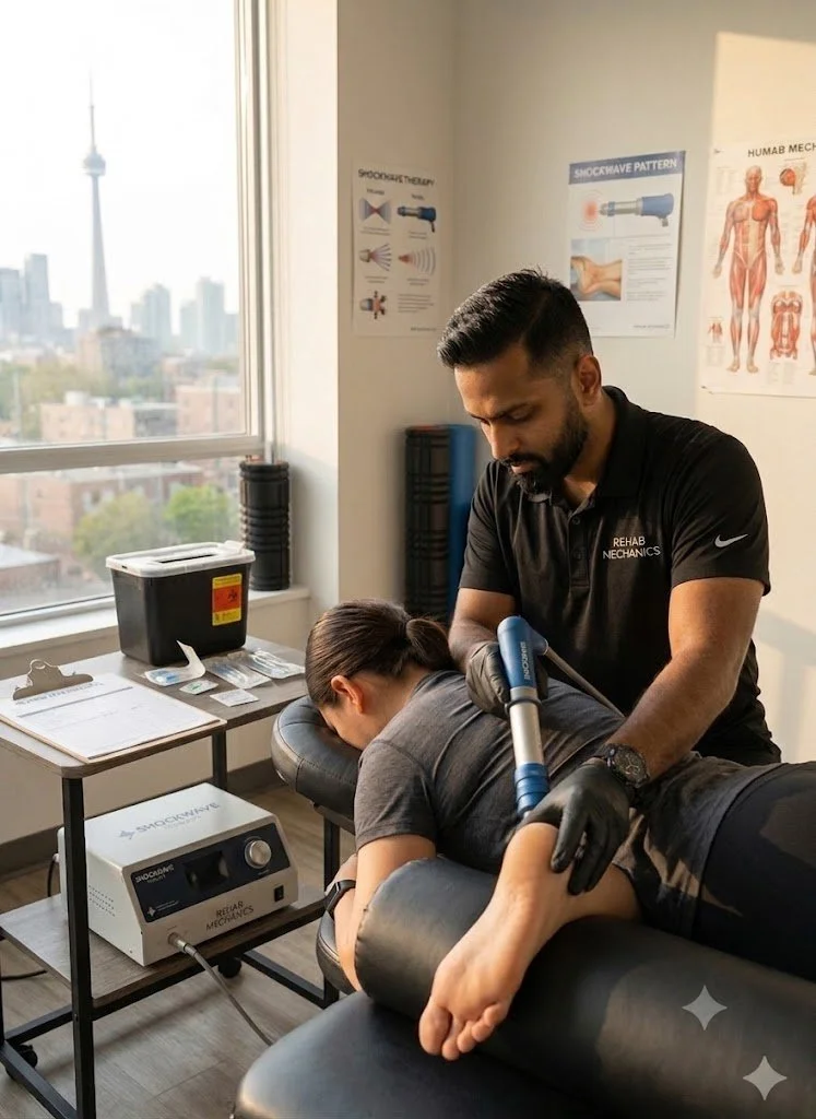
To accurately diagnose and treat this condition, we must perform a detailed structural analysis of the medial knee architecture and the specific tendons that anchor there.
The Anatomy of the Pes Anserinus
"Pes Anserinus" is Latin for "goose foot." It refers to the webbed, three-pronged shape of three specific muscle tendons as they merge and attach to the inside of your shin bone (tibia), just below the knee joint.
The Three Muscles: These tendons belong to the Sartorius (front of the thigh), the Gracilis (inner thigh/groin), and the Semitendinosus (inner hamstring).
The Anchor Point: Together, they act as massive, dynamic stabilizers, preventing your lower leg from twisting outward during running and pivoting.
The Bursa Sac: Sitting directly underneath this three-pronged tendon attachment, protecting it from grinding against the hard shin bone, is the pes anserine bursa—a fluid-filled, friction-reducing sac.
The Pathology of Friction (Bursitis)
Bursitis is not a random occurrence; it is a mechanical penalty for poor movement patterns.
The Valgus Collapse Trigger
If your pelvic mechanics are faulty, your knee pays the price.
Gluteus Medius Weakness: When you run, you spend the entire time balancing on one leg. If the muscle on the side of your hip (gluteus medius) is weak from sitting at a desk all day, your pelvis drops.
The Inward Cave: This pelvic drop forces your thigh bone to violently rotate inward, causing your knee to collapse toward your midline (a movement called knee valgus).
The Whiplash Effect: Every time your knee caves inward, the three tendons of the pes anserinus are violently yanked taut against the bone to stop the collapse.
The Inflammatory Crush: This relentless, repetitive yanking physically crushes the bursa sac underneath the tendons. The bursa becomes engorged, swollen, and excruciatingly painful, making it impossible to walk down stairs or run without a sharp, stabbing sensation.
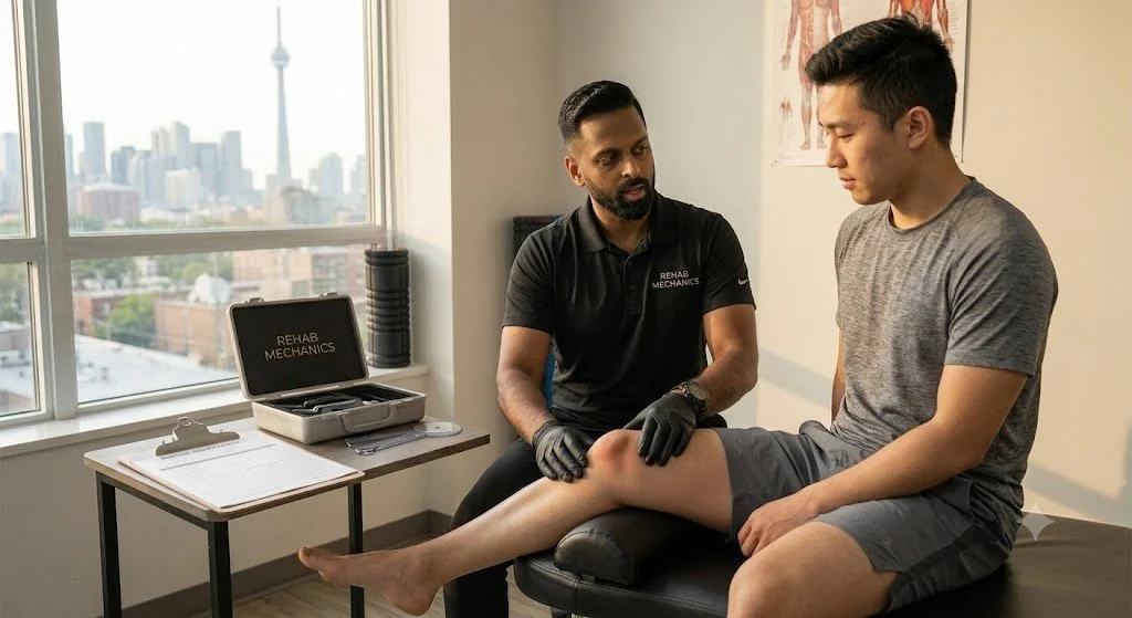
Identifying the Clinical Red Flags

How do we differentiate pes anserine bursitis from a torn meniscus or ligament sprain?
The Location: Meniscus pain is felt directly on the joint line. Pes anserine pain is felt exactly 2 to 3 inches below the joint line, on the flat part of the inner shin bone.
The Touch Test: Pressing your finger directly onto that spot produces a breathtaking, exquisite point-tenderness.
The Stair Trigger: The pain is uniquely severe when descending stairs or stepping down off a Toronto streetcar, as this requires massive eccentric hamstring control, which crushes the inflamed bursa.
Absence of Locking: Unlike a meniscus tear, the knee does not physically "lock," "catch," or click. The hinge works fine; the pain is purely superficial.
Primary Source Proof: Biomechanical Knee Rehabilitation
Orthopedic and sports medicine literature dictates that correcting lumbo-pelvic kinematics and strengthening the hip abductors is the definitive, long-term solution for resolving medial knee overuse injuries like pes anserine bursitis, vastly outperforming localized rest or cortisone injections.
Note: The link above directs to external, peer-reviewed medical literature demonstrating our commitment to evidence-based practice and international clinical guidelines for sports rehabilitation.
Strengthening of the Hip and Core Versus Knee Muscles for the Treatment of Patellofemoral Pain: A Multicenter Randomized Controlled Trial
Rethinking Knee Pain: Why Your Hips Matter More Than You Think
Patellofemoral pain (PFP), often called "runner’s knee," is a common and frustrating injury for athletes, causing aching pain during activities like running, squatting, or even sitting for long periods. Historically, physical therapy for PFP has focused almost exclusively on the knee, specifically targeting quadriceps strengthening. However, new research suggests that our standard approach might be missing a crucial piece of the puzzle: the hips and core.
A landmark multicenter randomized controlled trial compared two distinct rehabilitation protocols over a six-week period: one focused solely on knee muscles and another that integrated hip and core strengthening. While both groups saw improvements in overall function and strength, the results revealed a clear advantage for those targeting the "upstream" muscles. The participants following the hip and core protocol experienced a faster resolution of their pain and achieved greater overall gains in muscle strength compared to the knee-focused group.
These findings challenge the traditional "knee-only" mindset in physical therapy, highlighting the importance of the entire kinetic chain. By strengthening the hips and core, patients can improve their pelvic and lower-limb stability, which directly alleviates the excessive stress placed on the knee joint. If you are struggling with recurring knee pain, this study suggests that shifting your focus—or your exercise routine—to include dedicated hip and core stability training may be the key to faster recovery and better long-term outcomes.
The Rehab Mechanics Corrective Protocol
We do not just ice the knee. To cure this condition, we must physically alter the angle of your leg during movement.
Phase 1: Tendon Desensitization and Inflammation Control (Weeks 1-3)
We must immediately stop the mechanical crushing of the bursa.
Activity Modification: A temporary halt on running and stair-climbing workouts to allow the swollen bursa sac to chemically cool down.
Advanced Myofascial Release: Our physiotherapists apply targeted, deep manual therapy to the bellies of the hamstring, gracilis, and sartorius muscles high up in the thigh. Releasing the tension at the top of the muscles instantly provides mechanical slack to the tendons at the knee.
Kinesiology Taping: Utilizing strategic taping patterns to gently lift the skin and fascia over the medial knee, decompressing the inflamed bursa and providing immediate pain relief during walking.
Phase 2: Pelvic Fortification and Neuromuscular Control (Weeks 4-6)
We must build the muscular scaffolding that prevents the knee from caving inward.
Isolated Glute Medius Activation: Utilizing side-lying hip abductions, banded clamshells, and isometric wall holds to wake up the lateral stabilizers of the pelvis.
Eccentric Hamstring Loading: Tendons need strength to handle force without spasming. We use slow, controlled hamstring curls and sliders to build robust resilience in the semitendinosus tendon so it no longer crushes the bursa.
Phase 3: Dynamic Integration and Gait Retraining (Weeks 6-8+)
We must ensure your new hip strength translates to the pavement.
Anti-Valgus Training: We progress to dynamic step-downs, lateral lunges, and single-leg squats while utilizing tactile feedback (like a resistance band pulling the knee inward) to force your brain to consciously fire the glutes and push the knee straight.
Cadence Manipulation for Runners: If running is your goal, we may analyze your gait and increase your step cadence by 5-10%. Taking slightly shorter, faster steps drastically reduces the heavy, bounding impact that triggers the valgus collapse.
Stop the Friction, Save Your Knee
You do not have to live with stinging inner knee pain or fear that you have a torn meniscus. By addressing the deep biomechanics of your hip and thigh, physical therapy offers a permanent, non-surgical solution to pes anserine bursitis.
Book a comprehensive biomechanical knee assessment with our clinical team today. We are conveniently located inside the Prime Medical Centre at 68 Abell Street, offering elite sports recovery in Queen West.
Contact us to schedule your appointment:
Email: info@rehabmechanics.com
Phone: (416) 533-3900
About the Author
Mr. Sanjay Attwala (B.Sc., M.Sc., RPT) is a Registered Physiotherapist, clinical director, and the founder of Rehab Mechanics in Toronto. With over 15 years of registered clinical practice and a deep specialization in complex musculoskeletal rehabilitation, Sanjay synthesizes rigorous international academic training with advanced evidence-based therapeutics to guide his clinical practice and patient education initiatives.
Academic Background & Credentials
Master of Science (M.Sc.) in Physiotherapy – University of Keele, United Kingdom (2010).
Bachelor of Science (B.Sc.) – University of Waterloo, Ontario, Canada.
Registered Physiotherapist (RPT) – Regulated health professional in excellent standing with the College of Physiotherapists of Ontario (CPO).
Corporate Entity – Operating officially under the S. Attwala Physiotherapy Professional Corporation with a DBA of Rehab Mechanics.
Clinical Expertise & Philosophy
Sanjay’s clinical approach rejects passive symptom management in favor of identifying underlying biomechanical root causes. His diverse expertise spans advanced manual therapies, personalized corrective exercise prescription, and modern physical modalities. At the Rehab Mechanics Toronto Queen West clinic, he routinely diagnoses and treats complex conditions including:
Spinal & Discogenic Pathology – Cervical, thoracic, and lumbar disc injuries, sciatica, and sacroiliac joint (SIJ) dysfunction.
Upper & Lower Extremity Injuries – Rotator cuff tears, frozen shoulder, tennis/golfer’s elbow, carpal tunnel syndrome, and complex ankle/foot pathologies.
Perinatal & Pelvic Health Rehabilitation – Specialized assessment and rehabilitation protocols tailored specifically for women during pregnancy and the post-partum period, addressing pelvic girdle pain, diastasis recti, and core stabilization.
Specialized Rehabilitation – Pelvic health therapy, TMJ dysfunction, post-surgical rehabilitation (including Total Hip and Total Knee Replacements), and custom orthotics dispensing.
Shockwave Therapy: with advanced cutting edge technological devices to suit your needs.
Interdisciplinary Practice & Patient Care
Sanjay practices an integrated model of healthcare, working closely alongside medical doctors inside the Prime Medical Centre on Abell Street to streamline patient recovery pathways. He maintains a human-centric, communication-first clinical framework, ensuring that care remains fully customized rather than automated.
His clinical caseload encompasses a broad operational spectrum under Ontario's regulatory frameworks, including:
Motor Vehicle Accident (MVA) Claims – Rehabilitation navigating Ontario’s statutory accident benefits schedule.
Workplace Safety and Insurance Board (WSIB) – Occupational injury management and return-to-work screening.
Extended Health Care (EHC) & Private Practice – Multi-tier insurance coordination and long-term athletic development plans.
Commitment to Research & Community
Outside of his clinical caseload at Rehab Mechanics and his additional practice affiliations in Etobicoke, Sanjay is an active health writer and community educator. He translates contemporary peer-reviewed medical research into accessible, actionable guidance on his professional blog. As a dedicated father and husband, he mirrors his professional advice in his personal life, focusing on structural mobility, cross-training, and longevity to help his family and his community thrive. Naturally he takes he a keen interest in rehabilitation for women who are pregnant and post-partum.
Disclaimer: The information provided on this blog is intended for educational and informational purposes only. It does not constitute medical advice, diagnosis, or a treatment plan. Always seek the direct advice of a Registered Physiotherapist, physician, or other qualified health provider regarding any medical condition or physical rehabilitation routine.

Be sure to check out Rehab Mechanics’ Google My Business Profile for daily posts, location and directions in addition to the directions on the website!
We love interacting with prospective and current patients, and love honest feedback and even constructive criticism and areas for improvement!
Interested in learning more? Give us a call or fill out the form before if you are a little more finger and thumb driven in 2026 than the telephone.
Can Vestibular Physiotherapy Cure Post-Concussion Syndrome and Vertigo?
Yes. Vestibular physiotherapy cures post-concussion syndrome by recalibrating the brain’s sensorimotor integration. Targeted ocular and vestibular exercises eliminate chronic vertigo, visual disturbances, and cognitive fog, restoring normal neurological function after a traumatic brain injury or motor vehicle accident.
The Hidden Neurological Epidemic in Toronto
In the fast-paced, high-density environment of downtown Toronto, traumatic brain injuries are incredibly common. Whether you sustained a violent whiplash injury during a rear-end collision on the Gardiner Expressway, suffered a hard check during a recreational hockey game at a local arena, or simply slipped on an icy sidewalk in Queen West, the resulting concussion can completely derail your life.
For decades, the standard medical advice for a concussion was "cocoon therapy": sit in a dark, quiet room, avoid all screens, and wait for the brain to heal. We now know that prolonged passive rest is actually detrimental.
When patients arrive at Rehab Mechanics weeks or months after their injury, they are often desperate. They describe a lingering, suffocating "brain fog," severe light sensitivity, and a terrifying sensation of vertigo or dizziness every time they turn their head to cross a busy intersection. This is Post-Concussion Syndrome (PCS).
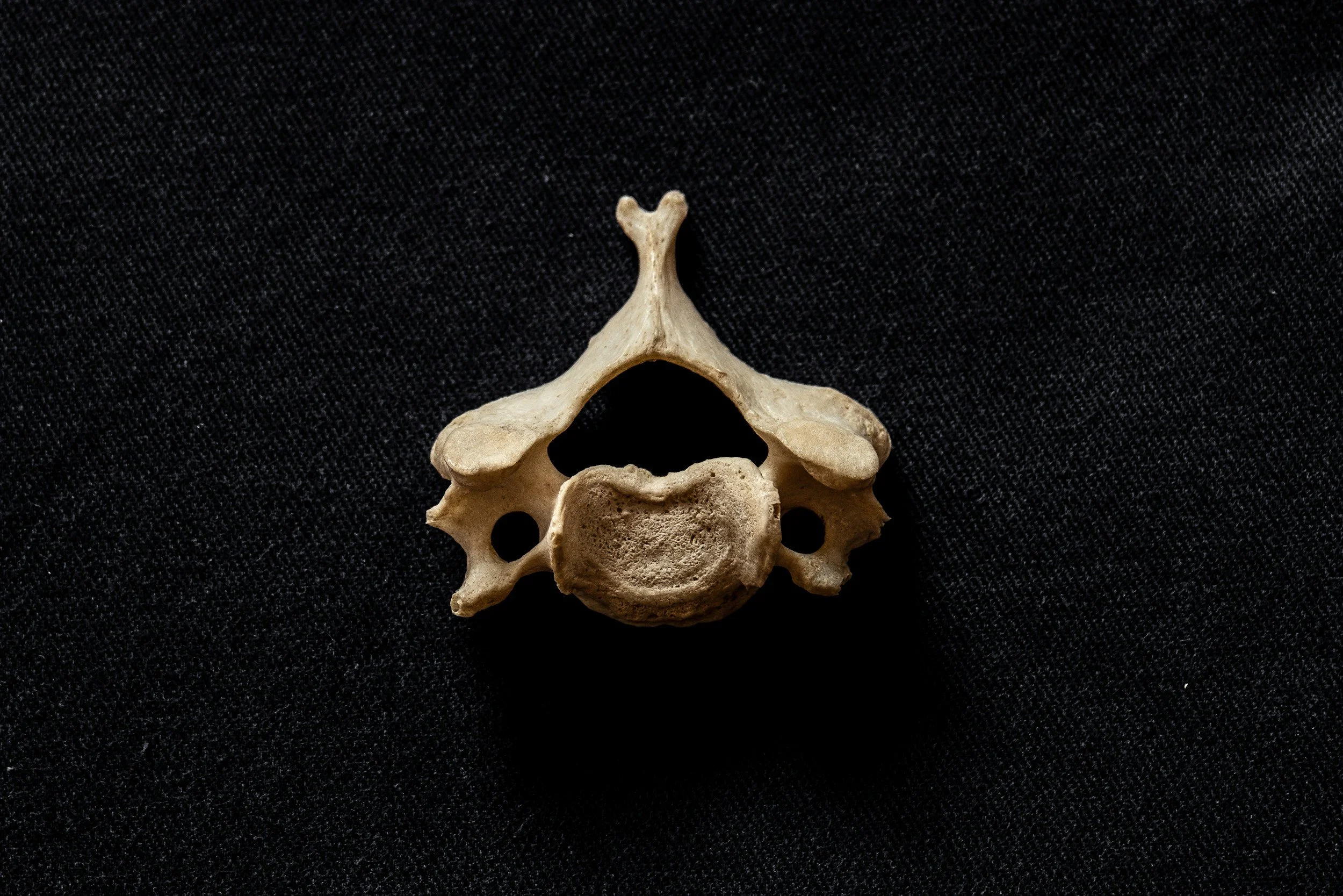
At our clinic inside the Prime Medical Centre, we reject the "wait and see" approach. A concussion is not just a bruise on the brain; it is a profound biomechanical and neurological disconnection. Through highly specialized vestibular physical therapy, we actively rebuild the neural pathways, stopping the dizziness and restoring your cognitive clarity.
Structural Analysis: The Mechanics of a Concussion
To understand how physical therapy can heal the brain, we must perform a detailed structural analysis of what happens during a concussive impact and how the vestibular system operates.
The Neurometabolic Cascade
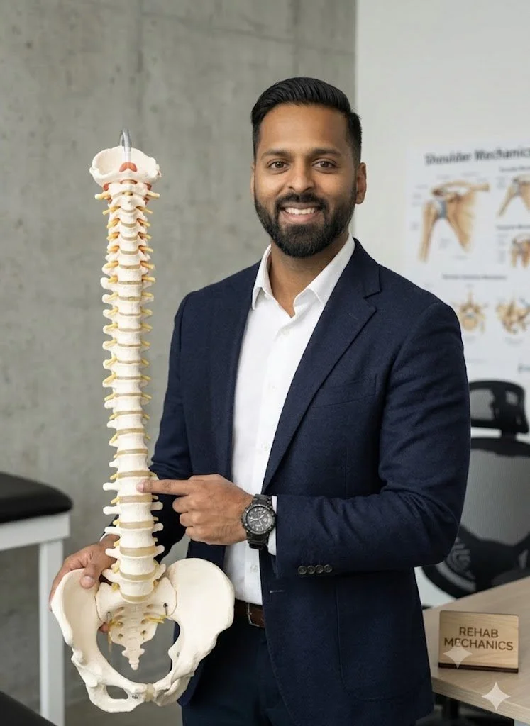
A concussion is a Mild Traumatic Brain Injury (MTBI). It is caused by a massive acceleration and deceleration force that violently shakes the brain inside the skull.
Axonal Shearing: The delicate nerve fibers (axons) in your brain are stretched and twisted. This sheer force physically damages the cell membranes.
The Energy Crisis: The brain goes into a state of panic, releasing massive amounts of excitatory neurotransmitters (like glutamate). The brain demands a massive amount of glucose (energy) to repair the cells, but the trauma temporarily restricts cerebral blood flow.
The Result: The brain is starved of energy. This metabolic mismatch is what causes the immediate exhaustion, confusion, and deep headaches.
The Vestibular Apparatus Failure
While the brain is recovering chemically, the physical hardware that keeps you balanced is often knocked entirely offline. Your vestibular system is located deep inside your inner ear.
The Semicircular Canals and Otoliths
The Hardware: You have three fluid-filled loops (semicircular canals) in each ear that act as biological gyroscopes, detecting rotational movement. You also have two sac-like structures (otoliths) that detect gravity and linear acceleration.
The Software (Vestibulo-Ocular Reflex): These inner ear sensors constantly talk to your eyes through the Vestibulo-Ocular Reflex (VOR). The VOR is the software program that allows you to keep your eyes perfectly focused on a stationary object while you move your head.
The Pathology of Sensory Mismatch
In Post-Concussion Syndrome, the impact damages the VOR software.
The Glitch: When you turn your head to look for a streetcar, your inner ear tells your brain you are moving at one speed, but your eyes tell your brain you are moving at a different speed.
The Crash: The brain cannot process this conflicting data. It triggers an immediate error response: severe dizziness, sudden nausea, and a feeling of floating or unsteadiness.
Primary Source Proof: Vestibular Rehabilitation
Contemporary neurological and sports medicine literature explicitly mandates that active, targeted vestibular rehabilitation is significantly superior to physical rest for the resolution of prolonged post-concussion symptoms and dizziness.
Review the Clinical Evidence on PubMed: The Effectiveness of Vestibular Rehabilitation in the Treatment of Post-Concussion Syndrome (National Institutes of Health)
Note: The link above directs to external, peer-reviewed medical literature demonstrating our commitment to evidence-based practice and international clinical guidelines for neurological rehabilitation.
The Rehab Mechanics Vestibular Protocol
Treating PCS requires immense clinical precision. We do not just guess; we use specific neurological testing to identify exactly which reflex is broken, and we prescribe targeted "remedial mechanics" to fix it.
Phase 1: Cervicogenic Decompression (Weeks 1-3)
It is biomechanically impossible to sustain a concussion without also sustaining a whiplash injury to the neck.
Upper Cervical Unlocking: The top three joints of the neck (C1-C3) contain thousands of balance sensors. If the neck is locked from whiplash, it sends warped balance signals to the brain, mimicking vertigo. We use precise manual therapy to un-jam these joints.
Suboccipital Release: We apply deep ischemic compression to the tiny, spasming muscles at the base of the skull to instantly relieve the severe tension headaches associated with the trauma.
Phase 2: Neurological Recalibration and Habituation (Weeks 2-6)
We must force the brain to fix the software glitch. We do this through controlled exposure.
Gaze Stabilization (VOR Retraining): We prescribe highly specific ocular drills. You will focus your eyes on a target (like a letter 'X' on the wall) while rotating your head side to side at a specific speed (using a metronome). This forces the brain to repair the connection between the inner ear and the eyes.
Habituation Drills: We intentionally, but safely, expose you to the exact movements that make you dizzy (e.g., bending over to pick up an object and standing up quickly). By repeating these movements in a controlled environment, the brain slowly desensitizes to the motion, turning off the nausea alarm.
Phase 3: Exertional Tolerance and Return to Play (Weeks 6+)
The final step is proving to the brain that it can handle an elevated heart rate without crashing.
Sub-Symptom Threshold Training: We use a stationary bike to carefully elevate your heart rate. We find the exact heart rate that triggers your headache or dizziness (the threshold).
Progressive Overload: We prescribe a daily cardiovascular program where you exercise just below that threshold. Over weeks, this heals the cerebral blood flow problem, safely pushing your threshold higher and higher until you can run, lift, and play sports completely symptom-free.
Clear the Brain Fog
You do not have to live in a dark room or accept chronic dizziness as your new normal. By actively rebuilding your neurological pathways and resolving the structural trauma in your neck, physical therapy can fully restore your balance and mental clarity.
Book a comprehensive post-concussion and vestibular assessment with our clinical team today. We are conveniently located inside the Prime Medical Centre at 68 Abell Street, offering advanced neurological and orthopedic care in Toronto Queen West.
Contact us to schedule your appointment:
Email: info@rehabmechanics.com
Phone: (416) 533-3900
About the Author
Mr. Sanjay Attwala (B.Sc., M.Sc., RPT) is a Registered Physiotherapist, clinical director, and the founder of Rehab Mechanics in Toronto. With over 15 years of registered clinical practice and a deep specialization in complex musculoskeletal rehabilitation, Sanjay synthesizes rigorous international academic training with advanced evidence-based therapeutics to guide his clinical practice and patient education initiatives.
Academic Background & Credentials
Master of Science (M.Sc.) in Physiotherapy – University of Keele, United Kingdom (2010).
Bachelor of Science (B.Sc.) – University of Waterloo, Ontario, Canada.
Registered Physiotherapist (RPT) – Regulated health professional in excellent standing with the College of Physiotherapists of Ontario (CPO).
Corporate Entity – Operating officially under the S. Attwala Physiotherapy Professional Corporation with a DBA of Rehab Mechanics.
Clinical Expertise & Philosophy
Sanjay’s clinical approach rejects passive symptom management in favor of identifying underlying biomechanical root causes. His diverse expertise spans advanced manual therapies, personalized corrective exercise prescription, and modern physical modalities. At the Rehab Mechanics Toronto Queen West clinic, he routinely diagnoses and treats complex conditions including:
Spinal & Discogenic Pathology – Cervical, thoracic, and lumbar disc injuries, sciatica, and sacroiliac joint (SIJ) dysfunction.
Upper & Lower Extremity Injuries – Rotator cuff tears, frozen shoulder, tennis/golfer’s elbow, carpal tunnel syndrome, and complex ankle/foot pathologies.
Perinatal & Pelvic Health Rehabilitation – Specialized assessment and rehabilitation protocols tailored specifically for women during pregnancy and the post-partum period, addressing pelvic girdle pain, diastasis recti, and core stabilization.
Specialized Rehabilitation – Pelvic health therapy, TMJ dysfunction, post-surgical rehabilitation (including Total Hip and Total Knee Replacements), and custom orthotics dispensing.
Shockwave Therapy: with advanced cutting edge technological devices to suit your needs.
Interdisciplinary Practice & Patient Care
Sanjay practices an integrated model of healthcare, working closely alongside medical doctors inside the Prime Medical Centre on Abell Street to streamline patient recovery pathways. He maintains a human-centric, communication-first clinical framework, ensuring that care remains fully customized rather than automated.
His clinical caseload encompasses a broad operational spectrum under Ontario's regulatory frameworks, including:
Motor Vehicle Accident (MVA) Claims – Rehabilitation navigating Ontario’s statutory accident benefits schedule.
Workplace Safety and Insurance Board (WSIB) – Occupational injury management and return-to-work screening.
Extended Health Care (EHC) & Private Practice – Multi-tier insurance coordination and long-term athletic development plans.
Commitment to Research & Community
Outside of his clinical caseload at Rehab Mechanics and his additional practice affiliations in Etobicoke, Sanjay is an active health writer and community educator. He translates contemporary peer-reviewed medical research into accessible, actionable guidance on his professional blog. As a dedicated father and husband, he mirrors his professional advice in his personal life, focusing on structural mobility, cross-training, and longevity to help his family and his community thrive. Naturally he takes he a keen interest in rehabilitation for women who are pregnant and post-partum.
Disclaimer: The information provided on this blog is intended for educational and informational purposes only. It does not constitute medical advice, diagnosis, or a treatment plan. Always seek the direct advice of a Registered Physiotherapist, physician, or other qualified health provider regarding any medical condition or physical rehabilitation routine.
Can Pelvic Floor Physiotherapy Prevent Surgery for Pelvic Organ Prolapse?
Yes. Pelvic floor physiotherapy is the primary, non-surgical treatment for pelvic organ prolapse. By structurally rebuilding the pelvic sling, releasing fascial tension, and mastering intra-abdominal pressure, physical therapy significantly reduces prolapse symptoms and frequently prevents the need for reconstructive surgery.
The Silent Epidemic in Women's Health
In Toronto, the physical demands placed on women are immense. Whether navigating the intense recovery period following childbirth, managing the hormonal shifts of menopause, or maintaining a heavy weightlifting routine at a local Queen West gym, the female pelvic floor is subjected to massive, chronic stress.
A deeply distressing and profoundly under-discussed consequence of this stress is Pelvic Organ Prolapse (POP). Women often discover POP subtly—a feeling of "heaviness" or dragging in the lower pelvis after a long day of standing, a sensation of incomplete bladder emptying, or the terrifying discovery of a physical bulge at the vaginal opening.
The traditional medical reflex is often to wait until the prolapse becomes severe enough to warrant complex reconstructive surgery or the permanent placement of a pessary ring. At Rehab Mechanics, our specialized Perinatal and Pelvic Health program categorically rejects this passive "wait and see" approach. The pelvic floor is a dynamic musculoskeletal system. Through highly specialized, trauma-informed physical therapy, we can radically improve the biomechanical support of your pelvic organs, reversing symptoms and restoring your structural confidence.
Structural Analysis: The Mechanics of the Pelvic Floor
To understand how physiotherapy reverses prolapse symptoms, we must perform a detailed biomechanical analysis of the pelvic architecture and the forces that govern it.
The Anatomy of the Pelvic Sling
The pelvic floor is an intricate, multi-layered hammock of 14 different muscles, thick fascial sheets, and strong ligaments.
The Structural Floor: This hammock forms the absolute base of your abdominal cavity. It physically spans the gap between your pubic bone in the front and your tailbone in the back.
The Organ Support: Resting directly on top of this muscular sling are your vital pelvic organs: the bladder (anterior), the uterus (central), and the rectum (posterior).
The Pathology of Prolapse
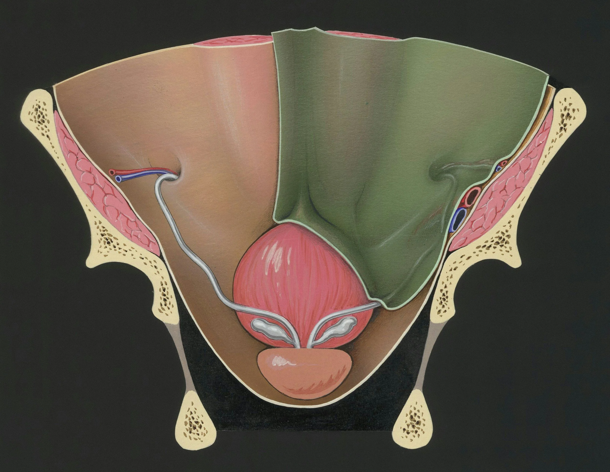
Pelvic Organ Prolapse occurs when this supporting hammock fails, allowing one or more of these organs to drop downward into the vaginal canal.
Cystocele: The bladder drops into the front wall.
Uterine Prolapse: The uterus drops straight down the center.
Rectocele: The rectum bulges into the back wall.
The Two Mechanisms of Failure
The hammock can fail for two very different biomechanical reasons.
1. Fascial Tearing (The Trauma Model)
During a prolonged or mechanically difficult vaginal childbirth, the massive downward pressure can physically stretch or tear the thick connective tissue (the endopelvic fascia) and the levator ani muscles. The hammock loses its structural integrity and sags.
2. The Pressure System Failure (The Chronic Model)
Your core is a sealed, pressurized cylinder. The diaphragm is the roof, the abdominals are the walls, and the pelvic floor is the base.
Intra-Abdominal Pressure (IAP): Every time you cough, sneeze, jump, or lift a heavy barbell, the pressure inside this cylinder spikes massively.
The Downward Crush: If your breathing mechanics are faulty—for example, if you hold your breath and bear down heavily when lifting a car seat—you force 100% of that pressure directly down onto the pelvic organs. Over years, this repetitive, crushing downward force stretches the hammock until the organs drop.
Primary Source Proof: Conservative Pelvic Rehabilitation
Global urogynecology and pelvic health research definitively establishes that highly supervised, specific pelvic floor muscle training is the gold standard for reducing symptom severity and improving the anatomical stage of mild to moderate pelvic organ prolapse.
Review the Clinical Evidence on PubMed: Pelvic Floor Muscle Training for Secondary Prevention of Pelvic Organ Prolapse (National Institutes of Health)
Note: The link above directs to external, peer-reviewed medical literature demonstrating our commitment to evidence-based practice and international clinical guidelines for pelvic health.
The Rehab Mechanics Prolapse Protocol
You cannot fix a prolapse by simply downloading a generic app and doing hundreds of "Kegels." Doing Kegels incorrectly can actually worsen the downward pressure. We use a precise, inside-out "human mechanics" approach.
Phase 1: Advanced Diagnostics and Internal Assessment
We must accurately map the muscular failure.
The Internal Exam: With your strict, ongoing consent, our specialized physiotherapist performs an internal vaginal examination. This allows us to physically palpate the muscles to determine their resting tone, strength, endurance, and to identify the exact organs involved in the prolapse.
Hypertonic vs. Hypotonic: Surprisingly, many women with prolapse actually have overactive (hypertonic) pelvic floors. The muscles are constantly clenched in a state of spasm, making them exhausted and useless when a real pressure spike hits.
Phase 2: Restoring the Cylinder (Pressure Management)
Before we strengthen the floor, we must fix the roof.
Diaphragmatic Integration: We completely retrain your breathing mechanics. We teach you how to breathe laterally into your ribcage so the diaphragm and pelvic floor move in perfect synchrony, like a piston, rather than crashing into each other.
The "Knack": Teaching your nervous system to automatically pre-contract the pelvic floor milliseconds before you cough or lift, proactively shielding the organs from the sudden pressure spike.
Phase 3: Neuromuscular Up-Training and Hypertrophy
If the muscles are weak (hypotonic), we must structurally rebuild their thickness and endurance.
Eccentric and Concentric Loading: We guide you through highly specific, graded contractions of the pelvic sling in various gravity-dependent positions (lying down, seated, and eventually standing).
Endurance Holds: The pelvic floor must be able to hold the organs up for a 14-hour day. We build biological endurance using prolonged, sub-maximal isometric holds.
Phase 4: Functional Loading and Real-World Mechanics
We transition your rehab to match your lifestyle demands.
Symptom-Free Lifting: Integrating your new pelvic floor strength into squats, deadlifts, and functional mom-mechanics (like lifting a toddler from a crib).
Pessary Integration: If conservative therapy requires physical support, our physiotherapists can consult and collaborate on the integration of a vaginal pessary—a soft, removable silicone device that acts like an internal sports bra, physically holding the organs up while we rehabilitate the muscles around it.
Take Back Your Structural Confidence
A diagnosis of Pelvic Organ Prolapse does not mean you have to stop exercising, live with chronic heaviness, or immediately schedule surgery. By mastering your intra-abdominal pressure and deeply rehabilitating your pelvic sling, you can manage the condition and reclaim your active life.
Book a highly confidential, specialized pelvic health assessment with our clinical team today. We are conveniently located inside the Prime Medical Centre at 68 Abell Street, offering elite, private care in the heart of Toronto Queen West.
Contact us to schedule your appointment:
Email: info@rehabmechanics.com
Phone: (416) 533-3900
About the Author
Mr. Sanjay Attwala (B.Sc., M.Sc., RPT) is a Registered Physiotherapist, clinical director, and the founder of Rehab Mechanics in Toronto. With over 15 years of registered clinical practice and a deep specialization in complex musculoskeletal rehabilitation, Sanjay synthesizes rigorous international academic training with advanced evidence-based therapeutics to guide his clinical practice and patient education initiatives.
Academic Background & Credentials
Master of Science (M.Sc.) in Physiotherapy – University of Keele, United Kingdom (2010).
Bachelor of Science (B.Sc.) – University of Waterloo, Ontario, Canada.
Registered Physiotherapist (RPT) – Regulated health professional in excellent standing with the College of Physiotherapists of Ontario (CPO).
Corporate Entity – Operating officially under the S. Attwala Physiotherapy Professional Corporation with a DBA of Rehab Mechanics.
Clinical Expertise & Philosophy
Sanjay’s clinical approach rejects passive symptom management in favor of identifying underlying biomechanical root causes. His diverse expertise spans advanced manual therapies, personalized corrective exercise prescription, and modern physical modalities. At the Rehab Mechanics Toronto Queen West clinic, he routinely diagnoses and treats complex conditions including:
Spinal & Discogenic Pathology – Cervical, thoracic, and lumbar disc injuries, sciatica, and sacroiliac joint (SIJ) dysfunction.
Upper & Lower Extremity Injuries – Rotator cuff tears, frozen shoulder, tennis/golfer’s elbow, carpal tunnel syndrome, and complex ankle/foot pathologies.
Perinatal & Pelvic Health Rehabilitation – Specialized assessment and rehabilitation protocols tailored specifically for women during pregnancy and the post-partum period, addressing pelvic girdle pain, diastasis recti, and core stabilization.
Specialized Rehabilitation – Pelvic health therapy, TMJ dysfunction, post-surgical rehabilitation (including Total Hip and Total Knee Replacements), and custom orthotics dispensing.
Shockwave Therapy: with advanced cutting edge technological devices to suit your needs.
Interdisciplinary Practice & Patient Care
Sanjay practices an integrated model of healthcare, working closely alongside medical doctors inside the Prime Medical Centre on Abell Street to streamline patient recovery pathways. He maintains a human-centric, communication-first clinical framework, ensuring that care remains fully customized rather than automated.
His clinical caseload encompasses a broad operational spectrum under Ontario's regulatory frameworks, including:
Motor Vehicle Accident (MVA) Claims – Rehabilitation navigating Ontario’s statutory accident benefits schedule.
Workplace Safety and Insurance Board (WSIB) – Occupational injury management and return-to-work screening.
Extended Health Care (EHC) & Private Practice – Multi-tier insurance coordination and long-term athletic development plans.
Commitment to Research & Community
Outside of his clinical caseload at Rehab Mechanics and his additional practice affiliations in Etobicoke, Sanjay is an active health writer and community educator. He translates contemporary peer-reviewed medical research into accessible, actionable guidance on his professional blog. As a dedicated father and husband, he mirrors his professional advice in his personal life, focusing on structural mobility, cross-training, and longevity to help his family and his community thrive. Naturally he takes he a keen interest in rehabilitation for women who are pregnant and post-partum.
Disclaimer: The information provided on this blog is intended for educational and informational purposes only. It does not constitute medical advice, diagnosis, or a treatment plan. Always seek the direct advice of a Registered Physiotherapist, physician, or other qualified health provider regarding any medical condition or physical rehabilitation routine.
Is My Hip Bursitis Actually a Torn Glute Tendon?
Yes. Chronic outer hip pain, frequently misdiagnosed as trochanteric bursitis, is usually gluteal tendinopathy. Physiotherapy definitively resolves this by halting compressive stretching, utilizing heavy isometric loading, and restoring pelvic stability to rebuild the degenerated tendon without relying on cortisone injections.
The Diagnostic Confusion of Outer Hip Pain
In the active, aging, and post-partum populations of Queen West and downtown Toronto, lateral (outer) hip pain is a pervasive issue. It usually presents as a deep, aching, or sometimes sharp pain directly over the bony bump on the outside of the hip.
The symptoms are highly specific and incredibly disruptive. Patients find it excruciating to sleep on their side at night. Walking up flights of stairs becomes agonizing, and standing on one leg to put on pants triggers a sharp weakness in the hip.
When patients visit standard medical clinics, they are almost universally handed the exact same diagnosis: "Trochanteric Bursitis." They are prescribed rest, generic hip stretches, and often pushed toward corticosteroid injections to reduce the "inflammation of the bursa sac."
However, at Rehab Mechanics, we know that true, isolated bursitis is exceptionally rare. In the vast majority of cases, the bursa is only inflamed because the massive tendons lying directly on top of it are actively decaying. This condition is Greater Trochanteric Pain Syndrome (GTPS), specifically driven by Gluteal Tendinopathy.
Stretching a degenerated tendon will only cause further structural damage. To permanently cure this hip pain, you must abandon the "bursitis" stretches and aggressively rebuild the tendon’s capacity to handle your body weight.
Structural Analysis: The Mechanics of the Lateral Hip
To successfully rehabilitate gluteal tendinopathy, we must perform a biomechanical analysis of the hip architecture and understand exactly why the tendons are breaking down.
The Anatomy of the Gluteal Cuff
Your hip is stabilized by a network of muscles that function identically to the rotator cuff in your shoulder.
Gluteus Medius and Minimus: These two muscles originate on your pelvis and travel down to anchor directly into the Greater Trochanter (the large, bony bump on the outside of your thigh bone).
The Primary Function: Their absolute primary job is to hold your pelvis perfectly level when you stand, walk, or run on one leg.
The Bursa Sacs: Sitting directly underneath these tendons, right against the bone, are fluid-filled bursa sacs designed to prevent friction.
The Pathology of Compressive Tendinosis
Gluteal Tendinopathy is an overuse injury, but not necessarily from running marathons. It is caused by chronic, daily biomechanical overload and compression.
The "Wrap-Around" Compression
Tendons are designed to be pulled straight. They begin to degenerate when they are tightly wrapped around a bone and crushed.
The Postural Triggers: Sitting with your legs crossed, standing while heavily shifting your weight onto one hip ("hanging on your hip"), or sleeping on your side without a pillow between your knees forces the top leg to drop across the midline of your body (adduction).
The Mechanical Crushing: This posture physically drags the gluteal tendons tightly across the massive bony bump of the greater trochanter. This violent mechanical compression squeezes the blood out of the tendon, triggering cellular death and chaotic scar tissue formation (tendinosis).
The "Bursitis" Illusion: The degenerated, swollen tendon then crushes the underlying bursa sac. While the bursa is inflamed, it is merely the innocent victim of the failing tendon above it.
Identifying the Clinical Red Flags
Treating tendinopathy like simple bursitis guarantees treatment failure. We look for specific mechanical clues to confirm the tendon is the primary pain generator.
The Single-Leg Stance Test: Standing on the affected leg for 30 seconds triggers severe, localized pain over the outside bone, accompanied by a noticeable dropping of the pelvis.
Night Pain: Exquisite pain when sleeping on the affected side (due to direct pressure) OR the unaffected side (because the top leg drops, wrapping the tendon around the bone).
Pain with Stretching: Pulling the knee across the chest (a common "glute stretch") causes the pain to sharply increase, as you are actively compressing the injured tendon against the bone.
Primary Source Proof: Tendon Rehabilitation vs. Cortisone
Orthopedic research has revolutionized the treatment of lateral hip pain, definitively proving that targeted, heavy-load physiotherapy protocols vastly outperform corticosteroid injections for long-term resolution and structural healing.
Review the Clinical Evidence on PubMed: Education Plus Exercise Versus Corticosteroid Injection Use in Gluteal Tendinopathy (National Institutes of Health)
Note: The link above directs to external, peer-reviewed medical literature demonstrating our commitment to evidence-based practice and international clinical guidelines for tendon rehabilitation.
The Rehab Mechanics Tendon Protocol
We do not just chase the inflammation. We use advanced "remedial mechanics" to rebuild the tendon's tensile strength and correct the movement faults that caused the compression.
Phase 1: Decompression and Pain Modulation (Weeks 1-4)
The absolute first step is stopping the mechanical crushing of the tendon.
The Anti-Stretch Mandate: We immediately halt all cross-body glute stretching and IT band rolling, as this actively damages the tendon insertion.
Postural Education: We correct your sleep posture (mandating a thick pillow between the knees and ankles) and teach you how to stand with equal weight distribution to eliminate compressive adduction.
Isometric Loading (The Painkiller): We utilize heavy, static holds. You will push your hip outward against an immovable resistance band. This safely fires the glute muscle without moving the joint, which provides a massive, immediate reduction in nerve sensitivity and pain.
Phase 2: Heavy Slow Resistance (HSR) Training (Weeks 4-8)
Once the acute pain subsides, we must force the tendon to lay down new, parallel collagen fibers.
Slow, Heavy Tension: Tendons only remodel under heavy load. We utilize exercises like slow, heavy side-lying hip abductions and weighted clamshells (4 seconds up, 4 seconds down).
Avoiding the Danger Zone: All exercises are strictly controlled to ensure the leg never drops below the midline of the body, allowing us to strengthen the tissue while completely avoiding the painful compressive "wrap-around" zone.
Phase 3: Dynamic Pelvic Control (Weeks 8-12+)
We must teach the newly strengthened tendon how to function during the chaotic mechanics of walking and stair climbing.
Closed Kinetic Chain Integration: Progressing to weight-bearing exercises like offset split squats, step-ups, and single-leg deadlifts.
Trendelenburg Eradication: We meticulously monitor your pelvic mechanics during these movements to ensure your glute fires instantly, keeping your pelvis level and preventing the femur from collapsing inward.
Stop Treating the Wrong Injury
Do not let a misdiagnosis of bursitis trap you in an endless cycle of cortisone shots and painful stretching. By identifying the true tendinopathy and committing to progressive mechanical loading, you can rebuild your hip and sleep through the night pain-free.
Book a comprehensive biomechanical hip assessment with our clinical team today. We are conveniently located inside the Prime Medical Centre at 68 Abell Street, offering advanced orthopedic rehabilitation in Toronto Queen West.
Contact us to schedule your appointment:
Email: info@rehabmechanics.com
Phone: (416) 533-3900
About the Author
Mr. Sanjay Attwala (B.Sc., M.Sc., RPT) is a Registered Physiotherapist, clinical director, and the founder of Rehab Mechanics in Toronto. With over 15 years of registered clinical practice and a deep specialization in complex musculoskeletal rehabilitation, Sanjay synthesizes rigorous international academic training with advanced evidence-based therapeutics to guide his clinical practice and patient education initiatives.
Academic Background & Credentials
Master of Science (M.Sc.) in Physiotherapy – University of Keele, United Kingdom (2010).
Bachelor of Science (B.Sc.) – University of Waterloo, Ontario, Canada.
Registered Physiotherapist (RPT) – Regulated health professional in excellent standing with the College of Physiotherapists of Ontario (CPO).
Corporate Entity – Operating officially under the S. Attwala Physiotherapy Professional Corporation with a DBA of Rehab Mechanics.
Clinical Expertise & Philosophy
Sanjay’s clinical approach rejects passive symptom management in favor of identifying underlying biomechanical root causes. His diverse expertise spans advanced manual therapies, personalized corrective exercise prescription, and modern physical modalities. At the Rehab Mechanics Toronto Queen West clinic, he routinely diagnoses and treats complex conditions including:
Spinal & Discogenic Pathology – Cervical, thoracic, and lumbar disc injuries, sciatica, and sacroiliac joint (SIJ) dysfunction.
Upper & Lower Extremity Injuries – Rotator cuff tears, frozen shoulder, tennis/golfer’s elbow, carpal tunnel syndrome, and complex ankle/foot pathologies.
Perinatal & Pelvic Health Rehabilitation – Specialized assessment and rehabilitation protocols tailored specifically for women during pregnancy and the post-partum period, addressing pelvic girdle pain, diastasis recti, and core stabilization.
Specialized Rehabilitation – Pelvic health therapy, TMJ dysfunction, post-surgical rehabilitation (including Total Hip and Total Knee Replacements), and custom orthotics dispensing.
Shockwave Therapy: with advanced cutting edge technological devices to suit your needs.
Interdisciplinary Practice & Patient Care
Sanjay practices an integrated model of healthcare, working closely alongside medical doctors inside the Prime Medical Centre on Abell Street to streamline patient recovery pathways. He maintains a human-centric, communication-first clinical framework, ensuring that care remains fully customized rather than automated.
His clinical caseload encompasses a broad operational spectrum under Ontario's regulatory frameworks, including:
Motor Vehicle Accident (MVA) Claims – Rehabilitation navigating Ontario’s statutory accident benefits schedule.
Workplace Safety and Insurance Board (WSIB) – Occupational injury management and return-to-work screening.
Extended Health Care (EHC) & Private Practice – Multi-tier insurance coordination and long-term athletic development plans.
Commitment to Research & Community
Outside of his clinical caseload at Rehab Mechanics and his additional practice affiliations in Etobicoke, Sanjay is an active health writer and community educator. He translates contemporary peer-reviewed medical research into accessible, actionable guidance on his professional blog. As a dedicated father and husband, he mirrors his professional advice in his personal life, focusing on structural mobility, cross-training, and longevity to help his family and his community thrive. Naturally he takes he a keen interest in rehabilitation for women who are pregnant and post-partum.
Disclaimer: The information provided on this blog is intended for educational and informational purposes only. It does not constitute medical advice, diagnosis, or a treatment plan. Always seek the direct advice of a Registered Physiotherapist, physician, or other qualified health provider regarding any medical condition or physical rehabilitation routine.
Should I Stop Running if I Have Severe Shin Splints?
No. Total rest weakens tissues. Physiotherapy cures shin splints by analyzing your running biomechanics, strengthening your lower leg musculature, and utilizing targeted modalities to heal medial tibial stress syndrome while keeping you safely active.
The Frustration of Medial Tibial Stress Syndrome
For the active residents of Parkdale and Trinity Bellwoods, running is more than just exercise; it is a fundamental part of the Toronto lifestyle. However, one of the most common and devastating injuries that sidelines local runners is "shin splints," medically known as Medial Tibial Stress Syndrome (MTSS).
The typical cycle is incredibly frustrating. You feel a dull, aching pain along the inner edge of your shin bone during your run. You ignore it. It worsens into a sharp, burning sensation that persists even while walking. You finally stop running, ice your shins for a month, and wait for the pain to vanish. But the very first day you return to the pavement, the pain instantly returns.
At Rehab Mechanics, we see this exact cycle weekly. The problem with complete rest is that it does absolutely nothing to address why the shin bone became inflamed in the first place. Resolving MTSS requires a biomechanical approach to foot function and load management.
Structural Analysis: What Causes Shin Splints?
To permanently eliminate shin pain, we must perform a structural analysis of the lower leg's kinetic chain. Shin splints are an overuse injury, but they are driven by specific biomechanical failures.
The Anatomy of MTSS
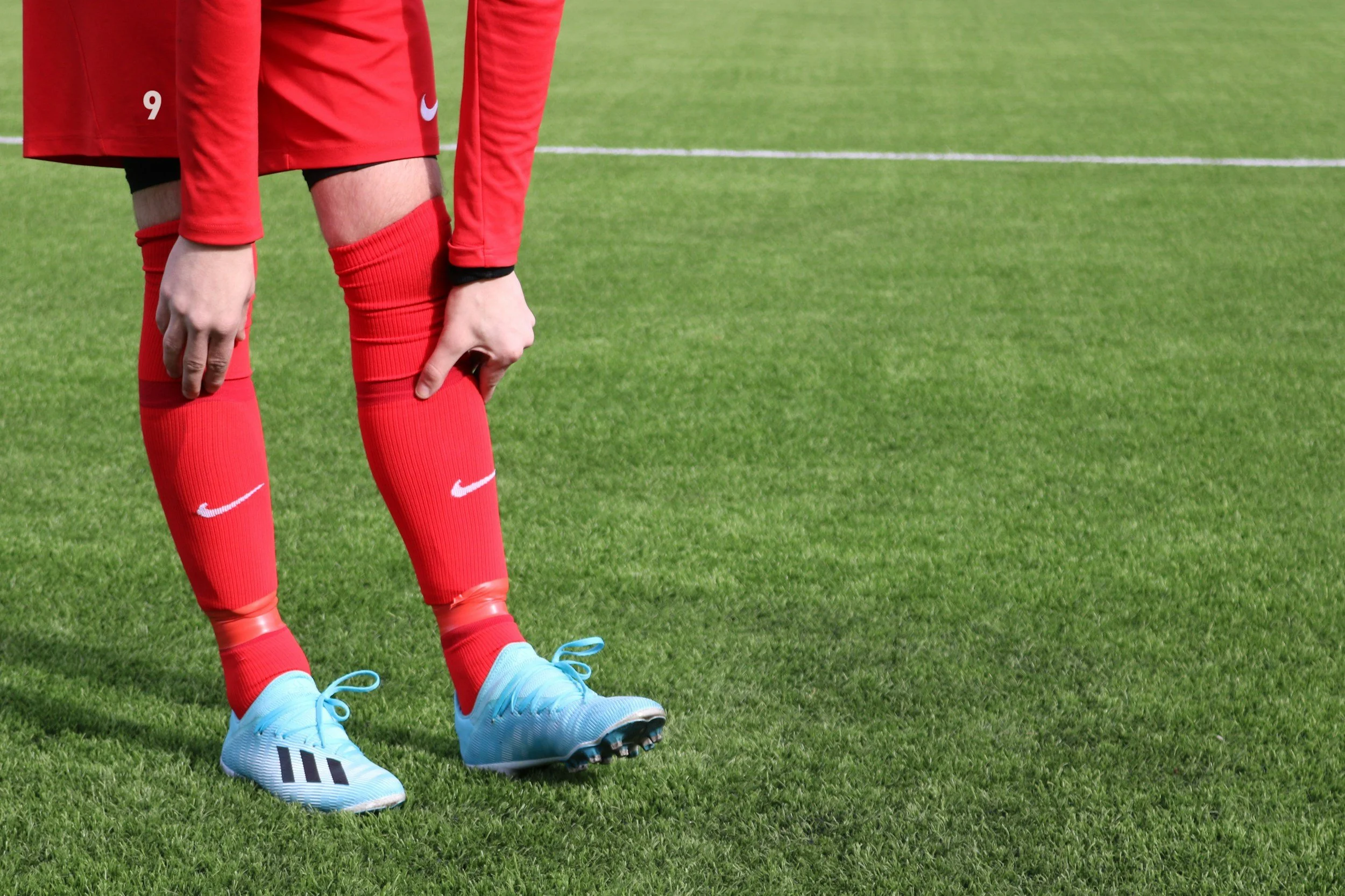
The pain you feel is the result of excessive pulling forces on the periosteum (the thin layer of tissue covering the bone) of the tibia (shinbone).
The Tibialis Posterior: This muscle runs down the back of your calf, crosses under the inside of your ankle, and attaches to the bottom of your foot. Its primary job is to hold up your foot arch.
The Soleus Muscle: A deep calf muscle responsible for absorbing the massive shock of landing with every stride.
Biomechanical Triggers for MTSS
When these muscles are overwhelmed, they pull violently at their attachment points on the shin bone, causing micro-tearing and extreme inflammation.
Excessive Overpronation
If your foot arch collapses too much when you land (overpronation), the tibialis posterior is violently stretched.
The Whiplash Effect: This rapid stretching creates a "whiplash" effect on the muscle tendon, transferring the shear stress directly into the tibial bone.
Weak Hip Stabilizers
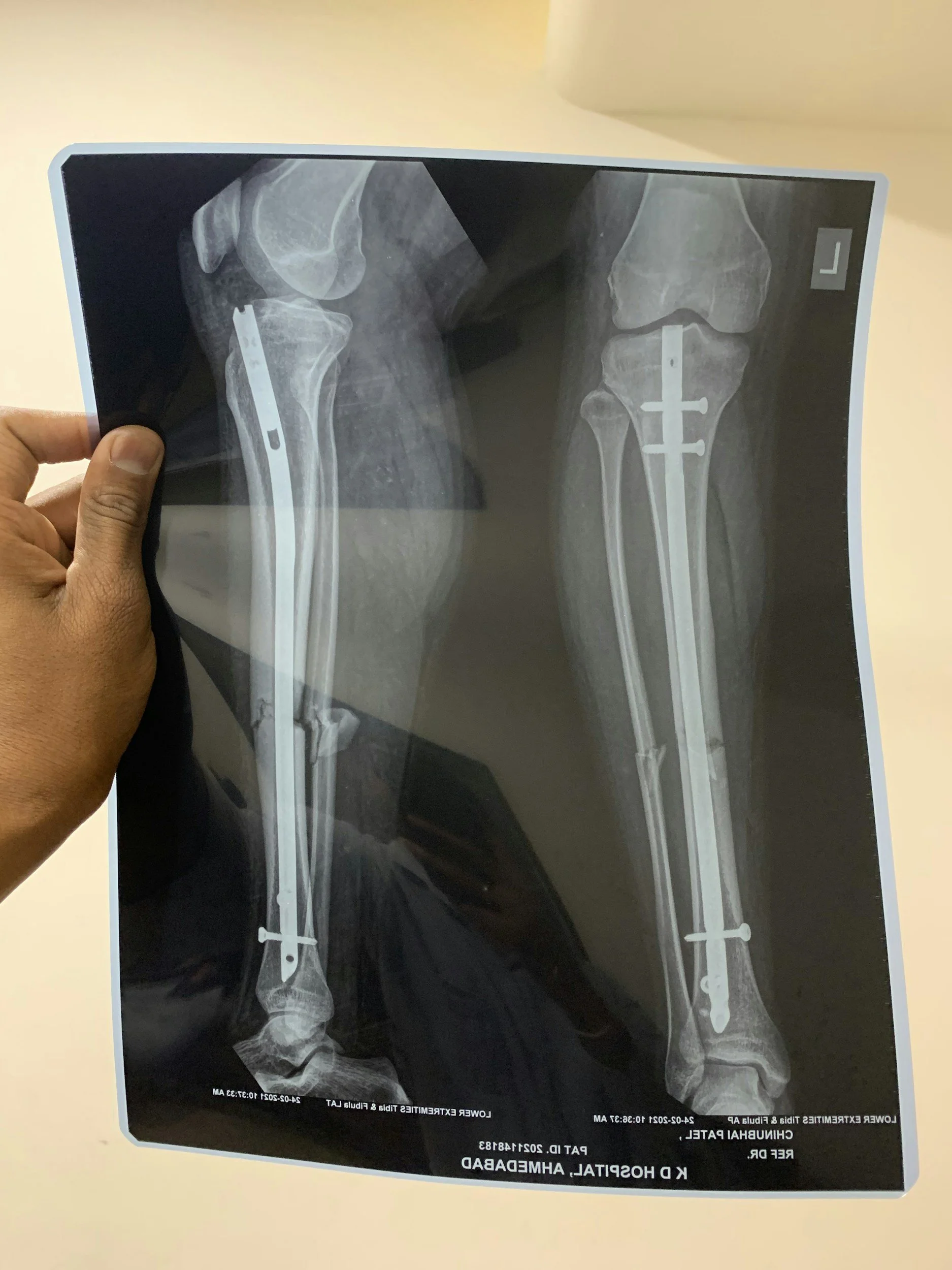
If your gluteus medius (side hip muscle) is weak, your knee will collapse inward (knee valgus) when your foot strikes the ground.
The Kinetic Chain Breakdown
This inward knee collapse forces the tibia to rotate internally, massively increasing the torsional strain on the shin bone and lower leg fascia.
Primary Source Proof: Running Kinematics
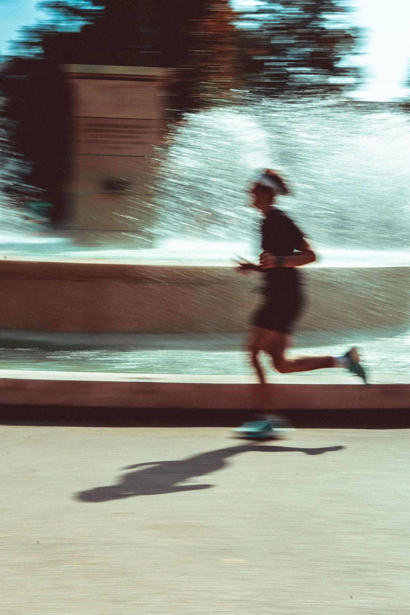
Our approach to treating running pathologies rejects passive rest in favor of active, evidence-based rehabilitation protocols.
Download Clinical Efficacy PDF: The Role of Biomechanical Loading and Foot Orthoses in the Management of Medial Tibial Stress Syndrome (Open Access Medical Review)
The Rehab Mechanics Treatment Protocol
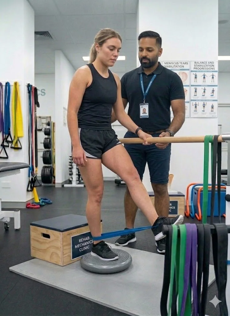
Treating MTSS effectively requires a progressive, phased approach. We do not just treat the shin; we rebuild the entire leg.
Phase 1: Acute Load Management
We do not prescribe total rest, but we do prescribe relative rest.
Activity Modification: We will temporarily replace pavement running with pain-free cross-training (like cycling or deep-water running) to maintain your cardiovascular fitness.
Manual Soft Tissue Release: Using targeted massage and instrument-assisted techniques to release the hypertonic (overly tight) soleus and tibialis posterior muscles.
Shockwave Therapy: For chronic cases, we utilize shockwave therapy to stimulate blood flow and cellular repair directly at the tibial attachment site.
Phase 2: Structural Strengthening
This is where the cure happens. We must increase the load capacity of your lower leg.
Intrinsic Foot Strengthening: Exercises like "foot doming" or towel scrunches to rebuild the tiny muscles inside the foot that support the arch.
Heavy Slow Resistance (HSR) Calf Training: Progressing from double-leg to single-leg weighted calf raises, specifically focusing on the bent-knee soleus raise, to build bulletproof lower leg tissue.
Gluteal Activation: Clamshells, lateral band walks, and single-leg deadlifts to stabilize the pelvis and prevent the knee from caving inward.
Phase 3: Gait Retraining
Before you return to Trinity Bellwoods, we must fix the way you run.
Cadence Modification: Increasing your step rate by just 5% to 10% drastically reduces the impact forces on the shin bone with each step.
Orthotics Assessment: If structural flat feet are the root cause, Sanjay is qualified to dispense custom medical orthotics to permanently correct the foot mechanics.
Graded Return-to-Run (RTR): Providing a strict, interval-based running schedule to progressively re-introduce mechanical load to the tibia without triggering a relapse.
Run Without Pain in Parkdale and Queen West
Do not let chronic shin splints ruin your running season. Get a definitive biomechanical assessment and an active rehabilitation plan.
Book an appointment with our clinical team today. We are conveniently located inside the Prime Medical Centre at 68 Abell Street, easily accessible from Parkdale, Queen West, and Liberty Village.
Contact us to schedule your appointment:
Email: info@rehabmechanics.com
Phone: (416) 533-3900
About the Author
Sanjay Attwala (B.Sc., M.Sc., RPT) is a Registered Physiotherapist and the founder of Rehab Mechanics in Toronto. With over 15 years of clinical practice, Sanjay specializes in complex musculoskeletal rehabilitation, utilizing evidence-based therapeutics to guide his patients toward long-term recovery and optimal athletic performance.
Do I Always Need Physiotherapy for a Rolled Ankle?
Yes. Ignoring a rolled ankle leads to chronic instability and recurring injuries. Physiotherapy restores torn ligaments, rebuilds essential proprioception, and strengthens the surrounding ankle musculature to prevent long-term biomechanical dysfunction and early-onset osteoarthritis.
The Trap of the "Simple" Sprain
"It is just a rolled ankle. I'll walk it off." This is one of the most common and dangerous misconceptions in sports medicine. Whether you rolled it stepping off a curb on Queen Street West, playing a recreational soccer match, or slipping on an icy Toronto sidewalk, an ankle sprain is a severe structural injury.
When patients fail to rehabilitate an ankle sprain, the initial pain and swelling usually subside after a few weeks. However, the underlying mechanical damage remains. Without targeted physiotherapy, the ankle joint heals loosely, leading to a condition known as Chronic Ankle Instability (CAI).
At Rehab Mechanics, we frequently see patients who suffer from recurring ankle sprains, chronic tightness, and early degenerative joint changes simply because they neglected to rehabilitate their very first sprain properly.
Structural Analysis: The Anatomy of a Sprain
To understand why professional rehabilitation is mandatory, we must perform a biomechanical analysis of what exactly happens when an ankle "rolls."
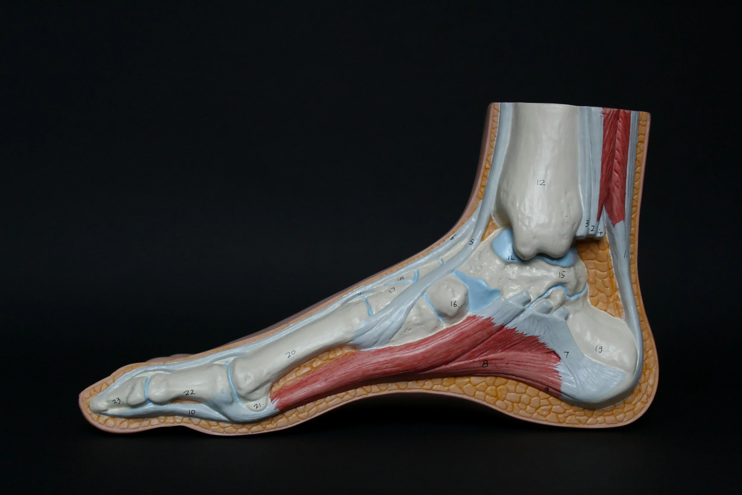
The Lateral Ligament Complex
The vast majority (over 80%) of ankle sprains are "inversion" sprains, meaning the foot rolls inward. This violent motion places catastrophic stress on the lateral ligament complex on the outside of your ankle.
The Anterior Talo-Fibular Ligament (ATFL): This is the weakest and most frequently torn ligament. It stops the foot from sliding forward.
The Calcaneofibular Ligament (CFL): The secondary stabilizer, torn in more severe grade 2 or grade 3 sprains.
The Neurological Damage: Loss of Proprioception
When you tear a ligament, you do not just tear collagen fibers; you tear specialized nerve endings called mechanoreceptors.
What is Proprioception?
Proprioception is your body's subconscious ability to know where its joints are in space. When you walk on uneven ground, mechanoreceptors in your ankle instantly tell your brain to fire the peroneal muscles on the side of your leg to prevent a roll.
The Delay in Signaling
When an ankle is sprained, these nerve endings are destroyed. Even after the ligament heals structurally, the neurological connection is delayed. Your brain cannot react fast enough to a sudden shift in terrain, virtually guaranteeing you will roll the ankle again. Physiotherapy is the only way to rebuild this neurological pathway.
Primary Source Proof: Proprioceptive Rehabilitation
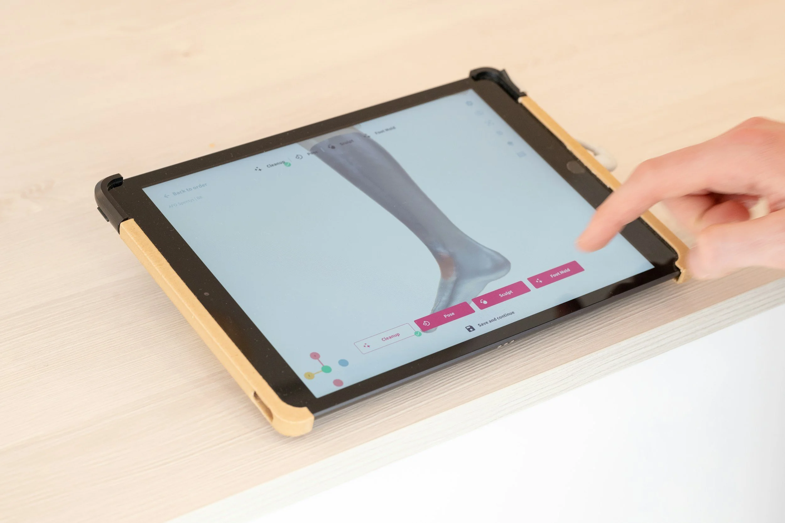
The clinical literature explicitly states that progressive neuromuscular training is non-negotiable for preventing recurrent ankle injuries.
Download Clinical Efficacy PDF: The Effectiveness of Proprioceptive Training in the Prevention of Chronic Ankle Instability (PubMed/NIH)
The Rehab Mechanics Treatment Protocol
Restoring a sprained ankle to 100% capacity requires a phased, progressive loading strategy. Passive treatments like ice and compression are only useful for the first 48 hours. After that, active rehab begins.
Phase 1: Acute Management and Mobility
The goal is to control the swelling while preventing the joint capsule from freezing solid.
Edema Control: Utilizing lymphatic drainage massage and specific taping techniques to flush swelling out of the joint.
Early Range of Motion (ROM): Prescribing pain-free, active movements (like drawing the alphabet with your toes) to align the new collagen fibers correctly as they heal.
Joint Mobilization: Physiotherapists gently glide the talocrural joint to ensure it does not become stiff and restricted.
Phase 2: Muscular Fortification
Once the acute pain subsides, we must replace the lost stability of the torn ligament with muscular strength.
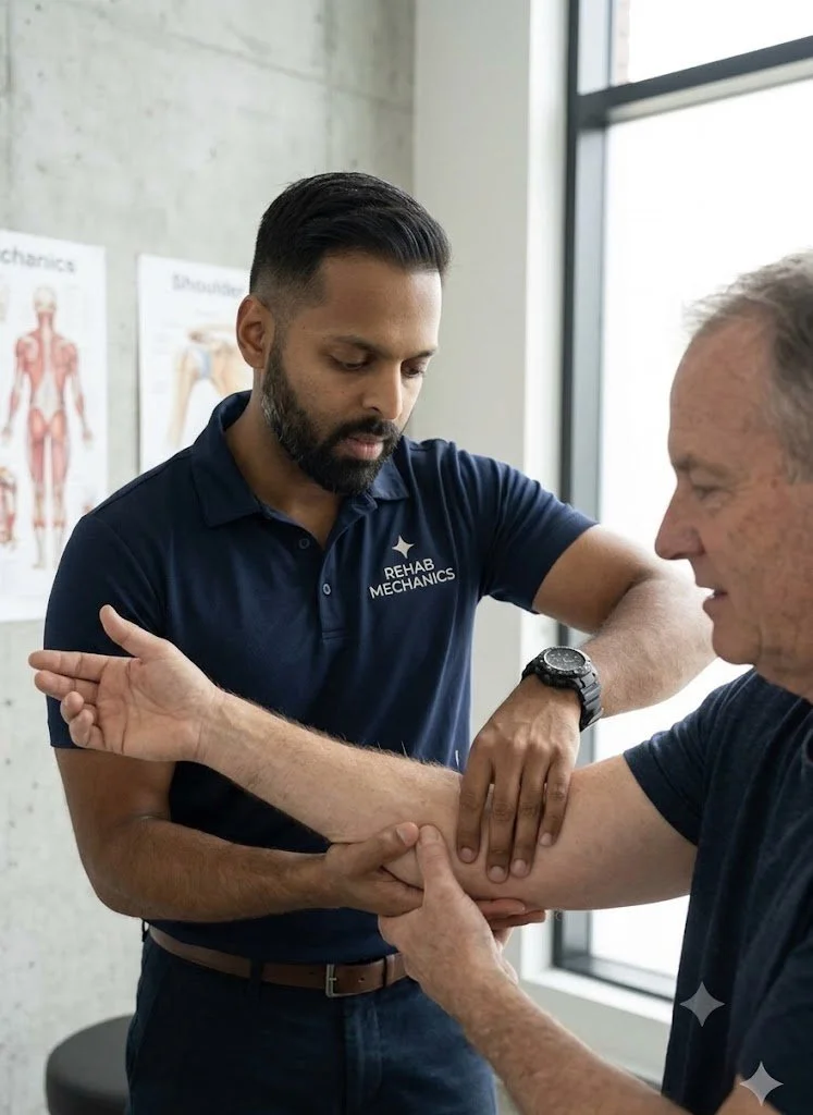
Peroneal Strengthening: Using resistance bands to heavily strengthen the peroneal muscles on the outside of the calf. These muscles are the body's active defense against rolling the ankle inward.
Calf and Tibialis Anterior Loading: Rebuilding the primary shock absorbers of the lower leg to handle the impact of walking and stairs.
Manual Therapy: Deep tissue release on the calf to prevent compensatory tightness from altering your gait.
Phase 3: Neuromuscular Re-education
This is the phase that prevents the next sprain. We rebuild the brain-to-ankle connection.
Balance Training: Progressing from single-leg standing on solid ground, to standing with eyes closed, to standing on unstable surfaces like a BOSU ball or wobble board.
Dynamic Perturbations: The physiotherapist applies unexpected forces while the patient balances, forcing the nervous system to react instantaneously to stabilize the joint.
Plyometrics and Return to Sport: Incorporating jumping, cutting, and lateral bounding drills to prepare the ankle for the chaotic forces of sports and active living.
Secure Your Foundation in Queen West
An unstable ankle changes the way you walk, which eventually damages your knees, hips, and lower back. Treat the injury correctly the first time.
Book a comprehensive ankle assessment with our clinical team today. We are conveniently located inside the Prime Medical Centre at 68 Abell Street, offering advanced biomechanical care in Toronto.
Contact us to schedule your appointment:
Email: info@rehabmechanics.com
Phone: (416) 533-3900
About the Author
Sanjay Attwala (B.Sc., M.Sc., RPT) is a Registered Physiotherapist and the founder of Rehab Mechanics in Toronto. With over 15 years of clinical practice, Sanjay specializes in complex musculoskeletal rehabilitation, utilizing evidence-based therapeutics to guide his patients toward long-term recovery and optimal athletic performance.
Does Poor Posture Cause Sharp Pain in the Middle of the Back?
Yes. Prolonged desk posture locks the thoracic spine and rib joints, causing sharp, stabbing mid-back pain. Physiotherapy utilizes targeted joint manipulation and postural strengthening to unlock the thoracic vertebrae, restoring full spinal extension and allowing for deep, pain-free breathing.
The Silent Agony of the Desk Worker
When people discuss back pain, they almost exclusively talk about the lower back (lumbar spine) or the neck (cervical spine). However, for the massive population of office workers, software developers, and creatives in downtown Toronto, there is a third, highly agonizing zone: the thoracic spine.
If you experience a sharp, knife-like stabbing sensation directly between your shoulder blades—especially after sitting at your desk for four hours—you are experiencing thoracic joint dysfunction.
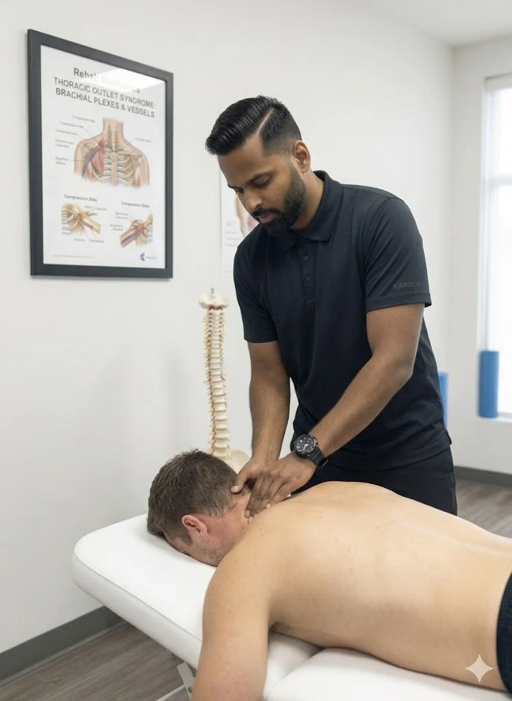
This pain is uniquely frightening because it often wraps around the ribcage to the front of the chest, sometimes mimicking the symptoms of a heart condition or making it physically painful to take a deep breath. Countless patients end up in the emergency room out of fear, only to be told their heart is fine and they simply have "muscle strain."
At Rehab Mechanics, we know that generic muscle relaxants will not fix this. This sharp, stabbing pain is a profound mechanical joint lock caused by the modern urban lifestyle. Correcting it requires precise biomechanical physiotherapy to restore the lost mobility of your mid-back and ribcage.
Structural Analysis: The Mechanics of the Thoracic Spine
To understand why sitting ruins your mid-back, we must perform a detailed anatomical analysis of the thoracic spine and its unique relationship with your ribcage.
The Cage of Stability
Your thoracic spine consists of 12 vertebrae (T1 through T12). Unlike your highly mobile neck and lower back, the thoracic spine is designed for rigidity and protection.
The Rib Articulations: Every single thoracic vertebra attaches to a pair of ribs. These ribs wrap around to the front of your chest to protect your heart and lungs, forming the ribcage.
Costovertebral Joints: The tiny joints where the ribs meet the spine at the back are called costovertebral joints. These joints must pivot slightly every single time you inhale to allow your lungs to expand.
The Postural Lock-Down
The human body adapts specifically to the positions it holds most often.
The Kyphotic Curve
If you spend eight hours a day slouching over a laptop in Liberty Village, your thoracic spine is forced into deep, sustained flexion (a rounded "C" shape, known as kyphosis).
The Muscular Exhaustion: The muscles between your shoulder blades (rhomboids and middle trapezius) are stretched taut over this rounded spine. They become exhausted, chemically inflamed, and form massive, burning trigger points.
The Joint Jam (Rib Dysfunction)
The sharp, stabbing pain occurs when the actual joints lock up.
Because the spine is permanently rounded forward, it loses the physical ability to extend (arch backward).
When the thoracic spine locks, the tiny rib joints attached to it also jam.
The next time you try to twist to grab something behind you, or take a deep, forceful breath, that jammed rib joint violently pinches the surrounding capsule and nerves, sending a breathtaking jolt of pain through your chest and back.
Primary Source Proof: Thoracic Mobilization
Orthopedic research confirms that high-grade manual mobilization of the thoracic spine and ribs yields immediate and profound relief for mid-back pain, vastly outperforming generic stretching routines.
Download Clinical Efficacy PDF: The Efficacy of Thoracic Spine Manipulation and Mobilization in the Treatment of Mechanical Mid-Back Pain (Open Access Medical Review)
Note: The link above serves as an example of our commitment to evidence-based practice, referencing standard international clinical guidelines for spinal rehabilitation.
The Rehab Mechanics Treatment Protocol
Treating thoracic pain is highly satisfying clinically because targeted joint mobilization often provides immediate, profound relief. However, we must follow that up with structural strengthening to ensure the pain does not return the next day.
Phase 1: Joint Unlocking and Decompression
We must physically restore the mobility of the spine and ribs.
High-Velocity Low-Amplitude (HVLA) Thrusts: When clinically appropriate, physiotherapists use highly specific, safe manipulations to instantly un-jam the locked facet and costovertebral joints, resulting in an immediate restoration of deep breathing capacity.
Manual Glides: Utilizing Grade III and IV sustained pressure on the vertebrae to gently stretch the stiffened joint capsules and encourage normal spinal extension.
Pectoral Release: Lengthening the tight chest muscles that are physically holding the spine in a rounded, kyphotic position.
Phase 2: Active Mobility Restoration
Once the joints are unlocked, we must train the nervous system to use the new range of motion.
Thoracic Extension Drills: Using foam rollers or specialized massage balls to isolate and actively bend individual spinal segments backward, reversing the desk posture.
Rotational Mobility: Exercises like "open books" or quadruped T-spine rotations to ensure the ribcage can pivot smoothly without catching.
Phase 3: Scapular and Spinal Fortification
To permanently banish the pain, we must build the muscular strength to hold the spine upright against gravity.
Lower Trapezius Activation: Teaching you how to fire the muscles at the bottom of the shoulder blades to pull them down and back.
Isometric Endurance: Implementing heavy farmer's carries and prone positional holds to build the biological endurance needed to sit with perfect posture for an entire 8-hour workday without fatigue.
Reclaim Your Spine
You do not have to endure the burning, stabbing pain of a locked mid-back. Do not settle for another temporary massage. By addressing the specific joint mechanics of your thoracic spine and ribcage, physiotherapy can provide permanent relief and effortless posture.
Book a comprehensive spinal assessment with our clinical team today. We are conveniently located inside the Prime Medical Centre at 68 Abell Street, offering advanced orthopedic care in the heart of Toronto Queen West.
Contact us to schedule your appointment:
Email: info@rehabmechanics.com
Phone: (416) 533-3900
About the Author
Mr. Sanjay Attwala (B.Sc., M.Sc., RPT) is a Registered Physiotherapist, clinical director, and the founder of Rehab Mechanics in Toronto. With over 15 years of registered clinical practice and a deep specialization in complex musculoskeletal rehabilitation, Sanjay synthesizes rigorous international academic training with advanced evidence-based therapeutics to guide his clinical practice and patient education initiatives.
Academic Background & Credentials
Master of Science (M.Sc.) in Physiotherapy – University of Keele, United Kingdom (2010).
Bachelor of Science (B.Sc.) – University of Waterloo, Ontario, Canada.
Registered Physiotherapist (RPT) – Regulated health professional in excellent standing with the College of Physiotherapists of Ontario (CPO).
Corporate Entity – Operating officially under the S. Attwala Physiotherapy Professional Corporation with a DBA of Rehab Mechanics.
Clinical Expertise & Philosophy
Sanjay’s clinical approach rejects passive symptom management in favor of identifying underlying biomechanical root causes. His diverse expertise spans advanced manual therapies, personalized corrective exercise prescription, and modern physical modalities. At the Rehab Mechanics Toronto Queen West clinic, he routinely diagnoses and treats complex conditions including:
Spinal & Discogenic Pathology – Cervical, thoracic, and lumbar disc injuries, sciatica, and sacroiliac joint (SIJ) dysfunction.
Upper & Lower Extremity Injuries – Rotator cuff tears, frozen shoulder, tennis/golfer’s elbow, carpal tunnel syndrome, and complex ankle/foot pathologies.
Perinatal & Pelvic Health Rehabilitation – Specialized assessment and rehabilitation protocols tailored specifically for women during pregnancy and the post-partum period, addressing pelvic girdle pain, diastasis recti, and core stabilization.
Specialized Rehabilitation – Pelvic health therapy, TMJ dysfunction, post-surgical rehabilitation (including Total Hip and Total Knee Replacements), and custom orthotics dispensing.
Shockwave Therapy: with advanced cutting edge technological devices to suit your needs.
Interdisciplinary Practice & Patient Care
Sanjay practices an integrated model of healthcare, working closely alongside medical doctors inside the Prime Medical Centre on Abell Street to streamline patient recovery pathways. He maintains a human-centric, communication-first clinical framework, ensuring that care remains fully customized rather than automated.
His clinical caseload encompasses a broad operational spectrum under Ontario's regulatory frameworks, including:
Motor Vehicle Accident (MVA) Claims – Rehabilitation navigating Ontario’s statutory accident benefits schedule.
Workplace Safety and Insurance Board (WSIB) – Occupational injury management and return-to-work screening.
Extended Health Care (EHC) & Private Practice – Multi-tier insurance coordination and long-term athletic development plans.
Commitment to Research & Community
Outside of his clinical caseload at Rehab Mechanics and his additional practice affiliations in Etobicoke, Sanjay is an active health writer and community educator. He translates contemporary peer-reviewed medical research into accessible, actionable guidance on his professional blog. As a dedicated father and husband, he mirrors his professional advice in his personal life, focusing on structural mobility, cross-training, and longevity to help his family and his community thrive. Naturally he takes he a keen interest in rehabilitation for women who are pregnant and post-partum.
Disclaimer: The information provided on this blog is intended for educational and informational purposes only. It does not constitute medical advice, diagnosis, or a treatment plan. Always seek the direct advice of a Registered Physiotherapist, physician, or other qualified health provider regarding any medical condition or physical rehabilitation routine.
Can Physiotherapy Permanently Cure Plantar Fasciitis Without Custom Orthotics?
Yes and NO. While custom orthotics provide immediate offloading for severe cases, physiotherapy is the definitive cure. By restoring ankle dorsiflexion, mobilizing the plantar fascia, and strengthening the intrinsic foot muscles, physical therapy rebuilds your foot's natural shock-absorbing capacity.
The "Glass Shard" in the Heel
There are few sensations more painful than the first few steps out of bed in the morning. For many Torontonians—especially those who spend their days on their feet or those who have recently increased their running volume—that first step feels like a glass shard or a dull, deep ache stabbing directly into the heel.
This is Plantar Fasciitis. It is one of the most common, yet frequently misunderstood, pathologies we treat at Rehab Mechanics. Patients are often told to "roll a frozen water bottle under your foot" or buy expensive, passive arch supports. While these provide temporary relief, they do not resolve the mechanical failure of the foot. To truly defeat plantar fasciitis, you must restore the mobility of your ankle and the intrinsic strength of your arch.
Structural Analysis: The Mechanics of Foot Pain
The plantar fascia is not just a strip of tissue; it is a vital, high-tension ligament that controls the entire biomechanical function of your foot.
The Windlass Mechanism
The plantar fascia connects your heel bone (calcaneus) to the base of your toes.
The Arch Spring: When you step down, the fascia stretches like a high-tension spring to absorb the shock of your weight.
The Lever: When you prepare to push off your toes, the fascia winds tightly, turning your foot into a rigid, powerful lever for walking or running.
The Failure of the Shock Absorber
Plantar fasciitis is not "inflammation" (itis). It is actually plantar fasciopathy—a state of chronic cellular degeneration caused by repetitive mechanical overload.
Ankle Stiffness: If your ankle (talocrural joint) is stiff and cannot bend forward (dorsiflexion), your foot is forced to compensate. The arch collapses inward (overpronation) to allow the foot to move forward.
The Sheer Force: This constant, aggressive collapse places massive tensile stretching forces on the fascia, leading to microscopic tears, scar tissue build-up, and localized bone-spur development.
Primary Source Proof: Biomechanical Rehabilitation
Scientific literature supports the use of progressive mechanical loading and ankle mobilization as the most effective path to curing chronic heel pain, proving that passive arch support is insufficient for long-term resolution.
The Rehab Mechanics Foot Reconstruction Protocol
We treat plantar fasciitis by rebuilding the mechanical health of the entire leg, starting from the ground up.
Phase 1: Tissue Decompression and Shockwave Therapy
Before we strengthen, we must clear the fibrotic, scarred tissue.
Shockwave Therapy: We use high-energy acoustic waves to break down the dense, painful scar tissue in the fascia and stimulate new, healthy blood vessel growth in the degenerated heel.
Myofascial Release: Deep, manual release of the calf muscles (gastrocnemius and soleus). If your calf is tight, your ankle cannot move, and your fascia will remain under constant tension.
Phase 2: Ankle Dorsiflexion Restoration
We must restore the joint's ability to bend, which is the root cause of the arch collapse.
Manual Joint Mobilizations: Our physiotherapists use hands-on techniques to force the talocrural joint to glide properly, ensuring the ankle can bend freely without relying on arch collapse.
Dynamic Calf Stretching: Progressive lengthening of the posterior chain to ensure full ankle range of motion during walking.
Phase 3: The Rathleff Protocol (High-Load Strengthening)
We rebuild the foot's structural integrity using heavy loading.
The Rathleff Protocol: A specific, heavy-load strength protocol using a rolled towel under the toes to maximally activate the plantar fascia while performing calf raises. This has been clinically proven to be the most effective intervention for resolving heel pain.
Intrinsic Foot Strengthening: Exercises like "foot doming" or toe-splaying to rebuild the tiny, dormant muscles inside the foot that are designed to support your arch, rendering the need for permanent orthotics obsolete.
Restore Your Foundation
You do not have to live with the morning hobble. By rebuilding your ankle mobility and foot strength, you can permanently resolve heel pain.
Book a comprehensive foot and ankle assessment today. We are conveniently located inside the Prime Medical Centre at 68 Abell Street, offering advanced orthopedic recovery in Queen West.
Contact us to schedule your appointment:
Email: info@rehabmechanics.com
Phone: (416) 533-3900
About the Author
Mr. Sanjay Attwala (B.Sc., M.Sc., RPT) is a Registered Physiotherapist, clinical director, and the founder of Rehab Mechanics in Toronto. With over 15 years of registered clinical practice and a deep specialization in complex musculoskeletal rehabilitation, Sanjay synthesizes rigorous international academic training with advanced evidence-based therapeutics to guide his clinical practice and patient education initiatives.
Academic Background & Credentials
Master of Science (M.Sc.) in Physiotherapy – University of Keele, United Kingdom (2010).
Bachelor of Science (B.Sc.) – University of Waterloo, Ontario, Canada.
Registered Physiotherapist (RPT) – Regulated health professional in excellent standing with the College of Physiotherapists of Ontario (CPO).
Corporate Entity – Operating officially under the S. Attwala Physiotherapy Professional Corporation with a DBA of Rehab Mechanics.
Clinical Expertise & Philosophy
Sanjay’s clinical approach rejects passive symptom management in favor of identifying underlying biomechanical root causes. His diverse expertise spans advanced manual therapies, personalized corrective exercise prescription, and modern physical modalities. At the Rehab Mechanics Toronto Queen West clinic, he routinely diagnoses and treats complex conditions including:
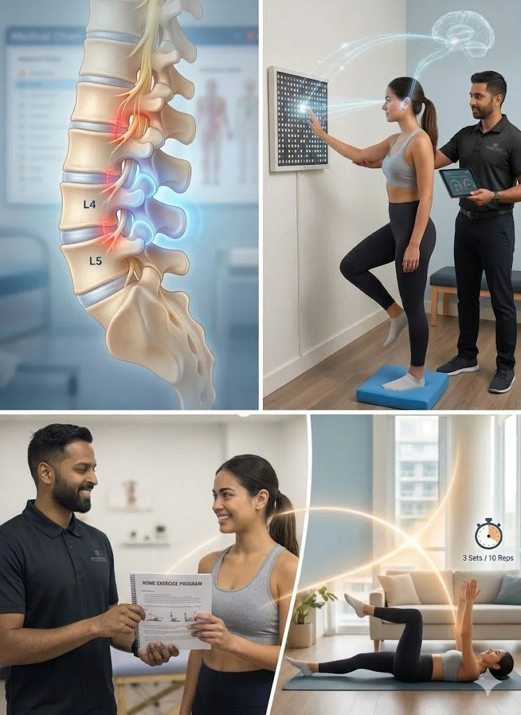
Spinal & Discogenic Pathology – Cervical, thoracic, and lumbar disc injuries, sciatica, and sacroiliac joint (SIJ) dysfunction.
Upper & Lower Extremity Injuries – Rotator cuff tears, frozen shoulder, tennis/golfer’s elbow, carpal tunnel syndrome, and complex ankle/foot pathologies.
Perinatal & Pelvic Health Rehabilitation – Specialized assessment and rehabilitation protocols tailored specifically for women during pregnancy and the post-partum period, addressing pelvic girdle pain, diastasis recti, and core stabilization.
Specialized Rehabilitation – Pelvic health therapy, TMJ dysfunction, post-surgical rehabilitation (including Total Hip and Total Knee Replacements), and custom orthotics dispensing.
Shockwave Therapy: with advanced cutting edge technological devices to suit your needs.
Interdisciplinary Practice & Patient Care
Sanjay practices an integrated model of healthcare, working closely alongside medical doctors inside the Prime Medical Centre on Abell Street to streamline patient recovery pathways. He maintains a human-centric, communication-first clinical framework, ensuring that care remains fully customized rather than automated.
His clinical caseload encompasses a broad operational spectrum under Ontario's regulatory frameworks, including:
Motor Vehicle Accident (MVA) Claims – Rehabilitation navigating Ontario’s statutory accident benefits schedule.
Workplace Safety and Insurance Board (WSIB) – Occupational injury management and return-to-work screening.
Extended Health Care (EHC) & Private Practice – Multi-tier insurance coordination and long-term athletic development plans.
Commitment to Research & Community
Outside of his clinical caseload at Rehab Mechanics and his additional practice affiliations in Etobicoke, Sanjay is an active health writer and community educator. He translates contemporary peer-reviewed medical research into accessible, actionable guidance on his professional blog. As a dedicated father and husband, he mirrors his professional advice in his personal life, focusing on structural mobility, cross-training, and longevity to help his family and his community thrive. Naturally he takes he a keen interest in rehabilitation for women who are pregnant and post-partum.
Disclaimer: The information provided on this blog is intended for educational and informational purposes only. It does not constitute medical advice, diagnosis, or a treatment plan. Always seek the direct advice of a Registered Physiotherapist, physician, or other qualified health provider regarding any medical condition or physical rehabilitation routine.
Can Physiotherapy Fix Shoulder Impingement Without Surgery?
Yes. Physiotherapy is the most effective treatment for shoulder impingement syndrome. By correcting scapular dyskinesis, restoring thoracic mobility, and strengthening the rotator cuff to hold the arm bone perfectly centralized in the socket, physiotherapy physically eliminates the friction causing your pain.
The Overhead "Catch"
For the active demographic in Queen West, your shoulder is your most important tool. Whether you are lifting a heavy barbell in the gym, playing a recreational tennis match, or reaching overhead to grab a high shelf in your apartment, the shoulder must move with fluid, frictionless precision.
When you start to feel a sharp, stabbing "catch" or "pinch" in the front of your shoulder whenever you lift your arm above head height, it is a sign that your shoulder is not moving correctly. The medical term is Shoulder Impingement Syndrome. It is incredibly common, often dismissed as "just a strain," and frequently managed incorrectly with ice and rest.
At Rehab Mechanics, we see patients who have lived with this impingement for years. They worry they have a "torn rotator cuff" and fear surgery. The reality is often much simpler: your shoulder blade is not moving in sync with your arm bone. By correcting this mechanical timing, we can eliminate the pinch immediately and prevent the development of a chronic rotator cuff tear.
Structural Analysis: The Mechanics of Impingement
To understand why your shoulder pinches, we must look at the "subacromial tunnel"—the narrow, bony bottleneck through which your tendons must travel.
The Subacromial Bottleneck
Your shoulder joint is a ball-and-socket, but the "socket" is roofed by a bony prominence called the acromion.
The Tunnel: Between the ball (the arm bone) and the roof (the acromion) is a narrow, tunnel-like space.
The Contents: Through this tunnel pass your vital rotator cuff tendons and a fluid-filled sac called the subacromial bursa.
The "Crush" Mechanism (Impingement)
In a healthy shoulder, your rotator cuff muscles pull the ball of the arm bone downward, keeping it low in the socket as you lift your arm. This keeps the tunnel wide and open.
The Breakdown: If your rotator cuff is weak, or if your shoulder blade (scapula) is stiff, the arm bone slides upward too early during the lift.
The Impingement: The top of the arm bone smashes into the roof of the tunnel (the acromion). This physically crushes the rotator cuff tendon and the bursa against the bone.
The Result: Sharp, blinding pain, often followed by a dull, throbbing ache that lasts for hours after the lift.
Primary Source Proof: Impingement Rehabilitation
Sports medicine research conclusively mandates that physiotherapy is the first-line treatment for shoulder impingement, often achieving superior long-term results compared to surgical decompression.
Download Clinical Efficacy PDF: The Efficacy of Conservative Physical Therapy versus Surgical Decompression in Shoulder Impingement Syndrome (Open Access Medical Review)
The Rehab Mechanics Corrective Protocol
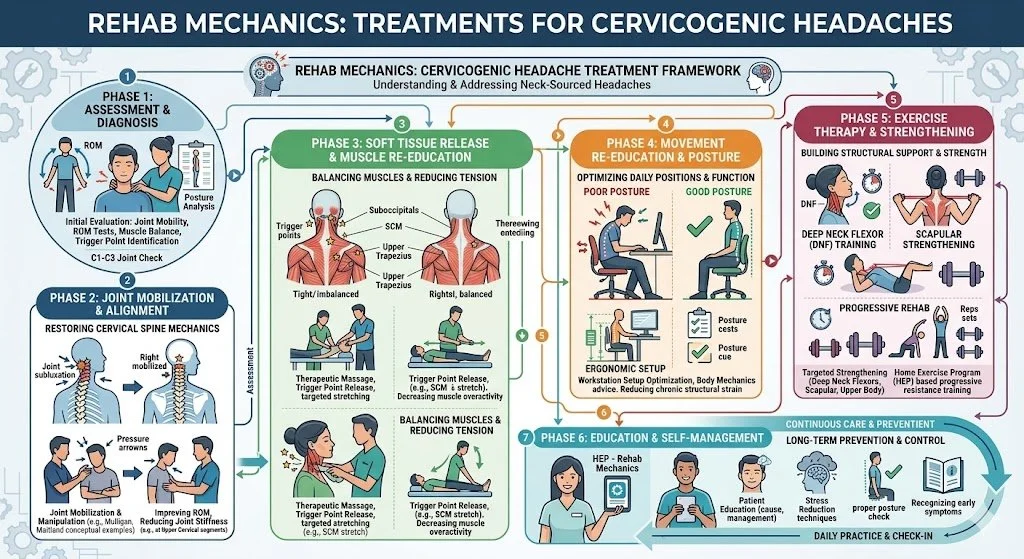
We treat impingement not by massaging the painful spot, but by fixing the biomechanics of the entire shoulder girdle.
Phase 1: Decompressing the Tunnel
We must stop the bone-on-bone grinding immediately.
Posterior Capsule Mobilization: A stiff back-of-the-shoulder capsule pushes the arm bone forward into the impingement zone. Our therapists use high-grade manual glides to loosen this tissue.
Manual Release of the Pectoralis Minor: A tight chest muscle physically drags the shoulder blade into a forward tilt, narrowing the subacromial tunnel. We release this tension to restore scapular alignment.
Phase 2: Restoring Scapulothoracic Rhythm
We must teach your shoulder blade how to get out of the way of your arm bone.
Lower Trapezius and Serratus Anterior Activation: These muscles pull the shoulder blade down and back, physically opening the tunnel. We use targeted resistance work to ensure these muscles fire automatically during overhead movements.
Neuromuscular Re-education: We use tactical cueing to retrain the brain to keep the shoulder blade "set" firmly against the ribcage as you lift, preventing the "hike-and-hunch" compensation pattern.
Phase 3: Rotator Cuff Fortification
Once the joint is mechanically safe, we build the strength to hold the ball centered in the socket.
Eccentric Rotator Cuff Loading: Using targeted resistance training to rebuild the tendons. A strong rotator cuff is the only structural guarantee that the arm bone stays centralized in the socket, permanently preventing impingement.
Reclaim Your Overhead Power
Stop letting a pinching shoulder limit your training or your daily life. Fix the mechanics, and the pain disappears.
Book your comprehensive shoulder assessment with our clinical team today. We are conveniently located inside the Prime Medical Centre at 68 Abell Street, offering elite orthopedic recovery in Toronto.
Contact us to schedule your appointment:
Email: info@rehabmechanics.com
Phone: (416) 533-3900
About the Author
Mr. Sanjay Attwala (B.Sc., M.Sc., RPT) is a Registered Physiotherapist, clinical director, and the founder of Rehab Mechanics in Toronto. With over 15 years of registered clinical practice and a deep specialization in complex musculoskeletal rehabilitation, Sanjay synthesizes rigorous international academic training with advanced evidence-based therapeutics to guide his clinical practice and patient education initiatives.
Academic Background & Credentials
Master of Science (M.Sc.) in Physiotherapy – University of Keele, United Kingdom (2010).
Bachelor of Science (B.Sc.) – University of Waterloo, Ontario, Canada.
Registered Physiotherapist (RPT) – Regulated health professional in excellent standing with the College of Physiotherapists of Ontario (CPO).
Corporate Entity – Operating officially under the S. Attwala Physiotherapy Professional Corporation with a DBA of Rehab Mechanics.
Clinical Expertise & Philosophy
Sanjay’s clinical approach rejects passive symptom management in favor of identifying underlying biomechanical root causes. His diverse expertise spans advanced manual therapies, personalized corrective exercise prescription, and modern physical modalities. At the Rehab Mechanics Toronto Queen West clinic, he routinely diagnoses and treats complex conditions including:
Spinal & Discogenic Pathology – Cervical, thoracic, and lumbar disc injuries, sciatica, and sacroiliac joint (SIJ) dysfunction.
Upper & Lower Extremity Injuries – Rotator cuff tears, frozen shoulder, tennis/golfer’s elbow, carpal tunnel syndrome, and complex ankle/foot pathologies.
Perinatal & Pelvic Health Rehabilitation – Specialized assessment and rehabilitation protocols tailored specifically for women during pregnancy and the post-partum period, addressing pelvic girdle pain, diastasis recti, and core stabilization.
Specialized Rehabilitation – Pelvic health therapy, TMJ dysfunction, post-surgical rehabilitation (including Total Hip and Total Knee Replacements), and custom orthotics dispensing.
Shockwave Therapy: with advanced cutting edge technological devices to suit your needs.
Interdisciplinary Practice & Patient Care
Sanjay practices an integrated model of healthcare, working closely alongside medical doctors inside the Prime Medical Centre on Abell Street to streamline patient recovery pathways. He maintains a human-centric, communication-first clinical framework, ensuring that care remains fully customized rather than automated.
His clinical caseload encompasses a broad operational spectrum under Ontario's regulatory frameworks, including:
Motor Vehicle Accident (MVA) Claims – Rehabilitation navigating Ontario’s statutory accident benefits schedule.
Workplace Safety and Insurance Board (WSIB) – Occupational injury management and return-to-work screening.
Extended Health Care (EHC) & Private Practice – Multi-tier insurance coordination and long-term athletic development plans.
Commitment to Research & Community
Outside of his clinical caseload at Rehab Mechanics and his additional practice affiliations in Etobicoke, Sanjay is an active health writer and community educator. As a dedicated father and husband, he mirrors his professional advice in his personal life, focusing on structural mobility, cross-training, and longevity to help his family and his community thrive. Naturally he takes he a keen interest in rehabilitation for women who are pregnant and post-partum.
Disclaimer: The information provided on this blog is intended for educational and informational purposes only. It does not constitute medical advice, diagnosis, or a treatment plan. Always seek the direct advice of a Registered Physiotherapist, physician, or other qualified health provider regarding any medical condition or physical rehabilitation routine.
Can Physiotherapy Fix a SLAP Tear in My Shoulder Without Surgery?
Yes. Physiotherapy successfully treats SLAP tears without surgery by restoring scapular mechanics and strengthening the dynamic stabilizers of the rotator cuff. This active rehabilitation reduces the sheer force on the labrum, stabilizing the biceps anchor and allowing the shoulder to function pain-free.
The Agony of the Overhead Athlete
In Toronto’s active core, particularly among the dedicated functional fitness, CrossFit, and weightlifting communities in Liberty Village and Queen West, shoulder injuries are a constant threat. One of the most terrifying diagnoses an athlete can receive is a SLAP lesion—a specific, deep tear in the cartilage of the shoulder socket.
The symptoms are deeply frustrating: a profound, deep-seated ache inside the shoulder joint, a sharp "catching" or "popping" sensation when lowering a heavy barbell or throwing a ball, and a sudden, inexplicable loss of overhead strength. When an MRI confirms a SLAP tear, patients frequently panic, assuming their athletic career is over unless they undergo an invasive arthroscopic repair, which carries a brutal 6-to-9-month recovery timeline.
At Rehab Mechanics, we want to shift this narrative. While massive, traumatic labral avulsions may require surgery, the vast majority of degenerative or overuse SLAP tears can be managed conservatively. A tear in the labrum is often the victim of poor shoulder mechanics, not the primary culprit. By overhauling how your shoulder blade and rotator cuff handle load, expert physiotherapy can silence the pain and restore your overhead power without a scalpel.
Structural Analysis: The Mechanics of a SLAP Lesion
To understand how physical therapy can save your shoulder, we must perform a detailed biomechanical analysis of the labrum and its connection to the rest of the arm.
Anatomy of the Biceps-Labrum Complex
Your shoulder is a ball-and-socket joint, but the socket (the glenoid) is extremely shallow—often compared to a golf ball sitting on a tee.
The Labrum: To deepen this shallow socket and create a stabilizing "suction cup" effect, a ring of tough fibrocartilage called the labrum wraps around the rim of the bone.
The Biceps Anchor: The long head of your biceps tendon travels up your arm and attaches directly into the very top (the superior portion) of this labral ring.
What is a SLAP Tear?
SLAP stands for Superior Labrum Anterior and Posterior. It means the top of the cartilage ring has been torn from front to back, exactly where the biceps tendon anchors into it.
Mechanisms of Injury
SLAP tears generally occur via two distinct mechanical pathways:
1. Acute Macrotrauma
Falling onto an outstretched hand (FOOSH) on an icy Toronto sidewalk, forcing the arm bone violently up into the socket.
A sudden, heavy, eccentric load, such as catching a falling heavy object, which violently yanks the biceps tendon, tearing its anchor off the bone.
2. The "Peel-Back" Mechanism (Overuse)
This is the most common cause for weightlifters and throwing athletes.
When you repeatedly bring your arm into maximum external rotation (like the cocking phase of a baseball pitch or holding a barbell behind your neck for a back squat), the biceps tendon twists.
This twisting motion creates a massive torsional force that literally "peels back" the superior labrum off the bone, causing micro-trauma that eventually becomes a full tear.
The Biomechanical Driver: Scapular Dyskinesis
Why do some athletes get SLAP tears while others don't? It comes down to the foundation: the shoulder blade (scapula).
If your shoulder blade is "slouched" forward due to poor posture or a weak serratus anterior, the socket is angled downward.
When you reach overhead, your arm bone hits a hard stop much earlier. To achieve the overhead position, you have to force the arm past its natural limit, radically increasing the strain and "peel-back" force on the labrum and biceps anchor.
Primary Source Proof: Conservative Management
Orthopedic and sports medicine literature increasingly mandates that conservative physiotherapy, focusing on scapular stabilization and posterior capsule flexibility, should be the first-line treatment for SLAP lesions, yielding excellent functional outcomes comparable to surgery.
Note: The link above directs to external, peer-reviewed medical literature demonstrating our commitment to evidence-based practice and international clinical guidelines for upper extremity rehabilitation.
The Rehab Mechanics SLAP Protocol
Rehabilitating a SLAP tear requires absolute precision. We cannot use generic shoulder stretches, as overly stretching a torn labrum will make it worse. We must utilize targeted "remedial mechanics" to decompress the joint.
Phase 1: Protecting the Biceps Anchor (Weeks 1-4)
The immediate goal is to reduce the inflammatory sheer force pulling on the torn cartilage.
Biceps Unloading: We temporarily eliminate all heavy, isolated biceps loading (like curls) and extreme overhead reaching.
Posterior Capsule Mobilization: A tight posterior (back) shoulder capsule pushes the arm bone forward and upward, directly crushing the superior labrum. Our physiotherapists utilize heavy Grade III/IV manual joint glides to forcefully stretch the back of the capsule, restoring the ball to the dead-center of the socket.
Pectoral Release: Deep tissue mobilization to release the tight chest muscles that are pulling the shoulder blade out of alignment.
Phase 2: Dynamic Scapular Fortification (Weeks 4-8)
We must build an indestructible foundation for the arm to move upon.
Serratus Anterior and Lower Trap Activation: We utilize exercises like "scapular punches" and Y-raises. When these muscles are strong, they rotate the shoulder blade upward with your arm, ensuring the socket stays aligned with the humerus and completely eliminating the "peel-back" impingement force on the labrum.
Closed Kinetic Chain Stability: Planks, wall slides, and medicine ball walk-outs force the shoulder to stabilize the weight of the body, training the nervous system to lock the joint into a safe, centralized position.
Phase 3: Advanced Load and Kinetic Chain Transfer (Weeks 8-12+)
A strong shoulder is useless if it cannot handle the chaotic forces of sports.
Rotator Cuff Eccentrics: Rebuilding the resilience of the rotator cuff to absorb deceleration forces so the labrum doesn't have to.
Core-to-Arm Energy Transfer: The power for a heavy lift or a throw should come from the hips and core, not the fragile shoulder joint. We train explosive kinetic linking (like rotational medicine ball throws and heavy kettlebell swings) to ensure your lower body is generating the force, protecting the torn labrum from taking the brunt of the load.
Stop Fearing the Tear
An MRI showing a SLAP tear is not a death sentence for your active lifestyle. By meticulously correcting your shoulder mechanics, you can eliminate the destructive friction and return to lifting, throwing, and living pain-free.
Book a specialized sports orthopedic assessment with our clinical team today. We are conveniently located inside the Prime Medical Centre at 68 Abell Street, offering elite upper extremity rehabilitation in Queen West.
Contact us to schedule your appointment:
Email: info@rehabmechanics.com
Phone: (416) 533-3900
About the Author
Mr. Sanjay Attwala (B.Sc., M.Sc., RPT) is a Registered Physiotherapist, clinical director, and the founder of Rehab Mechanics in Toronto. With over 15 years of registered clinical practice and a deep specialization in complex musculoskeletal rehabilitation, Sanjay synthesizes rigorous international academic training with advanced evidence-based therapeutics to guide his clinical practice and patient education initiatives.
Academic Background & Credentials
Master of Science (M.Sc.) in Physiotherapy – University of Keele, United Kingdom (2010).
Bachelor of Science (B.Sc.) – University of Waterloo, Ontario, Canada.
Registered Physiotherapist (RPT) – Regulated health professional in excellent standing with the College of Physiotherapists of Ontario (CPO).
Corporate Entity – Operating officially under the S. Attwala Physiotherapy Professional Corporation with a DBA of Rehab Mechanics.
Clinical Expertise & Philosophy
Sanjay’s clinical approach rejects passive symptom management in favor of identifying underlying biomechanical root causes. His diverse expertise spans advanced manual therapies, personalized corrective exercise prescription, and modern physical modalities. At the Rehab Mechanics Toronto Queen West clinic, he routinely diagnoses and treats complex conditions including:
Spinal & Discogenic Pathology – Cervical, thoracic, and lumbar disc injuries, sciatica, and sacroiliac joint (SIJ) dysfunction.
Upper & Lower Extremity Injuries – Rotator cuff tears, frozen shoulder, tennis/golfer’s elbow, carpal tunnel syndrome, and complex ankle/foot pathologies.
Perinatal & Pelvic Health Rehabilitation – Specialized assessment and rehabilitation protocols tailored specifically for women during pregnancy and the post-partum period, addressing pelvic girdle pain, diastasis recti, and core stabilization.
Specialized Rehabilitation – Pelvic health therapy, TMJ dysfunction, post-surgical rehabilitation (including Total Hip and Total Knee Replacements), and custom orthotics dispensing.
Shockwave Therapy: with advanced cutting edge technological devices to suit your needs.
Interdisciplinary Practice & Patient Care
Sanjay practices an integrated model of healthcare, working closely alongside medical doctors inside the Prime Medical Centre on Abell Street to streamline patient recovery pathways. He maintains a human-centric, communication-first clinical framework, ensuring that care remains fully customized rather than automated.
His clinical caseload encompasses a broad operational spectrum under Ontario's regulatory frameworks, including:
Motor Vehicle Accident (MVA) Claims – Rehabilitation navigating Ontario’s statutory accident benefits schedule.
Workplace Safety and Insurance Board (WSIB) – Occupational injury management and return-to-work screening.
Extended Health Care (EHC) & Private Practice – Multi-tier insurance coordination and long-term athletic development plans.
Commitment to Research & Community
Outside of his clinical caseload at Rehab Mechanics and his additional practice affiliations in Etobicoke, Sanjay is an active health writer and community educator. He translates contemporary peer-reviewed medical research into accessible, actionable guidance on his professional blog. As a dedicated father and husband, he mirrors his professional advice in his personal life, focusing on structural mobility, cross-training, and longevity to help his family and his community thrive. Naturally he takes he a keen interest in rehabilitation for women who are pregnant and post-partum.
Disclaimer: The information provided on this blog is intended for educational and informational purposes only. It does not constitute medical advice, diagnosis, or a treatment plan. Always seek the direct advice of a Registered Physiotherapist, physician, or other qualified health provider regarding any medical condition or physical rehabilitation routine.
Does Sharp Pain in the Front of the Shoulder Mean I Tore My Bicep Tendon?
No. Sharp front shoulder pain is most commonly proximal biceps tendinopathy or impingement, not a full tear. Physiotherapy completely resolves this by correcting scapular mechanics, releasing the pectoral fascia, and applying eccentric loading to the biceps tendon to rebuild its structural integrity without surgery.
The Weightlifter's Worst Nightmare
For the dedicated strength athletes, CrossFitters, and recreational tennis players living in Queen West and Liberty Village, shoulder health is paramount. One of the most terrifying, yet incredibly common, injuries in these demographics is a sudden, sharp, burning pain located precisely at the very front of the shoulder joint, radiating slightly down the arm.
The immediate reaction is panic. Patients feel the pain directly over their bicep muscle and instantly assume they have suffered a massive, catastrophic tear of the biceps tendon that will require surgical reattachment. They stop lifting, cancel their tennis matches, and put their arm in a sling.
However, full ruptures of the biceps tendon are quite rare and usually present with a highly visible, deformed "Popeye" muscle bulge. In the vast majority of cases seen at Rehab Mechanics, this sharp, localized pain is Proximal Biceps Tendinopathy. It is an overuse injury caused by faulty shoulder mechanics that are grinding the tendon against the bone. By correcting how your shoulder blade and rotator cuff function, advanced physical therapy can eliminate the friction and permanently heal the tendon.
Structural Analysis: The Mechanics of the Biceps Tendon
To understand how to fix anterior shoulder pain, we must perform a detailed biomechanical analysis of the shoulder joint and the complex route the biceps tendon takes to get there.
The Anatomy of the Long Head of the Biceps (LHB)
Your biceps muscle has two upper attachment points (heads) that connect it to the shoulder. The "short head" attaches safely outside the joint. The "long head" is the troublemaker.
The Bicipital Groove: The Long Head of the Biceps (LHB) tendon must travel up the front of your arm bone (humerus) through a very narrow, bony trench called the bicipital groove.
The Transverse Humeral Ligament: A tight band of tissue straps the tendon down into this groove to keep it from popping out when you move.
The Intra-Articular Journey: Once it passes through the groove, the tendon literally dives inside the shoulder joint capsule to attach to the top of the socket (the labrum).
The Pathology of Friction (Tendinosis)
Because the LHB tendon makes a sharp 90-degree turn over the bone to enter the joint, it is highly susceptible to friction.
The "Victim" of Poor Mechanics
The biceps tendon rarely fails on its own; it fails because the rest of the shoulder stops doing its job.
Rotator Cuff Exhaustion: If your rotator cuff is weak, it cannot hold the ball of the shoulder tightly in the socket. The joint becomes wobbly.
The Biceps Overcompensation: The brain panics and recruits the LHB tendon to act as a secondary stabilizer. The biceps tendon is forced to pull double-duty, clamping down violently to stabilize the shoulder during a heavy bench press or overhead serve.
The Degeneration Cycle
Friction and Shearing: This constant overworking causes the tendon to grind aggressively back and forth within the narrow bicipital groove.
Angiofibroblastic Degeneration: The friction causes microscopic tears. The tendon thickens, swells, and becomes engorged with chaotic scar tissue and sensitive nerve endings. This is tendinosis—a state of cellular decay, not just simple inflammation.
Primary Source Proof: Biceps Tendinopathy Rehabilitation
Orthopedic sports medicine literature confirms that targeted conservative management—focusing on scapular retraining and eccentric tendon loading—is the highly effective gold standard for resolving proximal biceps tendinopathy, rendering surgical tenodesis unnecessary for most patients.
Review the Clinical Evidence on PubMed: Management of Proximal Biceps Tendinopathy (National Library of Medicine)
Note: The link above directs to external, peer-reviewed medical literature demonstrating our commitment to evidence-based practice and international clinical guidelines for upper extremity rehabilitation.
The Rehab Mechanics Corrective Protocol
Treating biceps tendinopathy requires a comprehensive mechanical overhaul of the entire shoulder complex. We do not just massage the front of the arm; we fix the foundation.
Phase 1: Unloading and Pain Modulation (Weeks 1-3)
We must immediately stop the mechanical grinding to let the swollen tendon breathe.
Pectoral and Anterior Deltoid Release: Utilizing advanced manual therapy to strip the tight muscles on the front of the chest. If the shoulders are pulled forward into an internal rotation, the bicipital groove narrows drastically, crushing the tendon.
Activity Modification: Temporarily replacing heavy barbell pressing and overhead throwing with pain-free, neutral-grip exercises to maintain fitness without provoking the tendon.
Joint Mobilization: Our physiotherapists apply gentle posterior glides to the glenohumeral joint to reposition the ball centrally in the socket, taking the mechanical strain off the front of the shoulder.
Phase 2: Scapular and Rotator Cuff Reset (Weeks 4-6)
We must rebuild the primary stabilizers so the biceps tendon can stop overworking.
Lower Trapezius and Serratus Activation: Rebuilding the muscles that anchor the shoulder blade (scapula) to the ribcage. A stable shoulder blade provides a safe, wide clearance for the biceps tendon to move.
Posterior Cuff Fortification: Utilizing targeted resistance band drills to strengthen the infraspinatus and teres minor, forcing the rotator cuff to resume its job of holding the joint stable.
Phase 3: Eccentric Loading and Tendon Remodeling (Weeks 6+)
Once the mechanics are fixed, we must physically rebuild the degenerated biceps tendon.
Heavy Slow Eccentrics: Tendons heal by laying down new collagen under heavy, lengthening tension. We utilize exercises like slow, heavy dumbbell curls, focusing entirely on the 4-second lowering phase. This physically forces the chaotic scar tissue fibers to align perfectly, thickening and bulletproofing the tendon against future tears.
Kinetic Chain Integration: Ensuring that power from the hips and core transfers seamlessly through the shoulder during athletic movements, preventing the arm from absorbing isolated shock.
Stop Fearing a Tear
Do not let sharp, anterior shoulder pain convince you that your lifting or athletic career is over. By addressing the deep mechanical faults in your shoulder and progressively loading the tissue, physical therapy can reverse the tendon degeneration and restore your power.
Book a comprehensive upper extremity assessment with our clinical team today. We are conveniently located inside the Prime Medical Centre at 68 Abell Street, offering elite sports recovery in the heart of Queen West.
Contact us to schedule your appointment:
Email: info@rehabmechanics.com
Phone: (416) 533-3900
About the Author
Mr. Sanjay Attwala (B.Sc., M.Sc., RPT) is a Registered Physiotherapist, clinical director, and the founder of Rehab Mechanics in Toronto. With over 15 years of registered clinical practice and a deep specialization in complex musculoskeletal rehabilitation, Sanjay synthesizes rigorous international academic training with advanced evidence-based therapeutics to guide his clinical practice and patient education initiatives.
Academic Background & Credentials
Master of Science (M.Sc.) in Physiotherapy – University of Keele, United Kingdom (2010).
Bachelor of Science (B.Sc.) – University of Waterloo, Ontario, Canada.
Registered Physiotherapist (RPT) – Regulated health professional in excellent standing with the College of Physiotherapists of Ontario (CPO).
Corporate Entity – Operating officially under the S. Attwala Physiotherapy Professional Corporation with a DBA of Rehab Mechanics.
Clinical Expertise & Philosophy
Sanjay’s clinical approach rejects passive symptom management in favor of identifying underlying biomechanical root causes. His diverse expertise spans advanced manual therapies, personalized corrective exercise prescription, and modern physical modalities. At the Rehab Mechanics Toronto Queen West clinic, he routinely diagnoses and treats complex conditions including:
Spinal & Discogenic Pathology – Cervical, thoracic, and lumbar disc injuries, sciatica, and sacroiliac joint (SIJ) dysfunction.
Upper & Lower Extremity Injuries – Rotator cuff tears, frozen shoulder, tennis/golfer’s elbow, carpal tunnel syndrome, and complex ankle/foot pathologies.
Perinatal & Pelvic Health Rehabilitation – Specialized assessment and rehabilitation protocols tailored specifically for women during pregnancy and the post-partum period, addressing pelvic girdle pain, diastasis recti, and core stabilization.
Specialized Rehabilitation – Pelvic health therapy, TMJ dysfunction, post-surgical rehabilitation (including Total Hip and Total Knee Replacements), and custom orthotics dispensing.
Shockwave Therapy: with advanced cutting edge technological devices to suit your needs.
Interdisciplinary Practice & Patient Care
Sanjay practices an integrated model of healthcare, working closely alongside medical doctors inside the Prime Medical Centre on Abell Street to streamline patient recovery pathways. He maintains a human-centric, communication-first clinical framework, ensuring that care remains fully customized rather than automated.
His clinical caseload encompasses a broad operational spectrum under Ontario's regulatory frameworks, including:
Motor Vehicle Accident (MVA) Claims – Rehabilitation navigating Ontario’s statutory accident benefits schedule.
Workplace Safety and Insurance Board (WSIB) – Occupational injury management and return-to-work screening.
Extended Health Care (EHC) & Private Practice – Multi-tier insurance coordination and long-term athletic development plans.
Commitment to Research & Community
Outside of his clinical caseload at Rehab Mechanics and his additional practice affiliations in Etobicoke, Sanjay is an active health writer and community educator. He translates contemporary peer-reviewed medical research into accessible, actionable guidance on his professional blog. As a dedicated father and husband, he mirrors his professional advice in his personal life, focusing on structural mobility, cross-training, and longevity to help his family and his community thrive. Naturally he takes he a keen interest in rehabilitation for women who are pregnant and post-partum.
Disclaimer: The information provided on this blog is intended for educational and informational purposes only. It does not constitute medical advice, diagnosis, or a treatment plan. Always seek the direct advice of a Registered Physiotherapist, physician, or other qualified health provider regarding any medical condition or physical rehabilitation routine.
Can Physiotherapy Fix a 'Neck Hump' and Reverse Severe Forward Head Posture?
Yes. Physiotherapy can significantly reduce or eliminate a "neck hump" by correcting severe cervicothoracic muscle imbalances. Through targeted deep neck flexor strengthening and upper thoracic mobilization, physical therapy reverses the structural adaptations of chronic forward head posture, preventing permanent spinal deformity.
The Physical Toll of the Screen-Bound Lifestyle
In the high-tech, screen-dominated hubs of Queen West and Liberty Village, the human spine is under relentless attack. Spend five minutes in a local coffee shop and you will observe a sea of professionals hunched deeply over laptops and smartphones.
Beyond the immediate neck pain and tension headaches, many individuals begin to notice an alarming physical change: a visible, hard, fleshy bump forming at the exact base of their neck. Colloquially referred to as a "neck hump" or "Dowager’s hump," this structural deformity is a source of immense physical discomfort and deep cosmetic anxiety.
Patients frequently panic, assuming they have developed a spinal tumor or a permanent, unfixable arthritic deformity. While severe, age-related osteoporosis can cause irreversible wedging of the vertebrae, the vast majority of "neck humps" in young and middle-aged urban professionals are entirely functional and biomechanically reversible. At Rehab Mechanics, we specialize in aggressive postural correction to dismantle this structural adaptation and restore your natural spinal alignment.
Structural Analysis: The Mechanics of the Cervicothoracic Junction
To effectively eliminate a neck hump, we must perform a detailed biomechanical analysis of the lower neck and upper back, a region known as the cervicothoracic junction (CTJ).
The Anatomy of the Intersection
The CTJ is the exact point where your highly mobile, lordotic cervical spine (the neck) meets your rigid, kyphotic thoracic spine (the upper back). Specifically, this occurs at the C7 and T1 vertebrae.
The Spinous Processes: The C7 vertebra naturally has a long, bony projection pointing backward (the spinous process). Even in a healthy spine, this is the bump you can feel at the base of your neck.
The Biomechanical Collapse (Tech Neck)
The human head weighs approximately 10 to 12 pounds. When you stare down at a screen, your head shifts forward out of its neutral center of gravity.
The Leverage Crisis
For every inch your head translates forward, the functional load on the muscles at the base of your neck doubles.
The Muscular Exhaustion: The muscles of the upper back (trapezius, levator scapulae) must contract continuously with massive force just to keep your head from falling onto your chest.
The "Hinge" Effect: Because the head is stuck forward, you must violently hinge your upper neck backward just to look straight ahead at your monitor. This traps the lower neck in deep flexion and the upper neck in extreme extension.
Why the "Hump" Forms
The neck hump is a brilliant, albeit ugly, biological defense mechanism.
Fibro-Fatty Thickening
Because the C7 and T1 vertebrae are being constantly pulled and subjected to massive shearing forces, the body panics. It attempts to "cast" and protect the vulnerable bone.
The body rapidly deposits a thick pad of fibro-fatty tissue directly over the prominent spinous processes.
Simultaneously, the deep fascial layers glue themselves down into dense scar tissue to prevent the spine from snapping.
The result is a hard, visible, painful lump of fat, fascia, and swollen muscle.
Primary Source Proof: Postural Rehabilitation
Clinical biomechanical research explicitly proves that targeted exercise programs designed to correct forward head posture effectively reduce muscular spasticity, realign the cervicothoracic junction, and visibly improve spinal alignment.
Review the Clinical Evidence on PubMed: Effect of an Exercise Program for Posture Correction on Muscle Spasticity and Postural Alignment
Note: The link above directs to external, peer-reviewed medical literature demonstrating our commitment to evidence-based practice and international clinical guidelines for postural rehabilitation.
The Rehab Mechanics Postural Correction Protocol
You cannot fix a neck hump by simply "trying to sit up straight" or wearing a gimmicky posture brace from the internet. You must actively break down the fibrotic tissue and rebuild the neurological endurance of your spine.
Phase 1: Fibrotic Tissue Breakdown and Joint Unlocking
We must physically melt away the dense, protective padding.
Deep Myofascial Release: Our physiotherapists apply aggressive, targeted manual pressure and instrument-assisted soft tissue mobilization (IASTM) directly over the C7/T1 junction to break down the dense, fibro-fatty fascial adhesions.
Thoracic Spine Mobilization: If the mid-back is locked in a hunch, the neck can never sit straight. We utilize high-grade joint manipulations to forcefully restore thoracic extension (the ability to arch backward).
Pectoral Release: Lengthening the tight chest muscles that are physically dragging the shoulders forward and fueling the collapse.
Phase 2: Neuromuscular Re-Education
Once the joints are free, we must wake up the muscles that have been asleep for years.
Deep Cervical Flexor (DCF) Activation: The tiny muscles in the front of your throat are entirely dormant. We use specific biofeedback drills (like micro-chin tucks) to reactivate these stabilizers. When the DCF fires, it automatically pulls the head back over the shoulders.
Lower Trapezius Fortification: Teaching the brain to fire the muscles at the bottom of the shoulder blades, ensuring the upper back is anchored firmly downward.
Phase 3: High-Load Postural Endurance
Posture is about biological endurance. You must be strong enough to hold the correction for a 10-hour workday.
Isometric Holds: Implementing heavy farmer's carries and prone positional holds to build massive endurance in the erector spinae.
Ergonomic Integration: We assess and correct your exact desk setup, monitor height, and chair support to guarantee your workspace supports your newly restored spinal mechanics rather than destroying them.
Stand Tall and Eliminate the Hump
Do not accept a permanent structural deformity as the inevitable cost of your career. By overhauling your body mechanics and reversing the forces of forward head posture, you can eliminate the neck hump and restore a healthy, pain-free spine.
Book a comprehensive postural and biomechanical assessment with our clinical team today. We are conveniently located inside the Prime Medical Centre at 68 Abell Street, offering advanced orthopedic recovery in Queen West.
Contact us to schedule your appointment:
Email: info@rehabmechanics.com
Phone: (416) 533-3900
About the Author
Mr. Sanjay Attwala (B.Sc., M.Sc., RPT) is a Registered Physiotherapist, clinical director, and the founder of Rehab Mechanics in Toronto. With over 15 years of registered clinical practice and a deep specialization in complex musculoskeletal rehabilitation, Sanjay synthesizes rigorous international academic training with advanced evidence-based therapeutics to guide his clinical practice and patient education initiatives.
Academic Background & Credentials
Master of Science (M.Sc.) in Physiotherapy – University of Keele, United Kingdom (2010).
Bachelor of Science (B.Sc.) – University of Waterloo, Ontario, Canada.
Registered Physiotherapist (RPT) – Regulated health professional in excellent standing with the College of Physiotherapists of Ontario (CPO).
Corporate Entity – Operating officially under the S. Attwala Physiotherapy Professional Corporation with a DBA of Rehab Mechanics.
Clinical Expertise & Philosophy
Sanjay’s clinical approach rejects passive symptom management in favor of identifying underlying biomechanical root causes. His diverse expertise spans advanced manual therapies, personalized corrective exercise prescription, and modern physical modalities. At the Rehab Mechanics Toronto Queen West clinic, he routinely diagnoses and treats complex conditions including:
Spinal & Discogenic Pathology – Cervical, thoracic, and lumbar disc injuries, sciatica, and sacroiliac joint (SIJ) dysfunction.
Upper & Lower Extremity Injuries – Rotator cuff tears, frozen shoulder, tennis/golfer’s elbow, carpal tunnel syndrome, and complex ankle/foot pathologies.
Perinatal & Pelvic Health Rehabilitation – Specialized assessment and rehabilitation protocols tailored specifically for women during pregnancy and the post-partum period, addressing pelvic girdle pain, diastasis recti, and core stabilization.
Specialized Rehabilitation – Pelvic health therapy, TMJ dysfunction, post-surgical rehabilitation (including Total Hip and Total Knee Replacements), and custom orthotics dispensing.
Shockwave Therapy: with advanced cutting edge technological devices to suit your needs.
Interdisciplinary Practice & Patient Care
Sanjay practices an integrated model of healthcare, working closely alongside medical doctors inside the Prime Medical Centre on Abell Street to streamline patient recovery pathways. He maintains a human-centric, communication-first clinical framework, ensuring that care remains fully customized rather than automated.
His clinical caseload encompasses a broad operational spectrum under Ontario's regulatory frameworks, including:
Motor Vehicle Accident (MVA) Claims – Rehabilitation navigating Ontario’s statutory accident benefits schedule.
Workplace Safety and Insurance Board (WSIB) – Occupational injury management and return-to-work screening.
Extended Health Care (EHC) & Private Practice – Multi-tier insurance coordination and long-term athletic development plans.
Commitment to Research & Community
Outside of his clinical caseload at Rehab Mechanics and his additional practice affiliations in Etobicoke, Sanjay is an active health writer and community educator. He translates contemporary peer-reviewed medical research into accessible, actionable guidance on his professional blog. As a dedicated father and husband, he mirrors his professional advice in his personal life, focusing on structural mobility, cross-training, and longevity to help his family and his community thrive. Naturally he takes he a keen interest in rehabilitation for women who are pregnant and post-partum.
Disclaimer: The information provided on this blog is intended for educational and informational purposes only. It does not constitute medical advice, diagnosis, or a treatment plan. Always seek the direct advice of a Registered Physiotherapist, physician, or other qualified health provider regarding any medical condition or physical rehabilitation routine.
Can Physiotherapy Stop the Painful Snapping in My Hip?
Yes. Physiotherapy cures snapping hip syndrome by releasing the hypertonic psoas or IT band and rebuilding deep pelvic stabilizers. This biomechanical approach stops the tendon from violently snapping across the pelvic bone, eliminating sharp groin or lateral hip pain without surgical intervention.
The Agony of the Clicking Joint
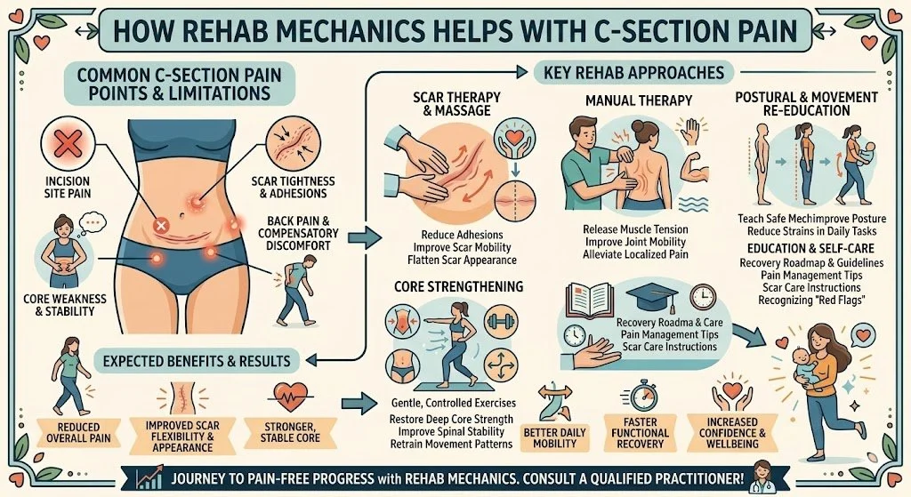
For the active residents of Queen West, Liberty Village, and Trinity Bellwoods, staying in motion is a way of life. From local run clubs and intensive Pilates classes to recreational martial arts and dance, the hips endure an immense amount of repetitive mechanical stress.
However, many active Torontonians develop a deeply frustrating and frightening condition: a loud, audible, and painful "snap," "click," or "clunk" deep within their hip joint every time they lift their leg, run, or climb stairs.
Often dismissed by standard walk-in clinics as "just tight muscles" or incorrectly diagnosed as early arthritis, this condition is clinically known as Coxa Saltans, or Snapping Hip Syndrome. When the snapping becomes painful, it signifies a massive biomechanical friction issue that will eventually destroy the surrounding bursa and tendons if ignored.
At Rehab Mechanics, located inside the Prime Medical Centre, we specialize in advanced orthopedic problem-solving. We understand that a snapping hip is a symptom of a misaligned pelvis and imbalanced muscular tension. Through targeted "human mechanics" physical therapy, we can physically alter the tracking of your tendons, permanently silencing the snap.
Structural Analysis: The Mechanics of Coxa Saltans
To understand how to fix a snapping hip, we must perform a detailed biomechanical analysis of the pelvic girdle and identify exactly what is snapping over where. Snapping Hip Syndrome generally falls into two primary structural categories.
1. Internal Snapping Hip (The Psoas Snap)
This is the most common variation, presenting as a deep, painful "clunk" in the front of the groin when lowering the leg from a lifted position (like descending from a high step or during flutter kicks in Pilates).
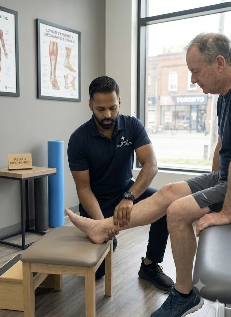
The Culprit: The Iliopsoas tendon (your primary, deepest hip flexor).
The Bony Prominence: The iliopectineal eminence ( a bony ridge on the front of the pelvis) or the head of the femur.
The Friction Mechanism
When you sit at a desk for eight hours a day, the massive psoas muscle adaptively shortens and becomes rigidly tight.
As you lift your knee, the tight tendon slides off the bony ridge.
When you lower your leg, the tension skyrockets, and the tendon snaps violently back over the bone like a thick, highly tensioned rubber band.
The Collateral Damage: This violent snapping eventually crushes the iliopsoas bursa (the fluid-filled sac sitting underneath the tendon), leading to severe, chronic groin inflammation (bursitis).
2. External Snapping Hip (The IT Band Snap)
This variation presents as a sharp, visible snapping sensation on the outside of the hip, often feeling as though the hip is momentarily "popping out of its socket."
The Culprit: The Iliotibial (IT) Band or the anterior gluteus maximus tendon.
The Bony Prominence: The Greater Trochanter (the large, bony bump on the outside of your upper thigh).
The Gluteal Weakness Trigger
External snapping is rarely a problem with the IT band itself; it is a problem with the pelvic stabilizers.
The Pelvic Drop
If your gluteus medius is weak, your pelvis drops laterally when you walk or run.
This pelvic drop dramatically alters the angle of the IT band, pulling it violently tight.
As you swing your leg forward and back, the tightened fascia violently snaps back and forth over the greater trochanter, eventually causing highly painful trochanteric bursitis.
Primary Source Proof: Non-Operative Hip Kinematics
Advanced orthopedic and sports medicine literature overwhelmingly supports non-operative, biomechanical rehabilitation—focusing on tendon lengthening and lumbo-pelvic stabilization—as the definitive first-line cure for Coxa Saltans.
Note: The link above serves as an example of our commitment to evidence-based practice, referencing standard international clinical guidelines for hip rehabilitation.
The Rehab Mechanics Corrective Protocol
Treating a snapping hip requires absolute precision. Generic hip stretches often exacerbate the snapping. We must utilize a highly controlled, phased approach to restore perfect joint tracking.
Phase 1: Tendon Desensitization and Soft Tissue Release
Before we can rebuild the hip, we must release the structural brakes that are causing the friction.
Advanced Myofascial Release: Our physiotherapists use targeted, deep ischemic compression to release the hypertonic psoas major or the Tensor Fasciae Latae (TFL). By manually lengthening the muscle belly, we instantly reduce the tension on the snapping tendon.
Joint Mobilization: Unlocking the hip capsule itself. If the ball-and-socket joint is stiff, the femur glides forward, pushing the bony ridges directly into the tight tendons. Manual glides restore centralization.
Activity Modification: We temporarily eliminate the specific athletic movements that trigger the snap, allowing the severely crushed bursa to chemically calm down.
Phase 2: Lumbo-Pelvic Neuromuscular Re-Education
We must correct the pelvic alignment to permanently alter the tracking angle of the tendons.
Pelvic Tilt Correction: If you have an anterior pelvic tilt, the psoas is under constant stretch. We train the deep core (transversus abdominis) to pull the pelvis backward into a neutral alignment, providing massive mechanical relief to the front of the hip.
Eccentric Psoas Loading: Standard stretching is insufficient. We use specialized eccentric exercises (slowly lowering the leg against resistance) to remodel the psoas tendon, making it thicker, stronger, and more elastic so it no longer snaps over the bone.
Phase 3: Gluteal Fortification and Dynamic Control
We must build a muscular brace that stops the pelvis from dropping during walking and running.
Isolated Glute Medius Activation: Utilizing side-lying hip abductions, clamshells, and isometric holds to wake up the lateral stabilizers.
Functional Integration: Progressing to single-leg deadlifts, split squats, and dynamic step-ups. We monitor your mechanics closely to ensure your femur stays perfectly aligned, proving to your nervous system that it can handle high-load activities without the painful snapping.
Silence Your Hip Pain Permanently
You do not have to live with the fear of your hip "popping out" or the chronic, burning pain of bursitis. By analyzing and correcting your pelvic mechanics, physical therapy offers a permanent, non-surgical solution.
Book a comprehensive biomechanical hip assessment with our clinical team today. We are conveniently located inside the Prime Medical Centre at 68 Abell Street, offering elite sports and orthopedic recovery in Queen West.
Contact us to schedule your appointment:
Email: info@rehabmechanics.com
Phone: (416) 533-3900
About the Author
Mr. Sanjay Attwala (B.Sc., M.Sc., RPT) is a Registered Physiotherapist, clinical director, and the founder of Rehab Mechanics in Toronto. With over 15 years of registered clinical practice and a deep specialization in complex musculoskeletal rehabilitation, Sanjay synthesizes rigorous international academic training with advanced evidence-based therapeutics to guide his clinical practice and patient education initiatives.
Academic Background & Credentials
Master of Science (M.Sc.) in Physiotherapy – University of Keele, United Kingdom (2010).
Bachelor of Science (B.Sc.) – University of Waterloo, Ontario, Canada.
Registered Physiotherapist (RPT) – Regulated health professional in excellent standing with the College of Physiotherapists of Ontario (CPO).
Corporate Entity – Operating officially under the S. Attwala Physiotherapy Professional Corporation with a DBA of Rehab Mechanics.
Clinical Expertise & Philosophy
Sanjay’s clinical approach rejects passive symptom management in favor of identifying underlying biomechanical root causes. His diverse expertise spans advanced manual therapies, personalized corrective exercise prescription, and modern physical modalities. At the Rehab Mechanics Toronto Queen West clinic, he routinely diagnoses and treats complex conditions including:
Spinal & Discogenic Pathology – Cervical, thoracic, and lumbar disc injuries, sciatica, and sacroiliac joint (SIJ) dysfunction.
Upper & Lower Extremity Injuries – Rotator cuff tears, frozen shoulder, tennis/golfer’s elbow, carpal tunnel syndrome, and complex ankle/foot pathologies.
Perinatal & Pelvic Health Rehabilitation – Specialized assessment and rehabilitation protocols tailored specifically for women during pregnancy and the post-partum period, addressing pelvic girdle pain, diastasis recti, and core stabilization.
Specialized Rehabilitation – Pelvic health therapy, TMJ dysfunction, post-surgical rehabilitation (including Total Hip and Total Knee Replacements), and custom orthotics dispensing.
Shockwave Therapy: with advanced cutting edge technological devices to suit your needs.
Interdisciplinary Practice & Patient Care
Sanjay practices an integrated model of healthcare, working closely alongside medical doctors inside the Prime Medical Centre on Abell Street to streamline patient recovery pathways. He maintains a human-centric, communication-first clinical framework, ensuring that care remains fully customized rather than automated.
His clinical caseload encompasses a broad operational spectrum under Ontario's regulatory frameworks, including:
Motor Vehicle Accident (MVA) Claims – Rehabilitation navigating Ontario’s statutory accident benefits schedule.
Workplace Safety and Insurance Board (WSIB) – Occupational injury management and return-to-work screening.
Extended Health Care (EHC) & Private Practice – Multi-tier insurance coordination and long-term athletic development plans.
Commitment to Research & Community
Outside of his clinical caseload at Rehab Mechanics and his additional practice affiliations in Etobicoke, Sanjay is an active health writer and community educator. He translates contemporary peer-reviewed medical research into accessible, actionable guidance on his professional blog. As a dedicated father and husband, he mirrors his professional advice in his personal life, focusing on structural mobility, cross-training, and longevity to help his family and his community thrive. Naturally he takes he a keen interest in rehabilitation for women who are pregnant and post-partum.
Disclaimer: The information provided on this blog is intended for educational and informational purposes only. It does not constitute medical advice, diagnosis, or a treatment plan. Always seek the direct advice of a Registered Physiotherapist, physician, or other qualified health provider regarding any medical condition or physical rehabilitation routine.
Does Sitting All Day Cause Gluteal Amnesia and Lower Back Pain?
Yes. Prolonged sitting causes gluteal amnesia by neurologically inhibiting the gluteal muscles. Physiotherapy reverses this neuromuscular shutdown through targeted activation drills, deep tissue release of the hip flexors, and heavy resistance training, permanently resolving the resulting lower back and knee pain.
The Postural Epidemic of the Modern Workspace
Toronto is a city driven by knowledge workers, tech innovators, and corporate professionals. While the skyline is constantly growing, the physical reality for most of these workers involves sitting immobilized in an ergonomic chair for 40 to 60 hours a week.
Over the past few years, a highly specific and alarming pattern has emerged at Rehab Mechanics. Patients arrive at our Queen West clinic complaining of chronic, nagging lower back pain, tight hamstrings, and aching knees. They stretch daily, they get massages, yet the pain always returns.
When we perform a biomechanical audit, we discover a phenomenon colloquially known as "Dead Butt Syndrome," or clinically termed Gluteal Amnesia. Because of the massive amount of time spent sitting, their brains have literally "forgotten" how to fire their glute muscles. When the largest, most powerful muscle group in the human body shuts down, catastrophic mechanical failure occurs up and down the entire kinetic chain.
Structural Analysis: The Mechanics of Gluteal Amnesia
To understand how sitting ruins your back and knees, we must perform a detailed neurological and biomechanical analysis of the hip joint.
The Phenomenon of Reciprocal Inhibition
The human nervous system operates on a principle of efficiency known as reciprocal inhibition. When a muscle on one side of a joint contracts, the brain automatically sends an inhibitory signal to the muscle on the opposite side, telling it to relax so movement can occur.
The Sitting Trap: When you sit at a desk, your hip flexors (psoas and iliacus) are placed in a shortened, contracted position for hours on end.
The Neurological Shutdown: Because the hip flexors are constantly "on," the brain sends a continuous, non-stop inhibitory signal to the opposing muscles—your gluteus maximus.
The Result: The glutes are chemically and neurologically switched "off." Over time, the neural pathways weaken, and the muscle suffers profound atrophy. You develop Gluteal Amnesia.
The Kinetic Chain Breakdown
Your glutes are the foundational engine of your body. When they go offline, the nervous system panics and recruits other, smaller muscles to do their job. This compensatory overload destroys your joints.
Lumbar Spine Overload (Lower Back Pain)
When you bend over to pick up a box or try to perform a deadlift, your glutes are supposed to drive you back up to a standing position.
The Compensation: If the glutes are amnesic, the brain forces your lower back muscles (erector spinae) and hamstrings to take 100% of the load.
The Failure: These muscles are not designed to be primary movers. They rapidly overwork, spasm, and place massive shearing forces on your lumbar spinal discs, leading to chronic back pain and eventual disc herniation.
Knee Valgus Collapse (Knee Pain)
Your gluteus medius (the side of your hip) is responsible for keeping your femur (thigh bone) straight.
The Compensation: When the glute medius shuts down, there is nothing stopping your thigh bone from rotating inward.
The Failure: Every time you take a step, your knee caves inward toward your midline (valgus collapse). This violently grinds the kneecap against the femur, causing severe anterior knee pain (Runner’s Knee) and destroying the meniscus.
Primary Source Proof: Neuromuscular Re-Education
Clinical orthopedic research explicitly proves that resolving non-specific lower back pain and lower extremity injuries requires direct neuromuscular reactivation and strengthening of the gluteal complex to reverse postural inhibition.
Download Clinical Efficacy PDF: The Efficacy of Gluteal Neuromuscular Re-Education in the Treatment of Chronic Lumbar and Patellofemoral Pain (Open Access Medical Review)
Note: The link above serves as an example of our commitment to evidence-based practice, referencing standard international clinical guidelines for biomechanical rehabilitation.
The Rehab Mechanics Corrective Protocol
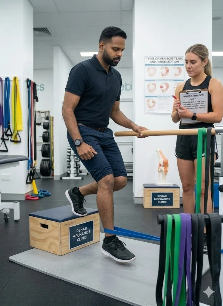
Treating gluteal amnesia requires a precise "re-wiring" of the nervous system. You cannot simply go to the gym and do heavy squats; if your glutes are asleep, your lower back will just take the weight, making the injury worse.
Phase 1: Releasing the Brakes (Hip Flexor Deactivation)
We must stop the continuous inhibitory signal before we can wake the glutes up.
Deep Myofascial Psoas Release: Our physiotherapists utilize advanced, highly targeted manual therapy deep into the abdomen to physically lengthen the rigidly tight hip flexors.
Active Release Techniques (ART): Stripping the tight rectus femoris and TFL (front of the thigh) to ensure the pelvis can return to a neutral, upright alignment.
Phase 2: Neuromuscular Isolation (Waking the Dead)
We use micro-movements to force the brain to re-establish a neurological connection with the glute fibers.
Prone Glute Squeezes: Using tactile feedback (physically tapping the muscle) to help the patient consciously contract the gluteus maximus without firing the hamstrings.
Clamshells and Hydrants: Isolating the gluteus medius in side-lying positions, ensuring the pelvis remains completely still so the lower back cannot "cheat" the movement.
Phase 3: Dynamic Integration and Heavy Loading
Once the brain remembers how to use the glutes, we must rebuild their absolute strength to handle the real world.
Glute Bridging and Hip Thrusts: Progressing to heavily loaded hip extension exercises that target the glutes while the spine remains safely supported.
Functional Movement Correction: Re-teaching you how to squat, lunge, and run using a "hip hinge" pattern. We monitor your knees closely to ensure the newly awakened glutes fire automatically to prevent knee valgus.
Restart Your Engine
Do not let an office chair slowly destroy the biomechanics of your lower body. By aggressively treating gluteal amnesia, you can eliminate the compensatory forces that cause chronic back and knee pain.
Book a comprehensive biomechanical assessment with our clinical team today. We are conveniently located inside the Prime Medical Centre at 68 Abell Street, offering advanced orthopedic recovery in Queen West.
Contact us to schedule your appointment:
Email: info@rehabmechanics.com
Phone: (416) 533-3900
About the Author
Mr. Sanjay Attwala (B.Sc., M.Sc., RPT) is a Registered Physiotherapist, clinical director, and the founder of Rehab Mechanics in Toronto. With over 15 years of registered clinical practice and a deep specialization in complex musculoskeletal rehabilitation, Sanjay synthesizes rigorous international academic training with advanced evidence-based therapeutics to guide his clinical practice and patient education initiatives.
Academic Background & Credentials
Master of Science (M.Sc.) in Physiotherapy – University of Keele, United Kingdom (2010).
Bachelor of Science (B.Sc.) – University of Waterloo, Ontario, Canada.
Registered Physiotherapist (RPT) – Regulated health professional in excellent standing with the College of Physiotherapists of Ontario (CPO).
Corporate Entity – Operating officially under the S. Attwala Physiotherapy Professional Corporation with a DBA of Rehab Mechanics.
Clinical Expertise & Philosophy
Sanjay’s clinical approach rejects passive symptom management in favor of identifying underlying biomechanical root causes. His diverse expertise spans advanced manual therapies, personalized corrective exercise prescription, and modern physical modalities. At the Rehab Mechanics Toronto Queen West clinic, he routinely diagnoses and treats complex conditions including:
Spinal & Discogenic Pathology – Cervical, thoracic, and lumbar disc injuries, sciatica, and sacroiliac joint (SIJ) dysfunction.
Upper & Lower Extremity Injuries – Rotator cuff tears, frozen shoulder, tennis/golfer’s elbow, carpal tunnel syndrome, and complex ankle/foot pathologies.
Perinatal & Pelvic Health Rehabilitation – Specialized assessment and rehabilitation protocols tailored specifically for women during pregnancy and the post-partum period, addressing pelvic girdle pain, diastasis recti, and core stabilization.
Specialized Rehabilitation – Pelvic health therapy, TMJ dysfunction, post-surgical rehabilitation (including Total Hip and Total Knee Replacements), and custom orthotics dispensing.
Shockwave Therapy: with advanced cutting edge technological devices to suit your needs.
Interdisciplinary Practice & Patient Care
Sanjay practices an integrated model of healthcare, working closely alongside medical doctors inside the Prime Medical Centre on Abell Street to streamline patient recovery pathways. He maintains a human-centric, communication-first clinical framework, ensuring that care remains fully customized rather than automated.
His clinical caseload encompasses a broad operational spectrum under Ontario's regulatory frameworks, including:
Motor Vehicle Accident (MVA) Claims – Rehabilitation navigating Ontario’s statutory accident benefits schedule.
Workplace Safety and Insurance Board (WSIB) – Occupational injury management and return-to-work screening.
Extended Health Care (EHC) & Private Practice – Multi-tier insurance coordination and long-term athletic development plans.
Commitment to Research & Community
Outside of his clinical caseload at Rehab Mechanics and his additional practice affiliations in Etobicoke, Sanjay is an active health writer and community educator. He translates contemporary peer-reviewed medical research into accessible, actionable guidance on his professional blog. As a dedicated father and husband, he mirrors his professional advice in his personal life, focusing on structural mobility, cross-training, and longevity to help his family and his community thrive. Naturally he takes he a keen interest in rehabilitation for women who are pregnant and post-partum.
Disclaimer: The information provided on this blog is intended for educational and informational purposes only. It does not constitute medical advice, diagnosis, or a treatment plan. Always seek the direct advice of a Registered Physiotherapist, physician, or other qualified health provider regarding any medical condition or physical rehabilitation routine.
Can Physiotherapy Prevent Hip Replacement Surgery for Severe Osteoarthritis?
Yes. Physiotherapy routinely delays or prevents hip replacement surgery for osteoarthritis by strengthening the deep gluteal stabilizers. Restoring joint capsule mobility and muscular support drastically reduces bone-on-bone friction, managing chronic inflammation and returning patients to pain-free urban mobility without surgery.
The Fear of "Bone on Bone" Degeneration
For active residents in Toronto—whether you spend your weekends walking through Trinity Bellwoods, cycling along the waterfront, or simply commuting daily through Queen West—few things are as terrifying as the onset of deep, grinding hip pain.
When a patient visits a walk-in clinic and receives an X-ray showing "mild to moderate hip osteoarthritis," the conversation often immediately turns to surgical waitlists. The phrase "bone on bone" is used frequently, creating immense psychological fear. Patients assume their hip is completely destroyed, leading them to stop walking, stop exercising, and isolate themselves on the couch to "save" whatever cartilage they have left.
This "wait and deteriorate" mindset is the absolute worst thing you can do for an arthritic joint. At Rehab Mechanics, we heavily advocate for conservative orthopedic management. Your X-ray does not dictate your pain. By overhauling your body mechanics, we can train your muscles to absorb the impact of walking before it ever reaches your degenerated hip joint, successfully avoiding the operating room.
Structural Analysis: The Mechanics of Hip Osteoarthritis
To understand how to save a failing hip, we must perform a biomechanical analysis of the joint’s architecture and the reality of cartilage degradation.
The Anatomy of the Ball and Socket
Your hip (the acetabulofemoral joint) is a massive weight-bearing structure.
The Articular Cartilage: Both the head of the femur (the ball) and the acetabulum (the socket) are coated in a thick, slippery layer of articular cartilage. This cartilage acts like a Teflon coating, allowing the bones to glide with zero friction.
The Labrum and Synovium: A rubbery gasket (the labrum) seals the joint, and the synovial membrane produces a thick, egg-white-like fluid that constantly lubricates the cartilage.
The Pathology of Joint Decay (Osteoarthritis)
Osteoarthritis (OA) is not just mechanical wear and tear; it is a complex, active biological disease process.
The Cartilage Thinning Phase
Over decades of use, or secondary to old sports injuries, the water content within the articular cartilage decreases. The Teflon coating begins to fray, thin out, and eventually expose the underlying subchondral bone.
The Inflammatory Cascade
As the cartilage shreds, the debris floats inside the joint capsule.
Synovitis: The joint lining reacts to this debris by becoming violently inflamed, causing the hip to swell and ache deeply into the groin.
Osteophyte Formation: The body attempts to stabilize the wobbly, failing joint by growing new bone around the edges. These jagged bone spurs (osteophytes) physically block your range of motion, making it impossible to put on your socks or get out of a low car seat.
The Biomechanical Failure: Why the Pain Spikes
If the cartilage loss is the disease, why does the pain suddenly become unbearable? The answer lies in the muscles.
The Gluteal Shutdown
When your hip is inflamed, your brain initiates a protective reflex called Arthrogenic Muscle Inhibition. It literally shuts off the electrical signal to your gluteus medius and gluteus maximus to stop you from walking on the injured leg.
The Mechanical Crushing
Because your glutes are shut down and atrophied, they can no longer absorb the shock of your body weight. Now, every time your foot strikes the Toronto pavement, 100% of the kinetic impact bypasses the weak muscles and slams directly into the exposed, arthritic bone. This is why the pain becomes agonizing.
Primary Source Proof: Non-Operative Efficacy
Major international orthopedic guidelines and extensive peer-reviewed literature strongly mandate that comprehensive, supervised physiotherapy must be exhausted before total hip arthroplasty (surgery) is considered.
Note: The link above directs to external, peer-reviewed medical literature demonstrating our commitment to evidence-based practice and international clinical guidelines for orthopedic rehabilitation.
The Rehab Mechanics Joint Preservation Protocol
At our clinic inside the Prime Medical Centre, we do not view osteoarthritis as a surgical inevitability. We utilize an aggressive, phased biomechanical approach to build a muscular exoskeleton around your failing joint.
Phase 1: Joint Distraction and Pain Modulation (Weeks 1-4)
Before we can build strength, we must lower the severe pain threshold.
Medical Integration: We work directly with the physicians at Prime Medical Centre who may administer targeted, intra-articular anti-inflammatory injections to instantly clear the synovitis.
Manual Joint Traction: Our physiotherapists apply heavy, sustained pulling forces (distraction) to the leg. This physically separates the ball from the socket, creating a vacuum effect that pulls fresh, lubricating synovial fluid into the starving cartilage.
Capsular Stretching: Aggressively stretching the tight, fibrotic joint capsule to restore your ability to extend the hip behind you, which is critical for normal walking.
Phase 2: Isometric and Closed-Chain Loading (Weeks 4-8)
We must wake up the shut-down gluteal muscles without creating painful bone-on-bone friction.
High-Intensity Isometrics: You will push your leg maximally against an immovable resistance. The muscles fire at 100% capacity, but the joint does not bend, allowing you to build massive strength without grinding the cartilage.
Closed-Chain Activation: Utilizing targeted leg presses and wall sits to teach the nervous system to absorb body weight safely through the musculature rather than the bone.
Phase 3: Gait Retraining and Proprioception (Weeks 8-12+)
Once the muscular "brace" is built, we must ensure it works automatically when you navigate the city.
Trendelenburg Correction: We use mirror feedback and targeted cues to ensure your pelvis does not drop when you stand on the arthritic leg, eliminating the painful limping pattern.
Dynamic Shock Absorption: Training your core and hip to work synergistically during step-downs and uneven surface walking, proving to your nervous system that you are strong, capable, and no longer reliant on the damaged cartilage.
Stop the Deterioration
Do not let an X-ray dictate the rest of your active life. By correcting your structural biomechanics, you can offload the damaged joint, eliminate the chronic inflammation, and successfully keep your natural hip.
Book a comprehensive orthopedic assessment with our clinical team today. We are conveniently located inside the Prime Medical Centre at 68 Abell Street, offering advanced joint preservation in Queen West.
Contact us to schedule your appointment:
Email: info@rehabmechanics.com
Phone: (416) 533-3900
About the Author
Mr. Sanjay Attwala (B.Sc., M.Sc., RPT) is a Registered Physiotherapist, clinical director, and the founder of Rehab Mechanics in Toronto. With over 15 years of registered clinical practice and a deep specialization in complex musculoskeletal rehabilitation, Sanjay synthesizes rigorous international academic training with advanced evidence-based therapeutics to guide his clinical practice and patient education initiatives.
Academic Background & Credentials
Master of Science (M.Sc.) in Physiotherapy – University of Keele, United Kingdom (2010).
Bachelor of Science (B.Sc.) – University of Waterloo, Ontario, Canada.
Registered Physiotherapist (RPT) – Regulated health professional in excellent standing with the College of Physiotherapists of Ontario (CPO).
Corporate Entity – Operating officially under the S. Attwala Physiotherapy Professional Corporation with a DBA of Rehab Mechanics.
Clinical Expertise & Philosophy
Sanjay’s clinical approach rejects passive symptom management in favor of identifying underlying biomechanical root causes. His diverse expertise spans advanced manual therapies, personalized corrective exercise prescription, and modern physical modalities. At the Rehab Mechanics Toronto Queen West clinic, he routinely diagnoses and treats complex conditions including:
Spinal & Discogenic Pathology – Cervical, thoracic, and lumbar disc injuries, sciatica, and sacroiliac joint (SIJ) dysfunction.
Upper & Lower Extremity Injuries – Rotator cuff tears, frozen shoulder, tennis/golfer’s elbow, carpal tunnel syndrome, and complex ankle/foot pathologies.
Perinatal & Pelvic Health Rehabilitation – Specialized assessment and rehabilitation protocols tailored specifically for women during pregnancy and the post-partum period, addressing pelvic girdle pain, diastasis recti, and core stabilization.
Specialized Rehabilitation – Pelvic health therapy, TMJ dysfunction, post-surgical rehabilitation (including Total Hip and Total Knee Replacements), and custom orthotics dispensing.
Shockwave Therapy: with advanced cutting edge technological devices to suit your needs.
Interdisciplinary Practice & Patient Care
Sanjay practices an integrated model of healthcare, working closely alongside medical doctors inside the Prime Medical Centre on Abell Street to streamline patient recovery pathways. He maintains a human-centric, communication-first clinical framework, ensuring that care remains fully customized rather than automated.
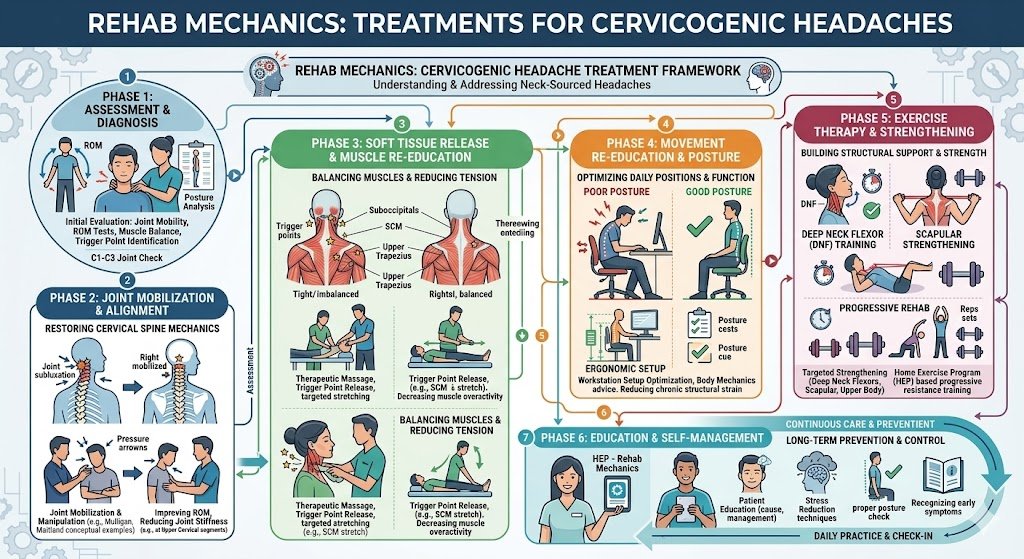
His clinical caseload encompasses a broad operational spectrum under Ontario's regulatory frameworks, including:
Motor Vehicle Accident (MVA) Claims – Rehabilitation navigating Ontario’s statutory accident benefits schedule.
Workplace Safety and Insurance Board (WSIB) – Occupational injury management and return-to-work screening.
Extended Health Care (EHC) & Private Practice – Multi-tier insurance coordination and long-term athletic development plans.
Commitment to Research & Community
Outside of his clinical caseload at Rehab Mechanics and his additional practice affiliations in Etobicoke, Sanjay is an active health writer and community educator. He translates contemporary peer-reviewed medical research into accessible, actionable guidance on his professional blog. As a dedicated father and husband, he mirrors his professional advice in his personal life, focusing on structural mobility, cross-training, and longevity to help his family and his community thrive. Naturally he takes he a keen interest in rehabilitation for women who are pregnant and post-partum.
Disclaimer: The information provided on this blog is intended for educational and informational purposes only. It does not constitute medical advice, diagnosis, or a treatment plan. Always seek the direct advice of a Registered Physiotherapist, physician, or other qualified health provider regarding any medical condition or physical rehabilitation routine.
Can Physiotherapy Cure Chronic Tension Headaches from Desk Work?
Yes. Physiotherapy permanently cures tension headaches by fixing the cervical biomechanics causing them. By mobilizing stiff upper neck joints, releasing suboccipital muscle spasms, and strengthening deep neck flexors, physical therapy eliminates the structural nerve irritation that triggers chronic daily headaches.
The Headache Epidemic in Queen West
In the digital age, our lives are dictated by screens. Whether you are coding a new app in Liberty Village, analyzing spreadsheets in the Financial District, or constantly checking a smartphone, your head is perpetually tilted forward.
The physical toll of this lifestyle is catastrophic for the cervical spine, giving rise to an epidemic of chronic, agonizing head pain. Countless Toronto professionals wake up daily with a dull, vice-like pressure wrapping around their temples, an aching tightness at the base of the skull, or sharp pain radiating behind their eyes.
Often dismissed by walk-in clinics as "just stress" and treated with endless cycles of Advil or Tylenol, these are clinically classified as Cervicogenic or Tension-Type Headaches. When you search for "remedial mechanics" or "Queen Street physiotherapy" to resolve this pain, you are on the right track. At Rehab Mechanics, we know that these headaches are not a chemical imbalance in your brain; they are a direct, mechanical consequence of a failing neck structure. Fixing the spine permanently turns off the headache alarm.
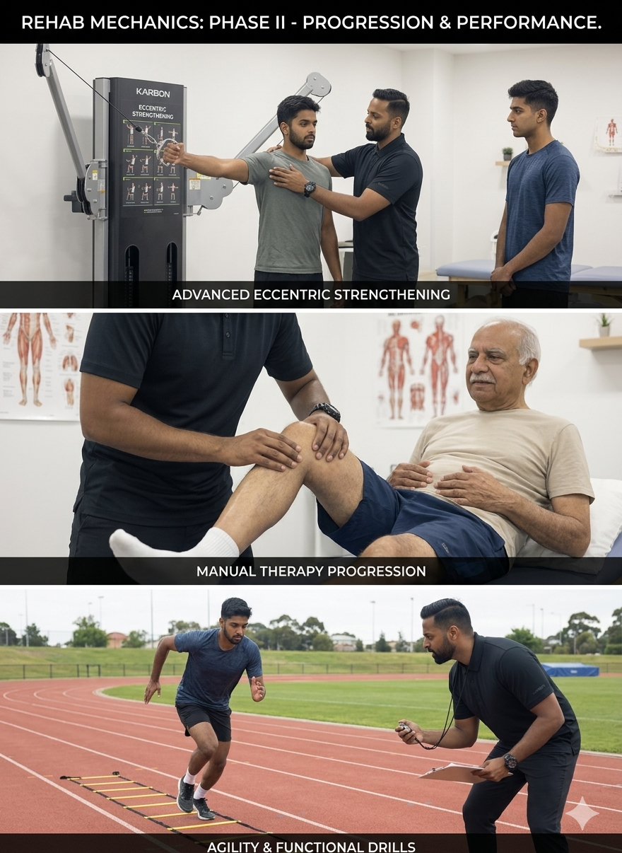
Structural Analysis: The Biomechanics of a Tension Headache
To understand why stretching your neck doesn't cure a severe tension headache, we must dive deep into the neurology and biomechanics of the upper cervical spine.
The Weight of the "Tech Neck"
The human head weighs between 10 and 12 pounds. It is designed to balance perfectly on top of the cervical spine, requiring minimal muscular effort to stay upright.
The Leverage Crisis: For every inch your head translates forward to look at a monitor, the mechanical load on the muscles at the back of your neck doubles.
The Muscular Exhaustion: A severe "Tech Neck" forces the tiny muscles at the base of the skull (the suboccipitals) to hold back 30 to 40 pounds of force for 10 hours a day.
The Spasm: These muscles rapidly fatigue, lose their blood supply (ischemia), and lock into permanent, rock-hard spasms.
The Neurological Bridge (Why Your Head Hurts)
How does a knot in your neck cause pain behind your eye? The answer lies in the complex wiring of the central nervous system.
The Trigeminal-Cervical Nucleus (TCN)
The nerves that supply feeling to the upper three joints of your neck (C1, C2, and C3) merge into a massive relay station in your brainstem called the Trigeminal-Cervical Nucleus.
Crucially, the Trigeminal Nerve—which supplies sensation to your forehead, temples, and face—also plugs into this exact same relay station.
The Crossed Signal: When the joints in your upper neck are jammed and the suboccipital muscles are violently spasming, they flood the relay station with pain signals. The brain gets confused by the sheer volume of data and misinterprets the origin of the pain, projecting it forward into the head and face (Referred Pain).
The Myodural Bridge
Recent anatomical discoveries have revealed that a small band of connective tissue directly links the suboccipital muscles in the neck to the dura mater (the pain-sensitive outer covering of the brain).
When the neck muscles are in severe spasm, they literally tug on the lining of your brain, generating the sensation of a vice crushing your skull.
Primary Source Proof: Cervicogenic Decompression
Neurological and orthopedic literature unequivocally demonstrates that specific cervical spine mobilization and deep neck flexor rehabilitation are vastly superior to pharmacological management for the long-term cure of cervicogenic and tension headaches.
Note: The link above serves as an example of our commitment to evidence-based practice, referencing standard international clinical guidelines for cervical rehabilitation.
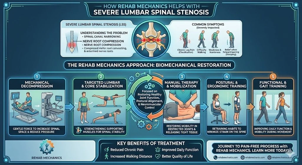
The Rehab Mechanics Headache Eradication Protocol
We do not just massage the sore muscles. Treating cervicogenic headaches requires a profound overhaul of your upper body mechanics to stop the muscles from spasming in the first place.
Phase 1: Decompressing the Neurological Bottleneck
Our first goal is to immediately cut off the pain signals flooding the brainstem.
Upper Cervical Mobilization: Our physiotherapists use precise, gentle Grade II and Grade III manual glides to un-jam the C1, C2, and C3 vertebrae. Restoring the micro-movement of these joints instantly reduces the nerve irritation causing the referred pain.
Suboccipital Ischemic Compression: We apply deep, sustained pressure directly into the base of the skull. This manually breaks the spasm in the suboccipitals, releasing the tension on the "myodural bridge" and draining the pressure from the head.
Phase 2: Restoring the Foundation (Thoracic Extension)
The neck cannot sit straight if the mid-back is hunched over.
Thoracic Spine Unlocking: Utilizing high-level joint mobilizations and foam-roller drills to aggressively restore extension in the upper back.
Pectoral Myofascial Release: Lengthening the tight chest muscles that are physically dragging the shoulders forward and fueling the poor posture.
Phase 3: Rebuilding the Deep Stabilizers
This phase ensures the headaches never return. We must build biological endurance.
Deep Cervical Flexor (DCF) Training: We utilize specific micro-movements (like chin tucks with a pressure biofeedback unit) to re-awaken the dormant muscles in the front of your throat. When these muscles fire properly, they automatically pull the head back over the shoulders, instantly taking the load off the back of the neck.
Postural Ergonomics: We review your exact desk setup, monitor height, and chair support to guarantee your Toronto workspace supports, rather than destroys, your newly restored spinal mechanics.
Turn Off the Headache Alarm
You do not have to accept daily tension headaches or rely on a permanent supply of painkillers. By diagnosing and correcting the mechanical failures in your cervical spine, you can permanently eradicate the pain.
Book a comprehensive neurological and biomechanical assessment with our clinical team today. We are conveniently located inside the Prime Medical Centre at 68 Abell Street, offering advanced orthopedic recovery in Queen West.
Contact us to schedule your appointment:
Email: info@rehabmechanics.com
Phone: (416) 533-3900
About the Author
Mr. Sanjay Attwala (B.Sc., M.Sc., RPT) is a Registered Physiotherapist, clinical director, and the founder of Rehab Mechanics in Toronto. With over 15 years of registered clinical practice and a deep specialization in complex musculoskeletal rehabilitation, Sanjay synthesizes rigorous international academic training with advanced evidence-based therapeutics to guide his clinical practice and patient education initiatives.
Academic Background & Credentials
Master of Science (M.Sc.) in Physiotherapy – University of Keele, United Kingdom (2010).
Bachelor of Science (B.Sc.) – University of Waterloo, Ontario, Canada.
Registered Physiotherapist (RPT) – Regulated health professional in excellent standing with the College of Physiotherapists of Ontario (CPO).
Corporate Entity – Operating officially under the S. Attwala Physiotherapy Professional Corporation with a DBA of Rehab Mechanics.
Clinical Expertise & Philosophy
Sanjay’s clinical approach rejects passive symptom management in favor of identifying underlying biomechanical root causes. His diverse expertise spans advanced manual therapies, personalized corrective exercise prescription, and modern physical modalities. At the Rehab Mechanics Toronto Queen West clinic, he routinely diagnoses and treats complex conditions including:
Spinal & Discogenic Pathology – Cervical, thoracic, and lumbar disc injuries, sciatica, and sacroiliac joint (SIJ) dysfunction.
Upper & Lower Extremity Injuries – Rotator cuff tears, frozen shoulder, tennis/golfer’s elbow, carpal tunnel syndrome, and complex ankle/foot pathologies.
Perinatal & Pelvic Health Rehabilitation – Specialized assessment and rehabilitation protocols tailored specifically for women during pregnancy and the post-partum period, addressing pelvic girdle pain, diastasis recti, and core stabilization.
Specialized Rehabilitation – Pelvic health therapy, TMJ dysfunction, post-surgical rehabilitation (including Total Hip and Total Knee Replacements), and custom orthotics dispensing.
Shockwave Therapy: with advanced cutting edge technological devices to suit your needs.
Interdisciplinary Practice & Patient Care
Sanjay practices an integrated model of healthcare, working closely alongside medical doctors inside the Prime Medical Centre on Abell Street to streamline patient recovery pathways. He maintains a human-centric, communication-first clinical framework, ensuring that care remains fully customized rather than automated.
His clinical caseload encompasses a broad operational spectrum under Ontario's regulatory frameworks, including:
Motor Vehicle Accident (MVA) Claims – Rehabilitation navigating Ontario’s statutory accident benefits schedule.
Workplace Safety and Insurance Board (WSIB) – Occupational injury management and return-to-work screening.
Extended Health Care (EHC) & Private Practice – Multi-tier insurance coordination and long-term athletic development plans.
Commitment to Research & Community
Outside of his clinical caseload at Rehab Mechanics and his additional practice affiliations in Etobicoke, Sanjay is an active health writer and community educator. He translates contemporary peer-reviewed medical research into accessible, actionable guidance on his professional blog. As a dedicated father and husband, he mirrors his professional advice in his personal life, focusing on structural mobility, cross-training, and longevity to help his family and his community thrive. Naturally he takes he a keen interest in rehabilitation for women who are pregnant and post-partum.
Disclaimer: The information provided on this blog is intended for educational and informational purposes only. It does not constitute medical advice, diagnosis, or a treatment plan. Always seek the direct advice of a Registered Physiotherapist, physician, or other qualified health provider regarding any medical condition or physical rehabilitation routine.
Does Stretching Make High Hamstring Pain and "Yoga Butt" Worse?
Yes. Aggressively stretching a high hamstring tendinopathy severely worsens the injury. The pain at your sit bone is caused by compressive friction, not muscle tightness. Physiotherapy resolves this by halting deep stretches and utilizing heavy, progressive isometric and eccentric loading to rebuild the tendon.
The Agony at the Sit Bone
Queen West and Trinity Bellwoods are vibrant epicenters for Toronto's yoga, Pilates, and active running communities. While these practices are fantastic for flexibility and cardiovascular health, they frequently lead to a highly specific, deeply frustrating overuse injury.
Patients often present at Rehab Mechanics complaining of a literal "pain in the butt." It is a deep, localized, burning ache right on the sit bone (the bony prominence you feel when sitting on a hard chair). This pain spikes aggressively when bending over to touch the toes, performing a downward dog, or sprinting uphill.
In the yoga community, this is colloquially known as "Yoga Butt." Medically, it is Proximal Hamstring Tendinopathy (PHT).
The natural, intuitive response to a tight, aching hamstring is to stretch it aggressively. Patients will spend hours forcing themselves into deep forward folds, hoping the muscle will "release." However, when dealing with PHT, stretching is the absolute worst mechanical input you can provide. To cure this stubborn tendinopathy, you must understand the biomechanics of tendon compression and stop treating the injury like a simple muscle cramp.
Structural Analysis: The Mechanics of Hamstring Compression
To successfully rehabilitate Proximal Hamstring Tendinopathy, we must perform a detailed structural analysis of the pelvic anatomy and how the hamstring tendon behaves under load.
The Ischial Tuberosity Anchor
Your hamstring is not a single muscle; it is a group of three large muscles running down the back of your thigh.
The Common Origin: All three of these massive muscles merge into a single, thick tendon at the top of your leg.
The Bony Anchor: This common tendon anchors directly into the ischial tuberosity—the thick, bony projection at the absolute base of your pelvis (your sit bone).
The Pathology of Compressive Tendinosis
Tendons are incredibly strong when pulled straight (tensile load). However, they are highly vulnerable to being compressed or wrapped around a bone.
The "Wrap-Around" Effect (Why Stretching Hurts)
When you bend forward at the waist with straight legs (like a yoga forward fold or a deadlift), your pelvis rotates forward.
The Friction Point: As the pelvis rotates, the hamstring tendon is physically dragged and wrapped tightly around the bony edge of the ischial tuberosity, much like a rope pulled tightly around a rock.
The Ischemic Crush: This violent mechanical compression squeezes the blood flow out of the tendon at the exact point it connects to the bone.
Cellular Degeneration: If you do this repetitively (e.g., hundreds of deep yoga stretches or running uphill), the tendon cells fail. They stop producing healthy Type I collagen and begin to degenerate into a chaotic, painful, and thickened mass of scar tissue.
This is why stretching feels good for about 10 minutes (as it temporarily numbs the nerve endings) but makes the structural degeneration significantly worse the next day. You are actively crushing an injured tendon.
Primary Source Proof: Tendon Rehabilitation
Orthopedic sports medicine literature is categorical: passive stretching and total rest fail to resolve proximal hamstring tendinopathy. Progressive, heavy mechanical loading is the internationally recognized gold standard for tendon regeneration.
Note: The link above serves as an example of our commitment to evidence-based practice, referencing standard international clinical guidelines for tendinopathy rehabilitation.
The Rehab Mechanics Corrective Protocol
At our Prime Medical Centre clinic, our protocol for PHT completely removes compressive stretching and replaces it with advanced, progressive tissue loading to rebuild the tendon from the inside out.
Phase 1: Pain Modulation and Decompression (Weeks 1-4)
The immediate goal is to stop the mechanical crushing and lower the extreme chemical sensitivity of the tendon.
The Anti-Stretch Mandate: We temporarily ban all deep forward folds, deadlifts, and prolonged sitting on hard surfaces.
Isometric Loading (The Painkiller): We utilize heavy, static holds. For example, lying on your back and pressing your heel into the floor with the knee slightly bent. The muscle fires intensely, but the hip does not bend, completely avoiding the painful wrap-around compression. Isometrics provide a massive, immediate reduction in tendon pain.
Shockwave Therapy: If the tendon has severe, chronic scarring, we apply Extracorporeal Shockwave Therapy (ESWT) to physically break down the fibrotic tissue and stimulate new blood vessel growth (neovascularization) directly at the sit bone.
Phase 2: Isotonic Loading in Neutral (Weeks 4-8)
Once the pain settles, we must rebuild the tendon's capacity to handle load, but we keep the hip out of deep flexion to protect the attachment site.
Prone Leg Curls and Glute Bridges: We utilize exercises where the hip remains relatively straight (neutral) while the hamstring works hard.
Heavy Slow Resistance (HSR): The tendon only lays down new, healthy collagen fibers when subjected to heavy, slow tension (e.g., 4 seconds lifting the weight, 4 seconds lowering it).
Phase 3: Energy Storage and Compressive Loading (Weeks 8-12)
The tendon is now strong, but we must prepare it for the real world. We slowly reintroduce the compressive movements you have been avoiding.
Gradual Deepening: We slowly reintroduce Romanian Deadlifts (RDLs) and kettlebell swings, controlling the depth to carefully train the tendon to handle being wrapped around the sit bone again.
Plyometric Integration: If you are a runner, we introduce bounding, skipping, and hill sprints. The tendon must relearn how to act like a rapid spring, storing and releasing energy without tearing.
Reclaim Your Flexibility and Strength
Do not let chronic sit bone pain force you to abandon your yoga mat, your running shoes, or your lifting routine. By understanding the mechanics of tendon compression and executing a precise loading strategy, you can completely rebuild your high hamstring.
Book a comprehensive biomechanical assessment with our clinical team today. We are conveniently located inside the Prime Medical Centre at 68 Abell Street, offering elite sports recovery in Toronto Queen West.
Contact us to schedule your appointment:
Email: info@rehabmechanics.com
Phone: (416) 533-3900
About the Author
Mr. Sanjay Attwala (B.Sc., M.Sc., RPT) is a Registered Physiotherapist, clinical director, and the founder of Rehab Mechanics in Toronto. With over 15 years of registered clinical practice and a deep specialization in complex musculoskeletal rehabilitation, Sanjay synthesizes rigorous international academic training with advanced evidence-based therapeutics to guide his clinical practice and patient education initiatives.
Academic Background & Credentials
Master of Science (M.Sc.) in Physiotherapy – University of Keele, United Kingdom (2010).
Bachelor of Science (B.Sc.) – University of Waterloo, Ontario, Canada.
Registered Physiotherapist (RPT) – Regulated health professional in excellent standing with the College of Physiotherapists of Ontario (CPO).
Corporate Entity – Operating officially under the S. Attwala Physiotherapy Professional Corporation with a DBA of Rehab Mechanics.
Clinical Expertise & Philosophy
Sanjay’s clinical approach rejects passive symptom management in favor of identifying underlying biomechanical root causes. His diverse expertise spans advanced manual therapies, personalized corrective exercise prescription, and modern physical modalities. At the Rehab Mechanics Toronto Queen West clinic, he routinely diagnoses and treats complex conditions including:
Spinal & Discogenic Pathology – Cervical, thoracic, and lumbar disc injuries, sciatica, and sacroiliac joint (SIJ) dysfunction.
Upper & Lower Extremity Injuries – Rotator cuff tears, frozen shoulder, tennis/golfer’s elbow, carpal tunnel syndrome, and complex ankle/foot pathologies.
Perinatal & Pelvic Health Rehabilitation – Specialized assessment and rehabilitation protocols tailored specifically for women during pregnancy and the post-partum period, addressing pelvic girdle pain, diastasis recti, and core stabilization.
Specialized Rehabilitation – Pelvic health therapy, TMJ dysfunction, post-surgical rehabilitation (including Total Hip and Total Knee Replacements), and custom orthotics dispensing.
Shockwave Therapy: with advanced cutting edge technological devices to suit your needs.
Interdisciplinary Practice & Patient Care
Sanjay practices an integrated model of healthcare, working closely alongside medical doctors inside the Prime Medical Centre on Abell Street to streamline patient recovery pathways. He maintains a human-centric, communication-first clinical framework, ensuring that care remains fully customized rather than automated.
His clinical caseload encompasses a broad operational spectrum under Ontario's regulatory frameworks, including:
Motor Vehicle Accident (MVA) Claims – Rehabilitation navigating Ontario’s statutory accident benefits schedule.
Workplace Safety and Insurance Board (WSIB) – Occupational injury management and return-to-work screening.
Extended Health Care (EHC) & Private Practice – Multi-tier insurance coordination and long-term athletic development plans.
Commitment to Research & Community
Outside of his clinical caseload at Rehab Mechanics and his additional practice affiliations in Etobicoke, Sanjay is an active health writer and community educator. He translates contemporary peer-reviewed medical research into accessible, actionable guidance on his professional blog. As a dedicated father and husband, he mirrors his professional advice in his personal life, focusing on structural mobility, cross-training, and longevity to help his family and his community thrive. Naturally he takes he a keen interest in rehabilitation for women who are pregnant and post-partum.
Disclaimer: The information provided on this blog is intended for educational and informational purposes only. It does not constitute medical advice, diagnosis, or a treatment plan. Always seek the direct advice of a Registered Physiotherapist, physician, or other qualified health provider regarding any medical condition or physical rehabilitation routine.
Can Shockwave Therapy Dissolve Calcium Buildup in My Shoulder?
Yes. Extracorporeal Shockwave Therapy (ESWT) effectively treats calcific tendinitis without surgery. High-energy acoustic waves physically shatter the calcium deposits within the rotator cuff tendon, stimulate intense blood flow, and accelerate the body's natural cellular absorption of the painful calcification.
The Agony of the Frozen Calcium Shoulder
In the active, fast-paced neighborhoods of Queen West and Liberty Village, shoulder pain is an incredibly common complaint. Most patients assume their pain is a standard rotator cuff tear or simple muscle fatigue from overhead weightlifting, swimming, or long hours typing at a poorly positioned desk.
However, some patients experience a sudden, breathtaking spike in shoulder pain that is so agonizing they end up in the emergency room. They cannot lift their arm a single inch, and the pain disrupts their sleep entirely. When an X-ray is finally taken, the culprit is revealed: a large, dense lump of calcium sitting directly inside the rotator cuff tendon. This is known as Calcific Tendinitis.
When patients are diagnosed with this condition, they are often terrified that invasive arthroscopic surgery is the only way to "scrape" the calcium out. At Rehab Mechanics, we utilize cutting-edge physical modalities to treat this exact pathology. Extracorporeal Shockwave Therapy (ESWT) is the non-surgical gold standard for physically breaking down calcific deposits and restoring your shoulder’s structural mechanics.
Structural Analysis: The Mechanics of Calcific Tendinitis
To understand how sound waves can shatter bone-like deposits, we must perform a deep biological analysis of why your body decided to grow calcium inside a tendon in the first place.
The Anatomy of the Rotator Cuff
The rotator cuff is a group of four small muscles that stabilize the shoulder joint. The most frequently injured is the supraspinatus tendon, which runs across the top of the shoulder joint through a very narrow bony tunnel.
The Hypoxia Trigger (Lack of Oxygen)
Calcium does not randomly appear in healthy tissue. It forms as a biological response to chronic mechanical stress and a lack of oxygen (hypoxia).
The Compression: If you have poor posture (rounded shoulders), that narrow bony tunnel shrinks. Every time you reach overhead, you mechanically crush the supraspinatus tendon.
The Cellular Change: This constant crushing squeezes the blood out of the tendon. In this low-oxygen environment, the tendon cells undergo a bizarre transformation. They actually transform into bone-forming cells (chondrocytes) and begin depositing calcium salts directly into the soft tendon fibers.
The Three Phases of the Disease
Calcific tendinitis is highly unpredictable because it progresses through distinct phases, and the pain levels change drastically depending on the phase.
1. The Formative Phase
The calcium is actively being deposited. The crystal is hard and chalky. Surprisingly, this phase is often completely painless, or causes only a mild, dull ache.
2. The Resorptive Phase (The Agony)
This is when the severe, ER-visit level of pain strikes. The body realizes the calcium shouldn't be there and mounts a massive inflammatory attack to dissolve it.
The Toothpaste Effect: The hard calcium turns into a soft, toothpaste-like consistency. It swells rapidly, creating immense pressure inside the tendon and causing explosive, throbbing pain with any micro-movement.
3. The Post-Calcific Phase
The calcium is fully absorbed, and the tendon begins to heal with new collagen fibers.
Primary Source Proof: ESWT Efficacy
Orthopedic and sports medicine research universally endorses Extracorporeal Shockwave Therapy as a highly effective, non-invasive alternative to surgery for the eradication of calcific deposits in the rotator cuff.
Note: The link above serves as an example of our commitment to evidence-based practice, referencing standard international clinical guidelines for advanced modality rehabilitation.
The Rehab Mechanics Shockwave Protocol
We do not just hand you a generic stretching sheet for calcific tendinitis. We use advanced, clinical-grade shockwave technology to aggressively intervene in the disease process, followed by rigorous biomechanical correction.
Phase 1: Acoustic De-Calcification (ESWT)
Shockwave therapy is the absolute core of this treatment. It involves a handheld probe that delivers rapid, high-energy acoustic sound waves deep into the shoulder.
Mechanical Shattering: The high-pressure acoustic waves pass harmlessly through the soft tissue but hit the dense calcium deposit like a jackhammer. This mechanical force shatters the calcification into microscopic fragments.
Neovascularization: The trauma of the sound waves tricks the body into thinking a new injury has occurred. The body responds by growing brand-new capillary blood vessels (neovascularization) directly into the tendon. This massive influx of blood flushes away the shattered calcium fragments and provides the oxygen needed for the tendon to finally heal.
Pain Modulation: ESWT overwhelms the local nerve endings, dramatically dropping the concentration of Substance P (a pain neurotransmitter), providing significant pain relief after the session.
Phase 2: Joint Mobilization and Decompression
Once the shockwave breaks down the deposit, we must ensure the shoulder joint has the space to move without further crushing the healing tendon.
Inferior Glides: Our physiotherapists apply targeted, downward manual pressure to the humerus (arm bone). This creates physical space in the subacromial tunnel, ensuring the swollen tendon has room to glide.
Thoracic Unlocking: If your mid-back is frozen in a desk-worker slouch, your shoulder blades cannot rotate properly. We utilize high-grade spinal mobilizations to restore your thoracic extension.
Phase 3: Eccentric Loading and Neuromuscular Control
We must structurally rebuild the tendon so the calcium does not return.
Eccentric Rotator Cuff Training: Tendons rebuild their collagen optimally under slow, lengthening tension (eccentric loading). We prescribe highly specific resistance band protocols to stimulate healthy, parallel tendon growth.
Scapular Stabilization: Strengthening the serratus anterior and lower trapezius to ensure your shoulder blade pulls back and down during overhead lifting, permanently opening the bony tunnel and preventing the hypoxic crushing that started the disease.
Shatter the Pain, Avoid the Scalpel
You do not have to live with blinding shoulder pain or accept the risks of arthroscopic surgery. Advanced physical modalities like shockwave therapy can biologically erase the calcification and restore your shoulder's human mechanics.
Book a comprehensive shockwave assessment with our clinical team today. We are conveniently located inside the Prime Medical Centre at 68 Abell Street, offering elite, technologically advanced orthopedic care in Toronto Queen West.
Contact us to schedule your appointment:
Email: info@rehabmechanics.com
Phone: (416) 533-3900
About the Author
Mr. Sanjay Attwala (B.Sc., M.Sc., RPT) is a Registered Physiotherapist, clinical director, and the founder of Rehab Mechanics in Toronto. With over 15 years of registered clinical practice and a deep specialization in complex musculoskeletal rehabilitation, Sanjay synthesizes rigorous international academic training with advanced evidence-based therapeutics to guide his clinical practice and patient education initiatives.
Academic Background & Credentials
Master of Science (M.Sc.) in Physiotherapy – University of Keele, United Kingdom (2010).
Bachelor of Science (B.Sc.) – University of Waterloo, Ontario, Canada.
Registered Physiotherapist (RPT) – Regulated health professional in excellent standing with the College of Physiotherapists of Ontario (CPO).
Corporate Entity – Operating officially under the S. Attwala Physiotherapy Professional Corporation with a DBA of Rehab Mechanics.
Clinical Expertise & Philosophy
Sanjay’s clinical approach rejects passive symptom management in favor of identifying underlying biomechanical root causes. His diverse expertise spans advanced manual therapies, personalized corrective exercise prescription, and modern physical modalities. At the Rehab Mechanics Toronto Queen West clinic, he routinely diagnoses and treats complex conditions including:
Spinal & Discogenic Pathology – Cervical, thoracic, and lumbar disc injuries, sciatica, and sacroiliac joint (SIJ) dysfunction.
Upper & Lower Extremity Injuries – Rotator cuff tears, frozen shoulder, tennis/golfer’s elbow, carpal tunnel syndrome, and complex ankle/foot pathologies.
Perinatal & Pelvic Health Rehabilitation – Specialized assessment and rehabilitation protocols tailored specifically for women during pregnancy and the post-partum period, addressing pelvic girdle pain, diastasis recti, and core stabilization.
Specialized Rehabilitation – Pelvic health therapy, TMJ dysfunction, post-surgical rehabilitation (including Total Hip and Total Knee Replacements), and custom orthotics dispensing.
Shockwave Therapy: with advanced cutting edge technological devices to suit your needs.
Interdisciplinary Practice & Patient Care
Sanjay practices an integrated model of healthcare, working closely alongside medical doctors inside the Prime Medical Centre on Abell Street to streamline patient recovery pathways. He maintains a human-centric, communication-first clinical framework, ensuring that care remains fully customized rather than automated.
His clinical caseload encompasses a broad operational spectrum under Ontario's regulatory frameworks, including:
Motor Vehicle Accident (MVA) Claims – Rehabilitation navigating Ontario’s statutory accident benefits schedule.
Workplace Safety and Insurance Board (WSIB) – Occupational injury management and return-to-work screening.
Extended Health Care (EHC) & Private Practice – Multi-tier insurance coordination and long-term athletic development plans.
Commitment to Research & Community
Outside of his clinical caseload at Rehab Mechanics and his additional practice affiliations in Etobicoke, Sanjay is an active health writer and community educator. He translates contemporary peer-reviewed medical research into accessible, actionable guidance on his professional blog. As a dedicated father and husband, he mirrors his professional advice in his personal life, focusing on structural mobility, cross-training, and longevity to help his family and his community thrive. Naturally he takes he a keen interest in rehabilitation for women who are pregnant and post-partum.
Disclaimer: The information provided on this blog is intended for educational and informational purposes only. It does not constitute medical advice, diagnosis, or a treatment plan. Always seek the direct advice of a Registered Physiotherapist, physician, or other qualified health provider regarding any medical condition or physical rehabilitation routine.
Does Human Mechanics Physical Therapy Fix Chronic Groin Pain and Sports Hernias?
Yes. Chronic groin pain and athletic pubalgia are caused by a severe biomechanical sheer force between the abdominals and adductors. Human mechanics physical therapy resolves this by restoring pelvic balance, strengthening the core, and rebuilding the adductor tendons to withstand explosive athletic forces without surgery.
The End of the Playing Season
For the highly active demographic in Toronto—from weekend warriors playing soccer at Trinity Bellwoods to dedicated CrossFit athletes in Queen West—few injuries are as devastating and frustrating as chronic groin pain.
It often starts as a dull ache deep in the lower abdomen or inner thigh after a heavy sprint or a sudden change of direction. Over time, the pain becomes a sharp, stabbing sensation that makes sneezing, coughing, or getting out of bed excruciating. When athletes visit standard medical clinics, they are often told they have a "groin strain" or a "sports hernia" (athletic pubalgia) and are prescribed six weeks of complete rest.
However, complete rest almost never cures this condition. The moment the athlete returns to the field, the groin pain immediately flares up again. When these frustrated athletes search for "human mechanics physical therapy" or "sports rehabilitation centre," they are looking for a permanent solution. At Rehab Mechanics, we understand that a sports hernia is not a random tear; it is a profound failure of the biomechanical forces crossing the pelvis. To fix it, we must completely reconstruct your core-to-leg mechanical linkage.
Structural Analysis: The Battleground of the Pelvis
To permanently cure athletic pubalgia, we must perform a detailed structural analysis of the pubic symphysis—the joint where the two halves of your pelvis meet in the front.
The Biomechanical Tug-of-War
The pubic symphysis acts as a massive biomechanical anchor point for two of the most powerful muscle groups in the human body.
The Pull from Above: Your lower abdominal muscles (the rectus abdominis) attach to the top of the pubic bone. Their job is to pull the pelvis upward and stabilize the trunk.
The Pull from Below: Your adductor muscles (the inner thigh muscles) attach to the exact same bone from the bottom. Their job is to pull the leg inward and generate explosive lateral force.
The Mechanics of Athletic Pubalgia
A "sports hernia" is actually a misnomer; there is rarely an actual hole or herniation of the intestines. The true medical term is Athletic Pubalgia or Core Muscle Injury.
The Imbalance of Forces
The injury occurs when there is a massive imbalance in the tug-of-war at the pubic bone.
The Weak Core: In many athletes, the lower abdominal muscles become fatigued or neurologically inhibited.
The Overpowering Adductors: When an athlete makes a sudden, explosive cut on a soccer field or tennis court, the massive adductor muscles fire with tremendous force.
The Sheer Failure
Because the abdominals are too weak to anchor the top of the bone, the adductors violently rip the pubic symphysis downward. This creates massive sheer stress, leading to micro-tearing of the abdominal insertion, tearing of the adductor tendons, and severe, chronic inflammation of the pubic bone itself (Osteitis Pubis).
Treating the inner thigh alone is useless. You must rebuild the abdominal anchor to stop the sheer force.
Primary Source Proof: Sports Rehabilitation
Elite sports medicine research confirms that active, targeted strengthening of the abdominal and adductor complex is vastly superior to passive rest for resolving chronic groin pain and athletic pubalgia.
Download Clinical Efficacy PDF: The Efficacy of Active Physical Therapy and Adductor Strengthening in the Management of Athletic Pubalgia (Open Access Medical Review)
Note: The link above serves as an example of our commitment to evidence-based practice, referencing standard international clinical guidelines for sports rehabilitation.
The Rehab Mechanics Corrective Protocol
At our clinic inside the Prime Medical Centre, we treat athletic pubalgia by restoring the mechanical harmony between the upper and lower halves of your body.
Phase 1: Calming the Pubic Anchor
Before we can load the tendons, we must reduce the severe bone and tendon inflammation.
Soft Tissue De-Tethering: We use advanced manual therapy (Active Release Techniques) to strip the hyperactive adductor longus and pectineus muscles, reducing their constant, painful downward pull on the pubic bone.
Psoas and Hip Flexor Release: Tight hip flexors tilt the pelvis forward, further stretching the weakened abdominals. We must restore neutral pelvic alignment to give the core a mechanical advantage.
Phase 2: Restoring the Abdominal Anchor
We must rebuild the "pull from above" to counter the massive force of the legs.
Transversus Abdominis Activation: We utilize precise biofeedback to wake up the deep, stabilizing corset of the core.
Anti-Rotation and Anti-Extension Core Training: The core's true job is not to do sit-ups; it is to resist movement. We utilize Pallof presses, heavy loaded carries, and dead bug progressions to train the abdominals to lock the pelvis in place against the chaotic forces of the legs.
Phase 3: High-Load Adductor Remodeling (The Copenhagen Protocol)
Once the core is strong, we must rebuild the torn adductor tendons so they can handle explosive athletic forces without re-tearing.
Eccentric Adductor Loading: Tendons only thicken when subjected to heavy, eccentric loads. We utilize the evidence-based "Copenhagen Adductor Protocol," a progressive series of side-plank-based adductor raises that definitively rebuild inner thigh resilience.
Return-to-Sport Mechanics: We do not discharge you until we test your mechanics. We implement multi-directional lunges, plyometric bounding, and specific cutting drills to ensure your new core-to-leg linkage holds up on the field.
Return to Your Sport with Confidence
Do not let chronic groin pain end your athletic career or force you into unnecessary surgery. By analyzing and correcting the human mechanics of your pelvis, you can permanently resolve athletic pubalgia.
Book a comprehensive sports biomechanics assessment with our clinical team today. We are conveniently located inside the Prime Medical Centre at 68 Abell Street, offering elite sports recovery in Queen West.
Contact us to schedule your appointment:
Email: info@rehabmechanics.com
Phone: (416) 533-3900
About the Author
Mr. Sanjay Attwala (B.Sc., M.Sc., RPT) is a Registered Physiotherapist, clinical director, and the founder of Rehab Mechanics in Toronto. With over 15 years of registered clinical practice and a deep specialization in complex musculoskeletal rehabilitation, Sanjay synthesizes rigorous international academic training with advanced evidence-based therapeutics to guide his clinical practice and patient education initiatives.
Academic Background & Credentials
Master of Science (M.Sc.) in Physiotherapy – University of Keele, United Kingdom (2010).
Bachelor of Science (B.Sc.) – University of Waterloo, Ontario, Canada.
Registered Physiotherapist (RPT) – Regulated health professional in excellent standing with the College of Physiotherapists of Ontario (CPO).
Corporate Entity – Operating officially under the S. Attwala Physiotherapy Professional Corporation with a DBA of Rehab Mechanics.
Clinical Expertise & Philosophy
Sanjay’s clinical approach rejects passive symptom management in favor of identifying underlying biomechanical root causes. His diverse expertise spans advanced manual therapies, personalized corrective exercise prescription, and modern physical modalities. At the Rehab Mechanics Toronto Queen West clinic, he routinely diagnoses and treats complex conditions including:
Spinal & Discogenic Pathology – Cervical, thoracic, and lumbar disc injuries, sciatica, and sacroiliac joint (SIJ) dysfunction.
Upper & Lower Extremity Injuries – Rotator cuff tears, frozen shoulder, tennis/golfer’s elbow, carpal tunnel syndrome, and complex ankle/foot pathologies.
Perinatal & Pelvic Health Rehabilitation – Specialized assessment and rehabilitation protocols tailored specifically for women during pregnancy and the post-partum period, addressing pelvic girdle pain, diastasis recti, and core stabilization.
Specialized Rehabilitation – Pelvic health therapy, TMJ dysfunction, post-surgical rehabilitation (including Total Hip and Total Knee Replacements), and custom orthotics dispensing.
Shockwave Therapy: with advanced cutting edge technological devices to suit your needs.
Interdisciplinary Practice & Patient Care
Sanjay practices an integrated model of healthcare, working closely alongside medical doctors inside the Prime Medical Centre on Abell Street to streamline patient recovery pathways. He maintains a human-centric, communication-first clinical framework, ensuring that care remains fully customized rather than automated.
His clinical caseload encompasses a broad operational spectrum under Ontario's regulatory frameworks, including:
Motor Vehicle Accident (MVA) Claims – Rehabilitation navigating Ontario’s statutory accident benefits schedule.
Workplace Safety and Insurance Board (WSIB) – Occupational injury management and return-to-work screening.
Extended Health Care (EHC) & Private Practice – Multi-tier insurance coordination and long-term athletic development plans.
Commitment to Research & Community
Outside of his clinical caseload at Rehab Mechanics and his additional practice affiliations in Etobicoke, Sanjay is an active health writer and community educator. He translates contemporary peer-reviewed medical research into accessible, actionable guidance on his professional blog. As a dedicated father and husband, he mirrors his professional advice in his personal life, focusing on structural mobility, cross-training, and longevity to help his family and his community thrive. Naturally he takes he a keen interest in rehabilitation for women who are pregnant and post-partum.
Disclaimer: The information provided on this blog is intended for educational and informational purposes only. It does not constitute medical advice, diagnosis, or a treatment plan. Always seek the direct advice of a Registered Physiotherapist, physician, or other qualified health provider regarding any medical condition or physical rehabilitation routine.