
Can Physiotherapy Assist in stopping the Spinning? Vestibular Therapy for Concussions and Vertigo
Mild Traumatic Brain Injuries (mTBI) and concussions frequently disrupt the inner ear and visual tracking systems, causing severe dizziness and brain fog. Vestibular physiotherapy assists in managing this by utilizing specific neuroplasticity exercises and graded aerobic exposure to rebuild sensorimotor pathways and restore balance.
The Patient’s Story
In a fast-paced city like Toronto, concussions happen in an instant. Whether you suffer a whiplash injury in a rear-end collision on the Gardiner Expressway, take a hard fall on an icy sidewalk in Trinity Bellwoods, or catch an accidental elbow playing rec hockey in Liberty Village, the aftermath is incredibly disorienting.
Patients frequently visit walk-in clinics and are given the outdated advice to adopt "cocoon therapy"—sitting in a dark, quiet room, avoiding screens, and waiting for the brain to heal. Weeks later, they attempt to walk down Queen West or look at a laptop in their Parkdale office, and the room violently spins. They experience severe nausea, heavy brain fog, and visual fatigue.
At Rehab Mechanics, we understand that prolonged rest actually harms a concussed brain. A concussion physically disrupts the neurological software connecting your eyes, your inner ear, and your neck. By utilizing advanced vestibular therapy and active neuroplasticity protocols, we safely guide your brain out of its dysfunctional state and restore your clarity.
Structural / Biomechanical Analysis
To understand why your head is spinning, we must perform a detailed neurological analysis of the body's balance systems and how a physical impact disrupts them.
The Vestibular Apparatus (The Hardware)
Deep inside your inner ear sits a complex system of fluid-filled loops (semicircular canals) and crystal-lined sacs (otoliths). These structures act as biological gyroscopes, constantly telling your brain exactly where your head is in space and how fast you are moving.
The Vestibulo-Ocular Reflex (The Software)
Your inner ear sensors must communicate perfectly with your eyes. This is the Vestibulo-Ocular Reflex (VOR). The VOR ensures that when you turn your head to the left, your eyes move to the right at the exact same speed, keeping your vision perfectly stable.
The Sensory Mismatch (The Concussion Effect)
During a rapid acceleration/deceleration injury (a concussion), the delicate nerve pathways facilitating the VOR are violently stretched or temporarily stunned.
The Glitch: When you turn your head to check a blind spot in traffic, your inner ear sends one speed signal, but your eyes send a slightly delayed signal.
The Brain Panic: The brain receives conflicting data. Unable to resolve the mismatch, the brain triggers a massive systemic alarm resulting in sudden vertigo, nausea, and disorientation.
Cervicogenic Overlap (The Whiplash Factor)
You cannot have a concussion without also experiencing whiplash in the cervical spine. The top joints of your neck are packed with balance sensors. If the neck muscles are locked in spasm from the impact, they send corrupted, stiff signals to the brain, further compounding the dizziness.
Clinical Red Flags
Identifying the exact trigger of the dizziness is crucial for creating an effective rehabilitation protocol:
Positional Vertigo: A sudden, violent spinning sensation triggered specifically when rolling over in bed or looking up at the ceiling (indicating BPPV / loose inner ear crystals).
Visual Motion Sensitivity: Feeling nauseous or overwhelmed in busy environments with moving patterns, like walking down the aisle of a grocery store or scrolling quickly on a smartphone.
Exercise Intolerance: A rapid spike in deep, throbbing headaches or dizziness when your heart rate elevates during light physical exertion.
Convergence Insufficiency: An inability to focus your eyes on an object moving close to your nose without experiencing double vision or eye pain.
The "Fog": A persistent, heavy feeling of being disconnected, delayed, or struggling to find words during conversations.
Primary Source Proof
Review the Clinical Evidence on PubMed supporting active vestibular and neuroplasticity interventions for post-concussion syndrome:
Targeted Vestibular and Visual Rehabilitation for Prolonged Postconcussion Symptoms (National Institutes of Health)
The Efficacy of Graded Aerobic Exercise in the Management of Post-Concussion Syndrome (National Institutes of Health)
Cervicogenic Dizziness: A Review of Diagnosis and Conservative Management (National Institutes of Health)
The Rehab Mechanics Corrective Protocol
We reject the "dark room" approach. We utilize highly objective, active testing to identify your exact symptom thresholds and systematically rebuild your neuro-mechanical pathways.
Phase 1 — Cervical Decompression and Acute Management
Suboccipital Release: Utilizing deep, targeted manual therapy at the base of the skull to melt the rigid muscle spasms caused by the whiplash mechanism, restoring clean proprioceptive signals from the neck.
Joint Mobilization: Gentle, sustained glides of the C1/C2 vertebrae to restore mechanical rotation and alleviate cervicogenic tension headaches.
Phase 2 — Vestibular and Ocular Habituation
VOR Retraining: We prescribe specific visual tracking drills. You will focus on a stationary target while turning your head to the rhythm of a metronome. This active repetition physically forces the brain to repair the broken software link between the eyes and the ears.
Habituation Drills: Safely and repetitively exposing you to the exact movements that trigger your dizziness, helping the nervous system to desensitize and stop overreacting to normal motion.
Phase 3 — Sub-Symptom Aerobic Conditioning
The Buffalo Concussion Treadmill Test (BCTT): We utilize this objective test to find the exact heart rate that triggers your symptoms.
Autonomic Reset: We prescribe a strict, daily aerobic exercise program (e.g., stationary cycling) at just below that trigger heart rate. This safely forces healing blood flow into the brain and gradually raises your physiological tolerance.
Phase 4 — Return-to-Activity Strategy
Dual-Task Processing: Challenging you to perform complex balance tasks (like standing on a BOSU ball) while answering cognitive questions, simulating the chaotic demands of the real world.
Workplace Integration: Structuring a graduated return-to-work plan, advising on screen-time limits, lighting adjustments, and posture breaks to ensure a durable recovery.
Related Conditions We Treat
Post-Concussion Syndrome (PCS)
Benign Paroxysmal Positional Vertigo (BPPV)
Whiplash Associated Disorders (WAD)
Cervicogenic Dizziness
Tension-Type Headaches
Vestibular Neuritis
Related Blogs
"Can Physiotherapy Fix Both My Neck Pain and My Numb Hands at the Same Time?"
"Should I See a Physiotherapist Right After a Car Accident?"
"Are Your Chronic Migraines Actually Being Caused by Your Neck?"
Services Used in Treatment
Manual Therapy
Neuromuscular Re-Education
Soft Tissue Release
Myofascial Release
Gait Retraining
Custom Orthotics
Strengthening Programs
Shockwave Therapy
FAQ Section
1. Can physiotherapy assist in managing post-concussion dizziness?
Yes. Physiotherapy supports recovery by utilizing targeted vestibular and visual exercises to recalibrate the brain's balance centers, helping to reduce the frequency and severity of dizziness.
2. Should I just rest in a dark room until my concussion heals?
No. While brief rest (24-48 hours) is appropriate immediately after injury, prolonged rest weakens the brain's ability to adapt. We utilize active, graded exposure to support recovery and promote neuroplasticity.
3. Why do I feel dizzy when I scroll on my phone?
This is visual motion sensitivity. The connection between your eyes and your balance system is disrupted. We help address contributing factors by retraining the vestibulo-ocular reflex (VOR) to process visual motion smoothly.
4. How does my neck impact my concussion symptoms?
The upper neck is packed with balance sensors. A whiplash injury causes muscle spasms that send warped signals to the brain, contributing heavily to dizziness. We optimize movement in the cervical spine to correct this.
5. Can exercise actually help my concussed brain?
Yes. Specifically prescribed, sub-symptom aerobic exercise increases blood flow to the brain, which helps reduce tissue irritation and supports cellular repair.
6. What is the Buffalo Concussion Treadmill Test?
It is a clinical assessment used to find the specific heart rate at which your symptoms increase. We use this data to create a safe, customized cardiovascular program to optimize your recovery.
7. How long does it take for concussion symptoms to resolve?
Every brain injury is unique. While minor concussions may resolve in weeks, post-concussion syndrome requires consistent neuromuscular re-education over several months to fully support recovery.
8. Can physical therapy help with my "brain fog"?
Yes. Brain fog is often linked to the intense energy the brain uses trying to keep you balanced and focused. By rehabilitating the visual and vestibular systems, we help reduce this massive neurological strain.
How Physiotherapy Helps
Reducing tissue irritation through targeted cervical mobilizations
Correcting pelvic drop to optimize global posture and balance
Improving cadence and visual stability during walking
Strengthening stabilizers in the deep neck flexors
Reducing mechanical overload on the visual tracking system
Improving foot mechanics to enhance proprioceptive ground feedback
Contact Us Today — All you have to lose is the pain
Book a comprehensive biomechanical assessment with our clinical team today. Located inside the Prime Medical Centre at 68 Abell Street, Queen West.
Email: info@rehabmechanics.com Phone: (416) 533-3900
About the Author
Mr. Sanjay Attwala (B.Sc., M.Sc., RPT) is a Registered Physiotherapist, clinical director, and the founder of Rehab Mechanics in Toronto. With over 15 years of registered clinical practice and a deep specialization in complex musculoskeletal rehabilitation, Sanjay synthesizes rigorous international academic training with advanced evidence-based therapeutics to guide his clinical practice and patient education initiatives.
Academic Background & Credentials
Master of Science (M.Sc.) in Physiotherapy – University of Keele, United Kingdom (2010).
Bachelor of Science (B.Sc.) – University of Waterloo, Ontario, Canada.
Registered Physiotherapist (RPT) – Regulated health professional in excellent standing with the College of Physiotherapists of Ontario (CPO).
Corporate Entity – Operating officially under the S. Attwala Physiotherapy Professional Corporation with a DBA of Rehab Mechanics.
Clinical Expertise & Philosophy
Sanjay’s clinical approach rejects passive symptom management in favor of identifying underlying biomechanical root causes. His diverse expertise spans advanced manual therapies, personalized corrective exercise prescription, and modern physical modalities. At the Rehab Mechanics Toronto Queen West clinic, he routinely diagnoses and treats complex conditions including:
Spinal & Discogenic Pathology – Cervical, thoracic, and lumbar disc injuries, sciatica, and sacroiliac joint (SIJ) dysfunction.
Upper & Lower Extremity Injuries – Rotator cuff tears, frozen shoulder, tennis/golfer’s elbow, carpal tunnel syndrome, and complex ankle/foot pathologies.
Perinatal & Pelvic Health Rehabilitation – Specialized assessment and rehabilitation protocols tailored specifically for women during pregnancy and the post-partum period, addressing pelvic girdle pain, diastasis recti, and core stabilization.
Specialized Rehabilitation – Pelvic health therapy, TMJ dysfunction, post-surgical rehabilitation (including Total Hip and Total Knee Replacements), and custom orthotics dispensing.
Shockwave Therapy: with advanced cutting edge technological devices to suit your needs.
Interdisciplinary Practice & Patient Care
Sanjay practices an integrated model of healthcare, working closely alongside medical doctors inside the Prime Medical Centre on Abell Street to streamline patient recovery pathways. He maintains a human-centric, communication-first clinical framework, ensuring that care remains fully customized rather than automated.
His clinical caseload encompasses a broad operational spectrum under Ontario's regulatory frameworks, including:
Motor Vehicle Accident (MVA) Claims – Rehabilitation navigating Ontario’s statutory accident benefits schedule.
Workplace Safety and Insurance Board (WSIB) – Occupational injury management and return-to-work screening.
Extended Health Care (EHC) & Private Practice – Multi-tier insurance coordination and long-term athletic development plans.
Commitment to Research & Community
Outside of his clinical caseload at Rehab Mechanics and his additional practice affiliations in Etobicoke, Sanjay is an active health writer and community educator. He translates contemporary peer-reviewed medical research into accessible, actionable guidance on his professional blog. As a dedicated father and husband, he mirrors his professional advice in his personal life, focusing on structural mobility, cross-training, and longevity to help his family and his community thrive. Naturally he takes he a keen interest in rehabilitation for women who are pregnant and post-partum.
Disclaimer: The information provided on this blog is intended for educational and informational purposes only. It does not constitute medical advice, diagnosis, or a treatment plan. Always seek the direct advice of a Registered Physiotherapist, physician, or other qualified health provider regarding any medical condition or physical rehabilitation routine.
Be sure to check out Rehab Mechanics’ Google My Business Profile for daily posts, location and directions in addition to the directions on the website!
We love interacting with prospective and current patients, and love honest feedback and even constructive criticism and areas for improvement!
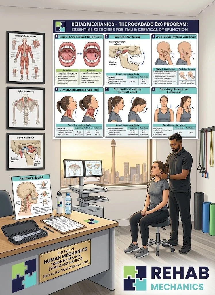















Is the Lateral Pterygoid Muscle Causing My Chronic Jaw Pain and Headaches?
Chronic jaw clicking and facial pressure are frequently driven by spasms in the lateral pterygoid muscle. Physiotherapy assists in managing TMJ dysfunction through intra-oral release and postural correction, addressing the contributing cervical factors to help reduce tissue irritation.
The Patient's Story
In the intense, screen-dominated work environments of Liberty Village and Toronto's Financial District, stress manifests physically. For countless professionals, the stress of looming deadlines and hours spent hunched over laptops translates directly into chronic, unconscious teeth clenching (bruxism).
Patients often arrive at Rehab Mechanics exhausted and frustrated. They complain of a constant, dull pressure deep in their cheeks, chronic tension headaches that wrap around their temples, and a jaw that loudly "clicks" or "pops" every time they try to eat a bagel or an apple. Many have visited dentists and received night guards, which protect their teeth from grinding but do not always address the deep muscular tension. Some have even been misdiagnosed with sinus issues.
The reality is that these patients are suffering from Temporomandibular Joint (TMJ) Dysfunction, and a primary culprit is often a tiny, highly overworked muscle hidden deep inside the skull: the Lateral Pterygoid. At our clinic in Queen West, we understand that you cannot easily access this muscle from the outside. By utilizing specialized intra-oral physical therapy and addressing the cervical mechanics driving the tension, we can assist in managing the jaw function and support the recovery of facial comfort.
Structural / Biomechanical Analysis
To understand why your jaw clicks and your head aches, we must perform a detailed biomechanical analysis of the TMJ architecture and the specific muscle responsible for opening your mouth.
The Anatomy of the TMJ
Your jaw (mandible) connects to your skull at the Temporomandibular Joint, located just in front of your ear.
The Articular Disc: Inside this joint space is a small, biconcave cartilaginous disc. It acts as a crucial shock absorber, allowing the jaw bone to hinge and glide smoothly when you open your mouth.
The Lateral Pterygoid Muscle
While the massive masseter and temporalis muscles close your jaw, the lateral pterygoid is heavily involved in opening it and moving it side-to-side.
The Hidden Location: It sits deep inside your skull, running horizontally from the sphenoid bone directly into the neck of the jawbone and—crucially—attaching directly to the articular disc itself.
The Biomechanical Breakdown (The Click)
When you clench your teeth from stress or adopt poor posture, the lateral pterygoid becomes exhausted, hypertonic, and locks into a spasm.
Disc Displacement: Because the spasming muscle is attached to the articular disc, it can pull the disc forward, out of its proper alignment.
The "Pop": When you try to open your mouth, the jaw bone has to shift over this displaced disc to open fully. This structural friction causes the painful "click" or "pop" you hear and feel.
The Restriction: If the muscle gets tight enough, the disc can become stuck in front of the joint, physically restricting the jaw from opening normally.
The "Tech Neck" Trigger
The jaw does not operate in isolation; it is heavily influenced by the posture of your neck.
When your head translates forward to look at a monitor ("Tech Neck"), it alters the resting tension of the muscles on the front of your throat.
This altered tension forces the closing muscles of your jaw to overwork just to keep your mouth balanced against gravity. The muscular system of the face exhausts itself, contributing to referred pressure into the temples and behind the eyes.
Clinical Red Flags
TMJ dysfunction driven by pterygoid spasm presents with highly specific mechanical signs:
Asymmetrical Opening (Deviation): When looking in a mirror, your jaw opens in a distinct "C" or "S" shape, zig-zagging rather than dropping perfectly straight down.
Painful Clicking: A sharp, audible pop localized just in front of the ear canal during chewing or yawning.
Deep Facial Aching: A dull, unrelenting pressure deep behind the cheekbones that is worse in the morning (from nighttime clenching) or at the end of a long workday.
Ear Fullness: Because the TMJ is millimeters from the ear canal, severe muscle tension can cause a sensation that the ear needs to "pop."
Primary Source Proof
Review the Clinical Evidence on PubMed supporting intra-oral manual therapy and postural correction for TMJ disorders:
The Effectiveness of Intra-Oral Manual Therapy in the Management of Temporomandibular Disorders (National Institutes of Health)
The Relationship Between Forward Head Posture and Temporomandibular Joint Dysfunction (National Institutes of Health)
Myofascial Trigger Points in the Lateral Pterygoid: Clinical Implications for Face and Jaw Pain (National Institutes of Health)
The Rehab Mechanics Corrective Protocol
Addressing the lateral pterygoid requires a highly specialized approach. We focus on internal joint mechanics alongside total-body postural correction.
Phase 1 — Intra-Oral Release and Decompression
Internal Pterygoid Release: Using sterile, medical-grade gloves, our specially trained physiotherapist will carefully access the muscles inside your mouth. We apply precise, sustained pressure to the lateral pterygoid belly to help release the dense spasm.
Joint Mobilization: Utilizing gentle, downward manual glides to the mandible to physically assist the TMJ capsule, supporting the articular disc space in returning to its proper anatomical position.
Phase 2 — Pelvic and Cervical Fortification
Fixing the Foundation: We cannot support the jaw if the head is slouched forward. We strengthen the deep core to stabilize the pelvis, providing an upright foundation for the entire spine.
Deep Cervical Flexor Activation: Utilizing specific biofeedback drills to re-awaken the tiny muscles in the front of the neck, helping to pull the head back over the shoulders and relieve downward tension on the jaw.
Phase 3 — Gait Retraining / Mechanics Correction
Jaw Tracking Re-Education: We utilize specific mirror-feedback techniques and tongue-positioning exercises to train your neuromuscular system to open the jaw symmetrically, helping to reduce the clicking.
Thoracic Spine Mobility: Using targeted manual therapy to support extension in the mid-back, ensuring fluid upper-body mechanics that assist in optimal head posture.
Phase 4 — Return-to-Activity Strategy
Habit Modification: Providing actionable coaching to address micro-traumas, including sleep posture correction and teaching the optimal "rest position" for the jaw (lips together, teeth apart, tongue resting lightly on the roof of the mouth).
Stress Load Management: Integrating diaphragmatic breathing techniques to actively support the nervous system, helping reduce the physiological drive to clench the jaw during high-stress scenarios.
Related Conditions We Treat
Temporomandibular Joint (TMJ) Dysfunction
Cervicogenic Headaches
Tension-Type Headaches
Myofascial Pain Syndrome
Cervical Radiculopathy
Bruxism (Teeth Grinding) Tension
Related Blogs
"Are Your Chronic Migraines Actually Being Caused by Your Neck?"
"The 'Tech Neck' Trigger Point: How Posture Creates Unbreakable Knots"
"Can Physiotherapy Cure Chronic Tension Headaches from Desk Work?"
Services Used in Treatment
Manual Therapy
Soft Tissue Release
Myofascial Release
Shockwave Therapy
Gait Retraining
Neuromuscular Re-Education
Custom Orthotics
Strengthening Programs
FAQ Section
1. Can physiotherapy assist in managing TMJ pain and clicking?
Yes. Physiotherapy supports recovery by utilizing specialized manual therapy to address the muscles controlling the jaw, and providing exercises to help optimize movement and tracking of the joint.
2. How do you address a muscle inside the mouth?
Our specially trained physiotherapists use a sterile glove to gently access the lateral pterygoid muscle from inside the cheek. We apply sustained, careful pressure to help reduce tissue irritation and release chronic tension.
3. Will addressing my neck posture help my jaw pain?
Yes. Forward head posture alters the tension of the muscles attached to your jaw, forcing it to overwork. We address contributing factors by strengthening cervical stabilizers to help improve head alignment.
4. Is jaw clicking always a sign of TMJ dysfunction?
Painless clicking can be common, but if the clicking causes discomfort or restriction, it indicates a mechanical tracking issue. We assist in managing this to optimize joint function.
5. Can tight jaw muscles cause referred pressure in my ears?
Yes. The muscles and ligaments of the TMJ are anatomically very close to the ear structures. Severe tension can lead to referred symptoms. We focus on soft tissue release to help reduce this pressure.
6. Do I still need to wear my dental night guard?
Yes. A night guard is crucial for protecting your teeth from the physical damage of grinding. Physiotherapy complements this by helping address the muscular factors that drive the clenching behavior.
7. How does stress affect my TMJ?
Stress often leads to unconscious clenching. We support recovery by integrating diaphragmatic breathing and neuromuscular re-education to help address contributing factors and relax the facial muscles.
8. How long does it take to see progress with TMJ physiotherapy?
Many patients experience relief from severe pressure after the first few sessions of intra-oral release, while building the postural endurance to maintain comfort requires consistent training over several weeks.
How Physiotherapy Helps
Reducing tissue irritation through internal myofascial release
Correcting pelvic drop to support global spinal posture
Improving cadence and upper body fluid dynamics
Strengthening stabilizers in the neck and mid-back
Reducing mechanical overload on the TMJ articular disc
Improving foot mechanics to anchor the kinetic chain
Contact Us Today — All you have to lose is the pain
Book a comprehensive biomechanical assessment with our clinical team today. Located inside the Prime Medical Centre at 68 Abell Street, Queen West.
Email: info@rehabmechanics.com Phone: (416) 533-3900
About the Author
Mr. Sanjay Attwala (B.Sc., M.Sc., RPT) is a Registered Physiotherapist, clinical director, and the founder of Rehab Mechanics in Toronto. With over 15 years of registered clinical practice and a deep specialization in complex musculoskeletal rehabilitation, Sanjay synthesizes rigorous international academic training with advanced evidence-based therapeutics to guide his clinical practice and patient education initiatives.
Academic Background & Credentials
Master of Science (M.Sc.) in Physiotherapy – University of Keele, United Kingdom (2010).
Bachelor of Science (B.Sc.) – University of Waterloo, Ontario, Canada.
Registered Physiotherapist (RPT) – Regulated health professional in excellent standing with the College of Physiotherapists of Ontario (CPO).
Corporate Entity – Operating officially under the S. Attwala Physiotherapy Professional Corporation with a DBA of Rehab Mechanics.
Clinical Expertise & Philosophy
Sanjay’s clinical approach rejects passive symptom management in favor of identifying underlying biomechanical root causes. His diverse expertise spans advanced manual therapies, personalized corrective exercise prescription, and modern physical modalities. At the Rehab Mechanics Toronto Queen West clinic, he routinely diagnoses and treats complex conditions including:
Spinal & Discogenic Pathology – Cervical, thoracic, and lumbar disc injuries, sciatica, and sacroiliac joint (SIJ) dysfunction.
Upper & Lower Extremity Injuries – Rotator cuff tears, frozen shoulder, tennis/golfer’s elbow, carpal tunnel syndrome, and complex ankle/foot pathologies.
Perinatal & Pelvic Health Rehabilitation – Specialized assessment and rehabilitation protocols tailored specifically for women during pregnancy and the post-partum period, addressing pelvic girdle pain, diastasis recti, and core stabilization.
Specialized Rehabilitation – Pelvic health therapy, TMJ dysfunction, post-surgical rehabilitation (including Total Hip and Total Knee Replacements), and custom orthotics dispensing.
Shockwave Therapy: with advanced cutting edge technological devices to suit your needs.
Interdisciplinary Practice & Patient Care
Sanjay practices an integrated model of healthcare, working closely alongside medical doctors inside the Prime Medical Centre on Abell Street to streamline patient recovery pathways. He maintains a human-centric, communication-first clinical framework, ensuring that care remains fully customized rather than automated.
His clinical caseload encompasses a broad operational spectrum under Ontario's regulatory frameworks, including:
Motor Vehicle Accident (MVA) Claims – Rehabilitation navigating Ontario’s statutory accident benefits schedule.
Workplace Safety and Insurance Board (WSIB) – Occupational injury management and return-to-work screening.
Extended Health Care (EHC) & Private Practice – Multi-tier insurance coordination and long-term athletic development plans.
Commitment to Research & Community
Outside of his clinical caseload at Rehab Mechanics and his additional practice affiliations in Etobicoke, Sanjay is an active health writer and community educator. He translates contemporary peer-reviewed medical research into accessible, actionable guidance on his professional blog. As a dedicated father and husband, he mirrors his professional advice in his personal life, focusing on structural mobility, cross-training, and longevity to help his family and his community thrive. Naturally he takes he a keen interest in rehabilitation for women who are pregnant and post-partum.
Disclaimer: The information provided on this blog is intended for educational and informational purposes only. It does not constitute medical advice, diagnosis, or a treatment plan. Always seek the direct advice of a Registered Physiotherapist, physician, or other qualified health provider regarding any medical condition or physical rehabilitation routine.
Be sure to check out Rehab Mechanics’ Google My Business Profile for daily posts, location and directions in addition to the directions on the website!
We love interacting with prospective and current patients, and love honest feedback and even constructive criticism and areas for improvement!
Can Physiotherapy Help Me Avoid Spinal Fusion Surgery for L5-S1 Degeneration?
L5-S1 degeneration causes severe lower back and radiating leg pain due to compressive disc narrowing. Physiotherapy supports recovery by correcting pelvic tilt and fortifying deep core stabilizers, reducing mechanical overload to help optimize movement and avoid surgical intervention.
The Patient's Story
Working in the Financial District or commuting heavily across Toronto takes a severe toll on the human spine. For many professionals, years of sitting in ergonomic chairs and standing on hard concrete eventually culminate in a terrifying episode: a sharp, paralyzing ache deep in the absolute base of the spine, often radiating heavy, dull pain into the glutes or legs.
Desperate for answers, patients undergo an MRI. The radiologist's report returns filled with frightening medical jargon: "Severe Degenerative Disc Disease," "Endplate Sclerosis," and "significant joint space narrowing at L5-S1."
For many patients, a surgical consultation follows, and the phrase "Spinal Fusion" is mentioned. The idea of having metal rods and screws permanently drilled into their spine creates massive anxiety. Patients often stop exercising entirely, terrified that any movement will further crush their crumbling discs.
At Rehab Mechanics in Queen West, we specialize in high-level spinal pathology, and we want to change this narrative. The L5-S1 segment naturally takes the brunt of human movement, and seeing degeneration on an X-ray is often a normal part of aging. Surgery should be an absolute last resort. By utilizing advanced human mechanics physical therapy, we can help you build an internal, muscular corset that supports the unstable joint, frequently assisting patients in avoiding the operating room and returning to an active lifestyle.
Structural / Biomechanical Analysis
To understand why your spine aches and how we can support its stability, we must perform a detailed biomechanical analysis of the very base of your spine.
The Anatomy of the L5-S1 Junction
The L5-S1 segment is the exact point where your lowest lumbar vertebra (L5) connects to your sacrum (S1, the tailbone structure of your pelvis).
The Shear Zone: Because the sacrum sits at a natural downward angle, gravity constantly tries to force the L5 vertebra to slide forward and down the slope.
The Anchor: The L5-S1 intervertebral disc and the surrounding ligaments must absorb massive shear forces every single time you stand, walk, or lift an object. It is the hardest-working joint in the human skeleton.
The Degenerative Cascade
We want you seeing this kind of cascade - one that regenerates! Rehab Mechanics!
Because this joint works so hard, it is usually the first to show signs of wear.
Disc Dehydration: The disc loses its water content and flattens, reducing its shock-absorbing capacity.
Endplate Sclerosis: As the disc flattens, the bones of the vertebrae experience more mechanical stress. In response, the body lays down extra calcium to thicken and harden the bone (Endplate Sclerosis). It is a biological defense mechanism, not a disease.
Facet Joint Overload: With the disc flattened, the tiny hinges at the back of the spine (facet joints) handle more load, leading to deep, localized back irritation.
The Biomechanical Tipping Point
Why does an aging spine suddenly become highly symptomatic? The answer is a loss of mechanical stability.
The Weak Foundation: If you have an anterior pelvic tilt (dumping the front of the pelvis downward) due to tight hip flexors from sitting all day, the angle of the L5-S1 joint becomes mechanically compromised.
Core Failure: If your deep abdominal muscles are weak, they cannot hold the L5 vertebra steady against gravity.
The Result: The degenerated joint wiggles and shifts microscopically under load. This constant micro-instability irritates the surrounding nerves and triggers protective muscle spasms as the body tries to stop the movement.
Clinical Red Flags
While degeneration is normal, certain symptoms indicate a severe loss of mechanical stability:
The Catching Sensation: A sharp, breathtaking "catch" in the extreme lower back when transitioning from sitting to standing.
Severe Extension Pain: Bending backward causes an immediate, sharp pinching sensation at the beltline.
Neurological Radiation: Dull, heavy, or aching pain that radiates deep into the buttocks or down the back of the thighs.
Morning Rigidity: The spine feels highly restricted and stiff for the first hour of the day until the joints warm up.
Primary Source Proof
Review the Clinical Evidence on PubMed supporting non-operative, biomechanically targeted stabilization for severe spinal degeneration:
The Efficacy of Specific Lumbar Stabilization Exercises in the Management of Degenerative Spondylosis (National Institutes of Health)
Biomechanical Effects of Pelvic Tilt on Lumbar Spinal Loads and Disc Degeneration (National Institutes of Health)
Non-Operative Management and Clinical Outcomes of Severe Degenerative Disc Disease at L5-S1 (National Institutes of Health)
The Rehab Mechanics Corrective Protocol
We cannot alter the thickened bone on your X-ray, but we can fundamentally change the forces acting on that joint. Our protocol focuses entirely on creating impenetrable core support.
Phase 1 — Load Modification and Decompression
Utilizing targeted manual traction and flexion-biased positioning to gently help reduce the compressive pressure on the discs and facet joints.
Implementing immediate postural modifications to avoid extreme spinal extension and heavy vertical loading during the workday.
Providing soft tissue release to the massive, spasming erector spinae muscles trying to protect the unstable joint.
Phase 2 — Pelvic Fortification and Alignment
Releasing the Brakes: Utilizing deep manual therapy to help reduce tension in the hip flexors (psoas) that are constantly pulling the spine into a deep arch.
Posterior Pelvic Tilt Training: Teaching the nervous system how to actively tuck the tailbone. This helps optimize the L5-S1 angle, assisting in reducing the forward shear force on the degenerated disc.
Phase 3 — Gait Retraining / Mechanics Correction
Transversus Abdominis (TvA) Activation: Utilizing precise biofeedback to wake up the deepest abdominal layer, helping it function as a biological weight belt that supports the L5 vertebra.
Hip Hinge Mastery: We meticulously teach you how to bend forward by pushing your hips back (like a deadlift), rather than rounding your fragile lower spine. By relying on glute power, we help ensure the lumbar spine remains stable.
Phase 4 — Return-to-Activity Strategy
Progressing to anti-extension core exercises (like Pallof presses and loaded carries) to help build the biological endurance necessary to support the spine all day.
Integrating complex, functional movements under strict biomechanical supervision to help optimize movement and build structural confidence.
Related Conditions We Treat
Degenerative Disc Disease (DDD)
Spondylolisthesis
Lumbar Herniated Discs
Lumbar Facet Joint Syndrome
Sciatica / Lumbar Radiculopathy
Sacroiliac Joint (SIJ) Dysfunction
Related Blogs
"Can Physiotherapy Stop the Progression of Degenerative Disc Disease?"
"Is My 'Locked' Lower Back a Herniated Disc or a Jammed Facet Joint?"
"Can a Herniated Disc in My Lower Back Heal on Its Own Without Surgery?"
Services Used in Treatment
Manual Therapy
Soft Tissue Release
Myofascial Release
Shockwave Therapy
Gait Retraining
Neuromuscular Re-Education
Custom Orthotics
Strengthening Programs
FAQ Section
1. Can physiotherapy assist in managing severe L5-S1 degeneration?
Yes. Physiotherapy supports recovery by addressing the biomechanical faults—specifically poor pelvic alignment and core weakness—helping to optimize movement and drastically reduce mechanical stress on the joint.
2. What does endplate sclerosis mean on my MRI?
It simply means the bone has thickened in response to increased mechanical stress from a thinning disc. We address contributing factors by improving your movement mechanics to help reduce this localized stress.
3. Will strengthening my core really help me avoid spinal fusion?
In many cases, yes. A strong deep core acts as an internal muscular support system. By strengthening these stabilizers, we help reduce the micro-instability that causes pain, often assisting in managing the condition without surgery.
4. Why does my lower back ache more when I arch backward?
Arching backward (extension) physically compresses the facet joints at the back of the spine. If your L5-S1 joint is degenerated, this compression causes irritation. We focus on neutral spine mechanics to support recovery.
5. Are traditional sit-ups safe for severe degenerative disc disease?
Typically, no. Repetitive sit-ups place compressive forces on vulnerable discs. We assist in managing your core strength using safer, anti-extension exercises like planks and dead-bugs.
6. How does releasing tight hips support my lower back?
Tight hip flexors constantly pull the front of your pelvis downward, altering your lower back posture. We use targeted release to support a neutral pelvic posture, helping to reduce pressure on the spine.
7. Can manual therapy help decompress my L5-S1 joint?
Yes. Specific manual traction and joint mobilizations can assist in managing joint stiffness, helping to optimize the mechanical space and reduce tissue irritation.
8. How long does it take to stabilize the spine?
While posture corrections can offer quick relief, building the necessary neuromuscular endurance to keep the spine stable under load typically requires several weeks of consistent strengthening programs.
How Physiotherapy Helps
Reducing tissue irritation through targeted mechanical decompression
Correcting pelvic drop and anterior tilt
Improving cadence and shock absorption during walking
Strengthening stabilizers in the deep core and glutes
Reducing mechanical overload on the degenerated disc
Improving foot mechanics to balance the upward kinetic chain
Contact Us Today — All you have to lose is the pain
Book a comprehensive biomechanical assessment with our clinical team today. Located inside the Prime Medical Centre at 68 Abell Street, Queen West.
Email: info@rehabmechanics.com Phone: (416) 533-3900
About the Author
Mr. Sanjay Attwala (B.Sc., M.Sc., RPT) is a Registered Physiotherapist, clinical director, and the founder of Rehab Mechanics in Toronto. With over 15 years of registered clinical practice and a deep specialization in complex musculoskeletal rehabilitation, Sanjay synthesizes rigorous international academic training with advanced evidence-based therapeutics to guide his clinical practice and patient education initiatives.
Academic Background & Credentials
Master of Science (M.Sc.) in Physiotherapy – University of Keele, United Kingdom (2010).
Bachelor of Science (B.Sc.) – University of Waterloo, Ontario, Canada.
Registered Physiotherapist (RPT) – Regulated health professional in excellent standing with the College of Physiotherapists of Ontario (CPO).
Corporate Entity – Operating officially under the S. Attwala Physiotherapy Professional Corporation with a DBA of Rehab Mechanics.
Clinical Expertise & Philosophy
Sanjay’s clinical approach rejects passive symptom management in favor of identifying underlying biomechanical root causes. His diverse expertise spans advanced manual therapies, personalized corrective exercise prescription, and modern physical modalities. At the Rehab Mechanics Toronto Queen West clinic, he routinely diagnoses and treats complex conditions including:
Spinal & Discogenic Pathology – Cervical, thoracic, and lumbar disc injuries, sciatica, and sacroiliac joint (SIJ) dysfunction.
Upper & Lower Extremity Injuries – Rotator cuff tears, frozen shoulder, tennis/golfer’s elbow, carpal tunnel syndrome, and complex ankle/foot pathologies.
Perinatal & Pelvic Health Rehabilitation – Specialized assessment and rehabilitation protocols tailored specifically for women during pregnancy and the post-partum period, addressing pelvic girdle pain, diastasis recti, and core stabilization.
Specialized Rehabilitation – Pelvic health therapy, TMJ dysfunction, post-surgical rehabilitation (including Total Hip and Total Knee Replacements), and custom orthotics dispensing.
Shockwave Therapy: with advanced cutting edge technological devices to suit your needs.
Interdisciplinary Practice & Patient Care
Sanjay practices an integrated model of healthcare, working closely alongside medical doctors inside the Prime Medical Centre on Abell Street to streamline patient recovery pathways. He maintains a human-centric, communication-first clinical framework, ensuring that care remains fully customized rather than automated.
His clinical caseload encompasses a broad operational spectrum under Ontario's regulatory frameworks, including:
Motor Vehicle Accident (MVA) Claims – Rehabilitation navigating Ontario’s statutory accident benefits schedule.
Workplace Safety and Insurance Board (WSIB) – Occupational injury management and return-to-work screening.
Extended Health Care (EHC) & Private Practice – Multi-tier insurance coordination and long-term athletic development plans.
Commitment to Research & Community
Outside of his clinical caseload at Rehab Mechanics and his additional practice affiliations in Etobicoke, Sanjay is an active health writer and community educator. He translates contemporary peer-reviewed medical research into accessible, actionable guidance on his professional blog. As a dedicated father and husband, he mirrors his professional advice in his personal life, focusing on structural mobility, cross-training, and longevity to help his family and his community thrive. Naturally he takes he a keen interest in rehabilitation for women who are pregnant and post-partum.
Disclaimer: The information provided on this blog is intended for educational and informational purposes only. It does not constitute medical advice, diagnosis, or a treatment plan. Always seek the direct advice of a Registered Physiotherapist, physician, or other qualified health provider regarding any medical condition or physical rehabilitation routine.
Be sure to check out Rehab Mechanics’ Google My Business Profile for daily posts, location and directions in addition to the directions on the website!
We love interacting with prospective and current patients, and love honest feedback and even constructive criticism and areas for improvement!
What Does a "Boggy End Feel" in My Knee Actually Mean?
A boggy end feel indicates joint effusion, where trapped fluid physically blocks knee movement. It presents as squishy resistance rather than a hard stop. Physiotherapy assists in managing this by restoring pelvic mechanics and utilizing targeted modalities to address the contributing biomechanical factors.
Not that kind of boggy per se but you have the right idea. Contact us today and we will see you the same day to give you an initial consultation!
The Patient's Story
Toronto is a highly active city, and knee tweaks are incredibly common. Whether you are playing a pickup soccer game in Trinity Bellwoods, cycling along the Martin Goodman Trail, or simply slipping on a wet step in your Queen West apartment building, your knee can take a sudden, awkward twist.
Following the initial tweak, many patients experience a deeply unsettling sensation. The knee doesn't necessarily feel broken, but when they try to straighten their leg completely, it stops short. Instead of a hard, bony stop, the joint feels like it is pushing into a dense, water-filled balloon. It feels "squishy" or "boggy," and attempting to force it straight creates an intense pressure deep inside the joint capsule.
Patients often attempt to aggressively stretch their hamstrings or calves, assuming the limitation is just a tight muscle. However, a "boggy end feel" is a massive clinical red flag for Knee Joint Effusion (severe internal swelling). At Rehab Mechanics, we know that trying to forcefully stretch through this specific type of mechanical block can cause further structural irritation. We use advanced diagnostic testing and targeted manual therapy to assist in managing the fluid and support the recovery of your joint’s range of motion.
Structural / Biomechanical Analysis
To understand why your knee feels blocked, we must break down the anatomy of the joint capsule and how it reacts to trauma.
The Synovial Joint Capsule
Your knee is entirely encased in a watertight, fibrous bag called the joint capsule.
The inner lining of this capsule is the synovial membrane.
In a healthy knee, this membrane produces a very small amount of thick, egg-white-like synovial fluid to lubricate the cartilage and allow the bones to glide smoothly.
The Mechanism of Effusion (Swelling)
When an internal structure of the knee is damaged—such as a minor meniscus tear, an MCL sprain, or a flare-up of underlying osteoarthritis—the joint experiences an acute trauma.
The Inflammatory Response: The body's defense mechanism kicks in, and the synovial membrane becomes hyperactive, overproducing synovial fluid in an attempt to protect and bathe the injured tissue.
The Pressurized Balloon: The joint capsule rapidly fills with this excess fluid, turning into a highly pressurized balloon.
The Biomechanics of the "Boggy End Feel"
In physical therapy, we test the "end feel" of a joint to determine what is limiting its movement.
Normal (Hard/Firm): When you straighten a healthy knee, the bones and ligaments eventually lock out, providing a firm, distinct endpoint.
The Boggy Block: When the capsule is severely swollen, the physical fluid gets trapped in the hinge. As you try to straighten the leg, you are physically compressing a pocket of pressurized liquid. The joint hits a soft, squishy, "boggy" block before the bones can actually lock out.
The Tipping Point (Arthrogenic Muscle Inhibition): The massive pressure inside the joint triggers a neurological reflex that actively shuts down your quadriceps muscle. Because you cannot fire your quad efficiently, you lose motor control, leading to a compensatory limp that alters your hip and lower back mechanics.
Clinical Red Flags
How do we differentiate internal effusion from a simple tight hamstring? We look for exact mechanical signs:
Visible Swelling (The Patellar Tap): The kneecap appears to be "floating" on a pocket of water. Pushing down on the kneecap causes it to bob up and down.
Loss of Terminal Extension: You physically cannot push the back of your knee flat against the floor or a treatment table.
A Feeling of "Fullness": A sensation of tight, bursting pressure behind the knee or deep inside the joint, especially when attempting to crouch or squat.
The Boggy Resistance: A soft, spongy resistance when a therapist attempts to passively straighten the leg.
Quadriceps Atrophy: A rapid, visible shrinking of the thigh muscle within days of the injury due to neurological inhibition.
Primary Source Proof
Review the Clinical Evidence on PubMed supporting advanced fluid management and biomechanical rehabilitation for knee joint effusion:
The Efficacy of Manual Lymphatic Drainage and Early Mobilization in the Management of Knee Joint Effusion (National Institutes of Health)
Arthrogenic Muscle Inhibition in Knee Osteoarthritis and Joint Effusion: Mechanisms and Management (National Institutes of Health)
Conservative Management of Meniscal Tears and Associated Joint Swelling (National Institutes of Health)
The Rehab Mechanics Corrective Protocol
We do not force a swollen joint. Our clinical protocol focuses on mechanical offloading and safely optimizing movement mechanics.
Phase 1 — Load Modification and Effusion Management
Utilizing advanced manual lymphatic drainage techniques and compressive wrapping to support the movement of stagnant fluid out of the joint capsule and into the lymphatic system of the thigh.
Implementing temporary activity modification to stop the mechanical friction that is contributing to fluid production.
Utilizing passive, pain-free range-of-motion drills (like heel slides) to gently assist the joint without triggering further irritation.
Phase 2 — Pelvic Fortification
While the knee is recovering, we strengthen the gluteus medius and deep core. A stable pelvis prevents the leg from collapsing inward when walking, protecting the vulnerable knee from sheer stress.
Activating the hip abductors to ensure proper weight distribution through the entire kinetic chain.
Phase 3 — Gait Retraining / Mechanics Correction
Restoring Terminal Extension: Using highly specific, gentle manual overpressure to carefully support the return of the final few degrees of knee extension as the fluid subsides.
Quadriceps Reactivation: Utilizing Neuromuscular Electrical Stimulation (NMES) and isometric holds to help reduce neuromuscular inhibition and assist the quadriceps in contracting again.
Correcting the compensatory limp to support healthy lower back mechanics.
Phase 4 — Return-to-Activity Strategy
Progressing to closed-kinetic-chain exercises like leg presses and mini-squats to safely reload the joint.
Integrating proprioceptive balance board training to help the knee's internal sensors react efficiently to unstable terrain.
Building a long-term self-management routine to maintain fluid dynamics and joint health.
Related Conditions We Treat
Meniscus Tears / Knee Derangement
ACL and MCL Sprains
Knee Osteoarthritis
Patellofemoral Pain Syndrome
Baker's Cyst
Post-Surgical Knee Rehabilitation
Related Blogs
"Is My Inner Knee Pain a Meniscus Tear or Pes Anserine Bursitis?"
"Can Physiotherapy Prevent Knee Replacement Surgery for Osteoarthritis?"
"Is the Bulge Behind My Knee a Baker's Cyst or a Torn Meniscus?"
Services Used in Treatment
Manual Therapy
Soft Tissue Release
Myofascial Release
Shockwave Therapy
Gait Retraining
Neuromuscular Re-Education
Custom Orthotics
Strengthening Programs
FAQ Section
1. Can physiotherapy assist in managing knee joint swelling?
Yes. Physiotherapy supports recovery by utilizing specific manual techniques to encourage fluid drainage and prescribing appropriate movements to optimize joint mechanics without causing further irritation.
2. Should I force my knee straight if it feels boggy?
No. Forcing a swollen, blocked joint can irritate the internal tissues further. We help reduce the swelling safely to gradually and naturally support the return of your range of motion.
3. Why does my thigh muscle look smaller after my knee swelled up?
Swelling inside the knee triggers a neurological reflex that inhibits the quadriceps muscle. We address contributing factors by using neuromuscular re-education to assist the muscle in firing properly.
4. How does pelvic strength help my swollen knee?
A stable pelvis helps ensure your leg stays properly aligned when you walk. We support recovery by strengthening the glutes, which helps reduce the twisting forces that can irritate a compromised knee.
5. Will custom orthotics help reduce knee swelling?
If excessive overpronation (flat feet) is causing your knee to cave inward and generate friction, orthotics can help optimize movement and reduce the mechanical stress on the joint capsule.
6. Is it safe to walk on a swollen knee?
It depends on the severity. We assist in managing your load by modifying your activities and assessing your gait, ensuring you remain as active as safely possible while the joint recovers.
7. Can shockwave therapy support joint recovery?
While shockwave therapy is primarily used for chronic tendon issues, it can support recovery by stimulating local blood flow and helping to reduce tissue irritation in the structures surrounding the joint.
8. How long does it take for a boggy end feel to resolve?
This varies based on the underlying injury. By actively managing the fluid and optimizing movement, many patients notice an improved ability to bend and straighten the joint within a few weeks of guided therapy.
How Physiotherapy Helps
Reducing tissue irritation through targeted fluid management
Correcting pelvic drop to support global leg alignment
Improving cadence and walking mechanics
Strengthening stabilizers in the quadriceps and hips
Reducing mechanical overload on the joint capsule
Improving foot mechanics to absorb shock efficiently
Contact Us Today — All you have to lose is the pain
Book a comprehensive biomechanical assessment with our clinical team today. Located inside the Prime Medical Centre at 68 Abell Street, Queen West.
Email: info@rehabmechanics.com Phone: (416) 533-3900
About the Author
Mr. Sanjay Attwala (B.Sc., M.Sc., RPT) is a Registered Physiotherapist, clinical director, and the founder of Rehab Mechanics in Toronto. With over 15 years of registered clinical practice and a deep specialization in complex musculoskeletal rehabilitation, Sanjay synthesizes rigorous international academic training with advanced evidence-based therapeutics to guide his clinical practice and patient education initiatives.
Academic Background & Credentials
Master of Science (M.Sc.) in Physiotherapy – University of Keele, United Kingdom (2010).
Bachelor of Science (B.Sc.) – University of Waterloo, Ontario, Canada.
Registered Physiotherapist (RPT) – Regulated health professional in excellent standing with the College of Physiotherapists of Ontario (CPO).
Corporate Entity – Operating officially under the S. Attwala Physiotherapy Professional Corporation with a DBA of Rehab Mechanics.
Clinical Expertise & Philosophy
Sanjay’s clinical approach rejects passive symptom management in favor of identifying underlying biomechanical root causes. His diverse expertise spans advanced manual therapies, personalized corrective exercise prescription, and modern physical modalities. At the Rehab Mechanics Toronto Queen West clinic, he routinely diagnoses and treats complex conditions including:
Spinal & Discogenic Pathology – Cervical, thoracic, and lumbar disc injuries, sciatica, and sacroiliac joint (SIJ) dysfunction.
Upper & Lower Extremity Injuries – Rotator cuff tears, frozen shoulder, tennis/golfer’s elbow, carpal tunnel syndrome, and complex ankle/foot pathologies.
Perinatal & Pelvic Health Rehabilitation – Specialized assessment and rehabilitation protocols tailored specifically for women during pregnancy and the post-partum period, addressing pelvic girdle pain, diastasis recti, and core stabilization.
Specialized Rehabilitation – Pelvic health therapy, TMJ dysfunction, post-surgical rehabilitation (including Total Hip and Total Knee Replacements), and custom orthotics dispensing.
Shockwave Therapy: with advanced cutting edge technological devices to suit your needs.
Interdisciplinary Practice & Patient Care
Sanjay practices an integrated model of healthcare, working closely alongside medical doctors inside the Prime Medical Centre on Abell Street to streamline patient recovery pathways. He maintains a human-centric, communication-first clinical framework, ensuring that care remains fully customized rather than automated.
His clinical caseload encompasses a broad operational spectrum under Ontario's regulatory frameworks, including:
Motor Vehicle Accident (MVA) Claims – Rehabilitation navigating Ontario’s statutory accident benefits schedule.
Workplace Safety and Insurance Board (WSIB) – Occupational injury management and return-to-work screening.
Extended Health Care (EHC) & Private Practice – Multi-tier insurance coordination and long-term athletic development plans.
Commitment to Research & Community
Outside of his clinical caseload at Rehab Mechanics and his additional practice affiliations in Etobicoke, Sanjay is an active health writer and community educator. He translates contemporary peer-reviewed medical research into accessible, actionable guidance on his professional blog. As a dedicated father and husband, he mirrors his professional advice in his personal life, focusing on structural mobility, cross-training, and longevity to help his family and his community thrive. Naturally he takes he a keen interest in rehabilitation for women who are pregnant and post-partum.
Disclaimer: The information provided on this blog is intended for educational and informational purposes only. It does not constitute medical advice, diagnosis, or a treatment plan. Always seek the direct advice of a Registered Physiotherapist, physician, or other qualified health provider regarding any medical condition or physical rehabilitation routine.
Be sure to check out Rehab Mechanics’ Google My Business Profile for daily posts, location and directions in addition to the directions on the website!
We love interacting with prospective and current patients, and love honest feedback and even constructive criticism and areas for improvement!
Is My Lingering Lower Back Pain Actually a Sacroiliac Joint Dysfunction?
Lingering, one-sided lower back pain that spikes when standing is frequently misdiagnosed as sciatica or a herniated disc, but is often Sacroiliac Joint (SIJ) Dysfunction. Physiotherapy assists in managing this condition by utilizing specific joint mobilizations to restore pelvic symmetry, and strengthening the deep myofascial slings to actively compress the joint. The biomechanical root cause is a failure of "force closure," where weak glutes and core muscles fail to stabilize the heavy pelvic keystone against the forces of gravity.
The Patient’s Story
For Toronto professionals enduring long commutes on the Gardiner Expressway, sitting in ergonomic chairs in the Financial District, or running the Martin Goodman Trail on weekends, lower back pain is incredibly common. Patients often present to the clinic deeply frustrated: they feel a sharp, nagging pain directly over the dimple on one side of their lower back. The pain typically starts as a dull ache after prolonged sitting and gradually worsens into a sharp, catching pain when transitioning from sitting to standing or when climbing the stairs at a TTC subway station. Often misdiagnosed by walk-in clinics as "sciatica" or a slipped disc, they spend months doing generic forward-bending stretches that completely fail to help. In reality, their lumbar spine is healthy; it is the massive joint anchoring their spine to their pelvis that has mechanically locked up or become dangerously unstable.
Structural / Biomechanical Analysis
The Pelvic Keystone (The SI Joint)
Your pelvis is the foundation of your skeleton. The Sacroiliac Joint is where the sacrum (the triangular bone at the base of the spine) connects to the two large iliac (hip) bones. It acts as a massive shock absorber, transferring the entire weight of your upper body down into your legs.
Form Closure vs. Force Closure
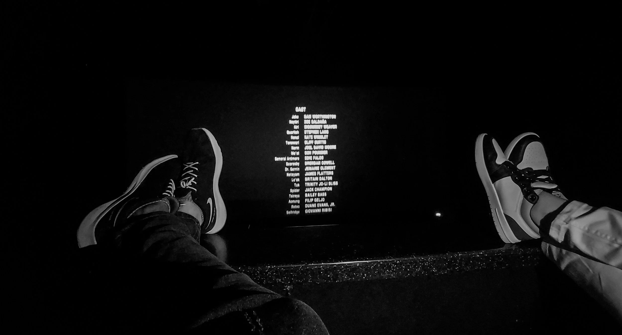
The end of the movie but not the end of your pain? Contact us today. All you have to lose is the pain!
The SI joint requires massive stability. "Form closure" refers to the interlocking shapes of the bones and the dense ligaments holding them together. "Force closure" refers to the dynamic, active compression provided by your muscles (like the glutes and lats) squeezing the joint shut during movement.
The Hypermobility Failure
Particularly common in post-partum women due to the hormone relaxin, the ligaments stretch and the joint moves too much. Without force closure from strong glutes to hold the bones together, the joint shears violently with every step, causing severe inflammation.
The Hypomobility Lock
Common in desk workers, the joint can become rigidly locked or slightly rotated out of alignment (an upslip or torsion). This physical locking jams the cartilaginous surfaces together, completely shutting down the natural shock-absorbing micro-movements and triggering massive spasms in the lower back muscles.
Clinical Red Flags
The Fortin Finger Test: The patient can point with one single finger directly to the PSIS (the bony dimple on the lower back) as the absolute epicenter of the pain.
Transitional Pain: A sharp, breathtaking jolt of pain when rolling over in bed, getting out of a low car, or standing up from a desk.
The Single-Leg Stance Test: Standing on one leg to put on pants or shoes is intensely painful or feels structurally weak.
Pseudosciatica: A deep, heavy ache that radiates into the groin or down the back of the thigh, but rarely travels below the knee into the calf.
Positive Provocation Cluster: Specific orthopedic tests (like the Thigh Thrust or Distraction Test) actively reproduce the familiar, sharp pelvic pain.
Primary Source Proof
Review the Clinical Evidence on PubMed supporting advanced differential diagnosis and stabilization for pelvic girdle pain:
Diagnosis and Treatment of Pelvic Girdle Pain (National Institutes of Health)
Motor Control and Myofascial Slings in Pelvic Stability (National Institutes of Health)
The Rehab Mechanics Corrective Protocol
Phase 1 — Load Modification
Utilizing Muscle Energy Techniques (MET)—gentle, patient-assisted isometric contractions—to coax a locked or rotated pelvic bone back into perfect, symmetrical alignment without forceful cracking.
Applying deep myofascial release to the piriformis and lower back erectors that have locked into a rigid, protective spasm around the failing joint.
Modifying daily habits, such as providing a specialized SIJ belt for hypermobile patients to mechanically lock the joint, allowing the inflamed ligaments to calm down.
Phase 2 — Pelvic Fortification
Activating the deep transverse abdominis (TvA) to act as a biological weight belt, violently compressing the SI joints together from the front.
Resurrecting the dormant gluteus maximus, ensuring the massive hip muscles actively squeeze the back of the pelvic ring shut (restoring Force Closure).
Correcting asymmetrical sitting habits (like crossing legs or sitting on a wallet) to prevent the pelvis from torquing out of alignment.
Phase 3 — Gait Retraining / Mechanics Correction
Re-training the Posterior Oblique Sling (the functional connection between the latissimus dorsi and the opposite glute). This fascial "X" across the back must fire synergistically to stabilize the SI joint during walking.
Addressing any functional leg-length discrepancies caused by a dropped arch or stiff ankle, which unevenly loads the pelvis.
Improving step cadence to reduce the vertical, jarring impact transferred through the SI joint with every foot strike.
Phase 4 — Return-to-Activity Strategy
Progressing to heavy, unilateral (one-sided) exercises like Bulgarian split squats and asymmetrical farmer's carries to build robust, real-world pelvic stability.
Reintegrating dynamic, rotational movements (like tennis or golf swings) under strict biomechanical supervision to ensure the SI joint remains locked down during athletic torque.
Establishing a permanent self-management routine focusing on gluteal power and core stiffness.
Related Conditions We Treat
Lumbar Disc Herniation
Sciatica / Pseudosciatica
Pelvic Girdle Pain (Perinatal)
Piriformis Syndrome
Lumbar Facet Joint Syndrome
Symphysis Pubis Dysfunction (SPD)
Related Blogs
"Why Does Stretching My Tight Hamstrings Make My Lower Back Pain Worse?"
"Does Sitting All Day Cause Gluteal Amnesia and Lower Back Pain?"
"Is Debilitating Pelvic Pain Just a Normal Part of Pregnancy?"
Services Used in Treatment
Manual Therapy
Soft Tissue Release
Myofascial Release
Shockwave Therapy
Gait Retraining
Neuromuscular Re-Education
Custom Orthotics
Strengthening Programs
FAQ Section
1. Can physiotherapy assist in managing sacroiliac joint pain?
Yes. Physiotherapy supports recovery by realigning the pelvic bones and strengthening the deep core and glutes to provide the dynamic compression (force closure) needed to optimize movement.
2. Is SI joint pain the same as sciatica?
No. While SI joint pain can radiate down the leg (pseudosciatica), it is caused by pelvic instability, whereas true sciatica is caused by a compressed nerve in the lumbar spine. We accurately diagnose and address contributing factors for both.
3. Will stretching help reduce my SI joint pain?
Aggressive stretching, especially deep forward folds or yoga twists, can often make a hypermobile SI joint worse by pulling the ligaments further. We focus on stabilization and strengthening programs instead.
4. Why does my lower back hurt more when I stand up from a chair?
Transitioning from sitting to standing requires a massive transfer of weight through the SI joint. If the joint is locked or unstable, this sudden sheer force triggers sharp pain.
5. How does core strength support my pelvis?
The deep core muscles wrap around your midsection like a corset. When contracted, they squeeze the SI joints tightly together, helping reduce mechanical overload and providing a stable foundation for the spine.
6. Do I need an SI joint belt?
For highly unstable or hypermobile joints (often seen in post-partum women), an SI belt can provide temporary mechanical compression, supporting recovery while we build your own muscular brace.
7. Can custom orthotics help balance my pelvis?
Yes. If poor foot mechanics (like a severely dropped arch on one side) are creating an uneven leg length, custom orthotics can help level the foundation and optimize movement through the pelvic ring.
8. How long does it take to stabilize the SI joint?
While manual therapy can often provide immediate relief for a locked joint, building the muscular "force closure" to keep it stable typically requires 6 to 8 weeks of consistent neuromuscular re-education.
How Physiotherapy Helps
Reducing tissue irritation through targeted joint alignment
Correcting pelvic drop and rotational torsion
Improving cadence and load transfer during walking
Strengthening stabilizers in the deep core and gluteal complex
Reducing mechanical overload on the pelvic ligaments
Improving foot mechanics to level the kinetic chain foundation
Contact Us Today — All you have to lose is the pain
Book a comprehensive biomechanical assessment with our clinical team today. Located inside the Prime Medical Centre at 68 Abell Street, Queen West.
Email: info@rehabmechanics.com Phone: (416) 533-3900
About the Author
Mr. Sanjay Attwala (B.Sc., M.Sc., RPT) is a Registered Physiotherapist, clinical director, and the founder of Rehab Mechanics in Toronto. With over 15 years of registered clinical practice and a deep specialization in complex musculoskeletal rehabilitation, Sanjay synthesizes rigorous international academic training with advanced evidence-based therapeutics to guide his clinical practice and patient education initiatives.
Academic Background & Credentials
Master of Science (M.Sc.) in Physiotherapy – University of Keele, United Kingdom (2010).
Bachelor of Science (B.Sc.) – University of Waterloo, Ontario, Canada.
Registered Physiotherapist (RPT) – Regulated health professional in excellent standing with the College of Physiotherapists of Ontario (CPO).
Corporate Entity – Operating officially under the S. Attwala Physiotherapy Professional Corporation with a DBA of Rehab Mechanics.
Clinical Expertise & Philosophy
Sanjay’s clinical approach rejects passive symptom management in favor of identifying underlying biomechanical root causes. His diverse expertise spans advanced manual therapies, personalized corrective exercise prescription, and modern physical modalities. At the Rehab Mechanics Toronto Queen West clinic, he routinely diagnoses and treats complex conditions including:
Spinal & Discogenic Pathology – Cervical, thoracic, and lumbar disc injuries, sciatica, and sacroiliac joint (SIJ) dysfunction.
Upper & Lower Extremity Injuries – Rotator cuff tears, frozen shoulder, tennis/golfer’s elbow, carpal tunnel syndrome, and complex ankle/foot pathologies.
Perinatal & Pelvic Health Rehabilitation – Specialized assessment and rehabilitation protocols tailored specifically for women during pregnancy and the post-partum period, addressing pelvic girdle pain, diastasis recti, and core stabilization.
Specialized Rehabilitation – Pelvic health therapy, TMJ dysfunction, post-surgical rehabilitation (including Total Hip and Total Knee Replacements), and custom orthotics dispensing.
Shockwave Therapy: with advanced cutting edge technological devices to suit your needs.
Interdisciplinary Practice & Patient Care
Sanjay practices an integrated model of healthcare, working closely alongside medical doctors inside the Prime Medical Centre on Abell Street to streamline patient recovery pathways. He maintains a human-centric, communication-first clinical framework, ensuring that care remains fully customized rather than automated.
His clinical caseload encompasses a broad operational spectrum under Ontario's regulatory frameworks, including:
Motor Vehicle Accident (MVA) Claims – Rehabilitation navigating Ontario’s statutory accident benefits schedule.
Workplace Safety and Insurance Board (WSIB) – Occupational injury management and return-to-work screening.
Extended Health Care (EHC) & Private Practice – Multi-tier insurance coordination and long-term athletic development plans.
Commitment to Research & Community
Outside of his clinical caseload at Rehab Mechanics and his additional practice affiliations in Etobicoke, Sanjay is an active health writer and community educator. He translates contemporary peer-reviewed medical research into accessible, actionable guidance on his professional blog. As a dedicated father and husband, he mirrors his professional advice in his personal life, focusing on structural mobility, cross-training, and longevity to help his family and his community thrive. Naturally he takes he a keen interest in rehabilitation for women who are pregnant and post-partum.
Disclaimer: The information provided on this blog is intended for educational and informational purposes only. It does not constitute medical advice, diagnosis, or a treatment plan. Always seek the direct advice of a Registered Physiotherapist, physician, or other qualified health provider regarding any medical condition or physical rehabilitation routine.
Be sure to check out Rehab Mechanics’ Google My Business Profile for daily posts, location and directions in addition to the directions on the website!
We love interacting with prospective and current patients, and love honest feedback and even constructive criticism and areas for improvement!
Can Physiotherapy Help Tennis Elbow If I Never Play Tennis?
Yes. Lateral epicondylitis (Tennis Elbow) is frequently caused by the repetitive micro-trauma of typing, mousing, and prolonged desk work. Physiotherapy assists in managing this condition by utilizing heavy eccentric loading, targeted shockwave therapy, and deep myofascial release to structurally rebuild the degenerated extensor tendons. The biomechanical root cause is often a combination of poor ergonomic wrist positioning and weak shoulder stabilizers, forcing the forearm to overwork and absorb massive, chronic strain.
The Patient’s Story
In Queen West and the tech hubs of Liberty Village, grip strength and wrist pain are silent epidemics. Patients often walk into the clinic bewildered, complaining of a sharp, burning pain on the outside of their elbow, yet they haven't picked up a tennis racket in their entire lives. The pain typically starts as a dull ache after ten-hour days coding or designing on a tablet, and gradually worsens into a sharp, stabbing sensation that makes picking up a coffee mug, turning a doorknob, or shaking hands excruciating. Often relying on bulky forearm braces and daily anti-inflammatories, these professionals quickly realize that passive rest isn't working. The chronic clicking of the mouse and typing on a flat keyboard relentlessly feeds the mechanical overload, leaving the forearm tendons in a constant state of decay.
Structural / Biomechanical Analysis
The Extensor Carpi Radialis Brevis (ECRB)
The muscles responsible for bending your wrist backward and keeping your hand stable while you type all merge into a single tendon. This common extensor tendon anchors directly to the lateral epicondyle—the bony bump on the outside of your elbow.
Angiofibroblastic Tendinosis (The Tendon Failure)
"Tennis elbow" is rarely an active, inflammatory "tendinitis." It is a degenerative "tendinosis." Repetitive typing causes microscopic tears in the ECRB tendon. Because tendons have a poor blood supply, the body fails to heal the tears with healthy collagen, instead patching it with chaotic, painful, disorganized scar tissue.
The Ergonomic Tipping Point
When your wrist rests heavily on the edge of a laptop and your hand is angled upward for hours, the extensor muscles are held in a state of continuous, exhausting isometric contraction. This chronic tension squeezes the blood out of the tendon, triggering cellular death.
The Kinetic Chain Breakdown
If your shoulder stabilizers (rotator cuff and mid-back) are weak from slumping, your elbow is forced to absorb the mechanical shock of every arm movement. A weak shoulder almost guarantees an overworked, painful forearm.
Clinical Red Flags
Exquisite Point Tenderness: Sharp, breathtaking pain when pressing directly on the bony bump on the outside of the elbow.
Pain with Resisted Extension: Severe pain triggered by trying to bend the wrist or middle finger backward against resistance.
Grip Weakness: A sudden, inexplicable loss of grip strength, often resulting in dropping light objects like a phone or a cup.
Morning Stiffness: The elbow joint feels incredibly rigid and painful for the first 30 minutes after waking up.
Double Crush Symptoms: Pain accompanied by numbness or tingling radiating from the neck down the arm, indicating a secondary nerve entrapment.
Primary Source Proof
Review the Clinical Evidence on PubMed supporting advanced tendon rehabilitation and loading protocols:
The Rehab Mechanics Corrective Protocol
Phase 1 — Load Modification
Utilizing Extracorporeal Shockwave Therapy (ESWT) to physically break down the chaotic scar tissue and stimulate new blood vessel growth (neovascularization) in the deadened tendon.
Implementing deep instrument-assisted soft tissue mobilization (IASTM) to melt trigger points in the forearm muscle bellies, providing mechanical slack to the tendon anchor.
Providing immediate ergonomic modifications, such as transitioning to a vertical mouse or split keyboard to place the wrist in a neutral position.
Phase 2 — Pelvic Fortification
You cannot have a strong arm without a strong base. Re-engaging the deep core and glutes to ensure you sit perfectly upright at your desk, un-hunching the upper body.
Correcting pelvic positioning in the office chair to align the lumbar and thoracic spine, allowing the arms to hang naturally rather than reaching aggressively forward.
Utilizing diaphragmatic breathing to release tension in the secondary breathing muscles of the neck.
Phase 3 — Gait Retraining / Mechanics Correction
Restoring full mobility to the thoracic spine and shoulder blades so they can act as the primary shock absorbers for the arm.
Strengthening the lower trapezius and rhomboids to anchor the shoulder blade securely against the ribcage, stopping the elbow from over-compensating.
Re-training basic lifting mechanics to ensure the larger muscles of the body initiate movement before the wrist extensors.
Phase 4 — Return-to-Activity Strategy
Progressing to Heavy Slow Resistance (HSR) training for the wrist extensors, focusing heavily on the eccentric (lowering) phase to structurally align new collagen fibers.
Reintegrating heavy, functional grip training (like loaded farmer's carries) to build absolute biological resilience in the forearm.
Establishing a 5-minute daily desk mobility routine to permanently address contributing factors and maintain fascial health.
Related Conditions We Treat
Medial Epicondylitis (Golfer's Elbow)
Carpal Tunnel Syndrome
Radial Tunnel Syndrome
De Quervain's Tenosynovitis
Thoracic Outlet Syndrome
Cubital Tunnel Syndrome
Related Blogs
"Is My Numb Arm Caused by Thoracic Outlet Syndrome or a Pinched Nerve?"
"Can Physiotherapy Cure 'Mommy Thumb' and Texting Wrist Without Cortisone?"
"The 'Tech Neck' Trigger Point: How Posture Creates Unbreakable Knots"
Services Used in Treatment
Manual Therapy
Soft Tissue Release
Myofascial Release
Shockwave Therapy
Gait Retraining
Neuromuscular Re-Education
Custom Orthotics
Strengthening Programs
FAQ Section
1. Can physiotherapy assist in managing tennis elbow from computer work? Yes. Physiotherapy supports recovery by addressing the biomechanical overload caused by poor ergonomics, utilizing targeted soft tissue release and strengthening to optimize movement in the forearm.
2. Why does my elbow hurt if I don't play sports? Lateral epicondylitis is an overuse injury. Typing and clicking a mouse for hours creates repetitive micro-trauma that can break down the tendon just as effectively as swinging a racket.
3. Will wearing a tight elbow brace fix the pain? A brace can help reduce immediate mechanical strain by changing the pull of the muscle, but it does not heal the tissue. Active strengthening and load modification are required to address contributing factors permanently.
4. Can shockwave therapy help reduce tendon pain? Yes. Shockwave therapy assists in managing chronic tendinopathies by breaking down rigid scar tissue and supporting local blood flow, which is crucial for cellular repair.
5. How does my shoulder strength affect my elbow? If your shoulder stabilizers are weak, your elbow and wrist must work twice as hard to control your arm. We strengthen stabilizers in the mid-back to reduce mechanical overload on the elbow.
6. Should I stop typing completely to let it heal? Total rest actually weakens tendons. We help you find safe, modified ways to continue working while introducing targeted mechanical load to support recovery and build tissue resilience.
7. Why are eccentric exercises important for tendons? Eccentric exercises (the slow lowering phase) specifically force the body to lay down strong, perfectly parallel collagen fibers, helping to reverse the chaotic scar tissue that causes chronic pain.
8. How long does it take to see progress with elbow pain? Tendon healing takes time. While manual therapy can provide immediate symptom relief, significant structural remodeling through strengthening programs typically occurs over 8 to 12 weeks.
How Physiotherapy Helps
Reducing tissue irritation through targeted shockwave therapy
Correcting pelvic drop and seated posture
Improving cadence and arm mechanics
Strengthening stabilizers in the shoulder and forearm
Reducing mechanical overload on the extensor tendons
Improving foot mechanics to support a neutral spine
Contact Us Today — All you have to lose is the pain
Book a comprehensive biomechanical assessment with our clinical team today. Located inside the Prime Medical Centre at 68 Abell Street, Queen West. Email: info@rehabmechanics.com Phone: (416) 533-3900
About the Author
Mr. Sanjay Attwala (B.Sc., M.Sc., RPT) is a Registered Physiotherapist, clinical director, and the founder of Rehab Mechanics in Toronto. With over 15 years of registered clinical practice and a deep specialization in complex musculoskeletal rehabilitation, Sanjay synthesizes rigorous international academic training with advanced evidence-based therapeutics to guide his clinical practice and patient education initiatives.
Academic Background & Credentials
Master of Science (M.Sc.) in Physiotherapy – University of Keele, United Kingdom (2010).
Bachelor of Science (B.Sc.) – University of Waterloo, Ontario, Canada.
Registered Physiotherapist (RPT) – Regulated health professional in excellent standing with the College of Physiotherapists of Ontario (CPO).
Corporate Entity – Operating officially under the S. Attwala Physiotherapy Professional Corporation with a DBA of Rehab Mechanics.
Clinical Expertise & Philosophy
Sanjay’s clinical approach rejects passive symptom management in favor of identifying underlying biomechanical root causes. His diverse expertise spans advanced manual therapies, personalized corrective exercise prescription, and modern physical modalities. At the Rehab Mechanics Toronto Queen West clinic, he routinely diagnoses and treats complex conditions including:
Spinal & Discogenic Pathology – Cervical, thoracic, and lumbar disc injuries, sciatica, and sacroiliac joint (SIJ) dysfunction.
Upper & Lower Extremity Injuries – Rotator cuff tears, frozen shoulder, tennis/golfer’s elbow, carpal tunnel syndrome, and complex ankle/foot pathologies.
Perinatal & Pelvic Health Rehabilitation – Specialized assessment and rehabilitation protocols tailored specifically for women during pregnancy and the post-partum period, addressing pelvic girdle pain, diastasis recti, and core stabilization.
Specialized Rehabilitation – Pelvic health therapy, TMJ dysfunction, post-surgical rehabilitation (including Total Hip and Total Knee Replacements), and custom orthotics dispensing.
Shockwave Therapy: with advanced cutting edge technological devices to suit your needs.
Interdisciplinary Practice & Patient Care
Sanjay practices an integrated model of healthcare, working closely alongside medical doctors inside the Prime Medical Centre on Abell Street to streamline patient recovery pathways. He maintains a human-centric, communication-first clinical framework, ensuring that care remains fully customized rather than automated.
His clinical caseload encompasses a broad operational spectrum under Ontario's regulatory frameworks, including:
Motor Vehicle Accident (MVA) Claims – Rehabilitation navigating Ontario’s statutory accident benefits schedule.
Workplace Safety and Insurance Board (WSIB) – Occupational injury management and return-to-work screening.
Extended Health Care (EHC) & Private Practice – Multi-tier insurance coordination and long-term athletic development plans.
Commitment to Research & Community
Outside of his clinical caseload at Rehab Mechanics and his additional practice affiliations in Etobicoke, Sanjay is an active health writer and community educator. He translates contemporary peer-reviewed medical research into accessible, actionable guidance on his professional blog. As a dedicated father and husband, he mirrors his professional advice in his personal life, focusing on structural mobility, cross-training, and longevity to help his family and his community thrive. Naturally he takes he a keen interest in rehabilitation for women who are pregnant and post-partum.
Disclaimer: The information provided on this blog is intended for educational and informational purposes only. It does not constitute medical advice, diagnosis, or a treatment plan. Always seek the direct advice of a Registered Physiotherapist, physician, or other qualified health provider regarding any medical condition or physical rehabilitation routine.
Be sure to check out Rehab Mechanics’ Google My Business Profile for daily posts, location and directions in addition to the directions on the website!
We love interacting with prospective and current patients, and love honest feedback and even constructive criticism and areas for improvement!
Is My Lingering Lower Back Pain Actually a Sacroiliac Joint Dysfunction?
Lingering, one-sided lower back pain that spikes when standing is frequently misdiagnosed as sciatica or a herniated disc, but is often Sacroiliac Joint (SIJ) Dysfunction. Physiotherapy assists in managing this condition by utilizing specific joint mobilizations to restore pelvic symmetry, and strengthening the deep myofascial slings to actively compress the joint. The biomechanical root cause is a failure of "force closure," where weak glutes and core muscles fail to stabilize the heavy pelvic keystone against the forces of gravity.
The Patient’s Story
For Toronto professionals enduring long commutes on the Gardiner Expressway, sitting in ergonomic chairs in the Financial District, or running the Martin Goodman Trail on weekends, lower back pain is incredibly common. Patients often present to the clinic deeply frustrated: they feel a sharp, nagging pain directly over the dimple on one side of their lower back. The pain typically starts as a dull ache after prolonged sitting and gradually worsens into a sharp, catching pain when transitioning from sitting to standing or when climbing the stairs at a TTC subway station. Often misdiagnosed by walk-in clinics as "sciatica" or a slipped disc, they spend months doing generic forward-bending stretches that completely fail to help. In reality, their lumbar spine is healthy; it is the massive joint anchoring their spine to their pelvis that has mechanically locked up or become dangerously unstable.
Structural / Biomechanical Analysis
The Pelvic Keystone (The SI Joint)
Your pelvis is the foundation of your skeleton. The Sacroiliac Joint is where the sacrum (the triangular bone at the base of the spine) connects to the two large iliac (hip) bones. It acts as a massive shock absorber, transferring the entire weight of your upper body down into your legs.
Form Closure vs. Force Closure
The SI joint requires massive stability. "Form closure" refers to the interlocking shapes of the bones and the dense ligaments holding them together. "Force closure" refers to the dynamic, active compression provided by your muscles (like the glutes and lats) squeezing the joint shut during movement.
The Hypermobility Failure
Particularly common in post-partum women due to the hormone relaxin, the ligaments stretch and the joint moves too much. Without force closure from strong glutes to hold the bones together, the joint shears violently with every step, causing severe inflammation.
The Hypomobility Lock
Common in desk workers, the joint can become rigidly locked or slightly rotated out of alignment (an upslip or torsion). This physical locking jams the cartilaginous surfaces together, completely shutting down the natural shock-absorbing micro-movements and triggering massive spasms in the lower back muscles.
Clinical Red Flags
The Fortin Finger Test: The patient can point with one single finger directly to the PSIS (the bony dimple on the lower back) as the absolute epicenter of the pain.
Transitional Pain: A sharp, breathtaking jolt of pain when rolling over in bed, getting out of a low car, or standing up from a desk.
The Single-Leg Stance Test: Standing on one leg to put on pants or shoes is intensely painful or feels structurally weak.
Pseudosciatica: A deep, heavy ache that radiates into the groin or down the back of the thigh, but rarely travels below the knee into the calf.
Positive Provocation Cluster: Specific orthopedic tests (like the Thigh Thrust or Distraction Test) actively reproduce the familiar, sharp pelvic pain.
Primary Source Proof
Review the Clinical Evidence on PubMed supporting advanced differential diagnosis and stabilization for pelvic girdle pain:
Diagnosis and Treatment of Pelvic Girdle Pain (National Institutes of Health)
Motor Control and Myofascial Slings in Pelvic Stability (National Institutes of Health)
The Rehab Mechanics Corrective Protocol
Phase 1 — Load Modification
Utilizing Muscle Energy Techniques (MET)—gentle, patient-assisted isometric contractions—to coax a locked or rotated pelvic bone back into perfect, symmetrical alignment without forceful cracking.
Applying deep myofascial release to the piriformis and lower back erectors that have locked into a rigid, protective spasm around the failing joint.
Modifying daily habits, such as providing a specialized SIJ belt for hypermobile patients to mechanically lock the joint, allowing the inflamed ligaments to calm down.
Phase 2 — Pelvic Fortification
Activating the deep transverse abdominis (TvA) to act as a biological weight belt, violently compressing the SI joints together from the front.
Resurrecting the dormant gluteus maximus, ensuring the massive hip muscles actively squeeze the back of the pelvic ring shut (restoring Force Closure).
Correcting asymmetrical sitting habits (like crossing legs or sitting on a wallet) to prevent the pelvis from torquing out of alignment.
Phase 3 — Gait Retraining / Mechanics Correction
Re-training the Posterior Oblique Sling (the functional connection between the latissimus dorsi and the opposite glute). This fascial "X" across the back must fire synergistically to stabilize the SI joint during walking.
Addressing any functional leg-length discrepancies caused by a dropped arch or stiff ankle, which unevenly loads the pelvis.
Improving step cadence to reduce the vertical, jarring impact transferred through the SI joint with every foot strike.
Phase 4 — Return-to-Activity Strategy
Progressing to heavy, unilateral (one-sided) exercises like Bulgarian split squats and asymmetrical farmer's carries to build robust, real-world pelvic stability.
Reintegrating dynamic, rotational movements (like tennis or golf swings) under strict biomechanical supervision to ensure the SI joint remains locked down during athletic torque.
Establishing a permanent self-management routine focusing on gluteal power and core stiffness.
Related Conditions We Treat
Lumbar Disc Herniation
Sciatica / Pseudosciatica
Pelvic Girdle Pain (Perinatal)
Piriformis Syndrome
Lumbar Facet Joint Syndrome
Symphysis Pubis Dysfunction (SPD)
Related Blogs
"Why Does Stretching My Tight Hamstrings Make My Lower Back Pain Worse?"
"Does Sitting All Day Cause Gluteal Amnesia and Lower Back Pain?"
"Is Debilitating Pelvic Pain Just a Normal Part of Pregnancy?"
Services Used in Treatment
Manual Therapy
Soft Tissue Release
Myofascial Release
Shockwave Therapy
Gait Retraining
Neuromuscular Re-Education
Custom Orthotics
Strengthening Programs
FAQ Section
1. Can physiotherapy assist in managing sacroiliac joint pain? Yes. Physiotherapy supports recovery by realigning the pelvic bones and strengthening the deep core and glutes to provide the dynamic compression (force closure) needed to optimize movement.
2. Is SI joint pain the same as sciatica? No. While SI joint pain can radiate down the leg (pseudosciatica), it is caused by pelvic instability, whereas true sciatica is caused by a compressed nerve in the lumbar spine. We accurately diagnose and address contributing factors for both.
3. Will stretching help reduce my SI joint pain? Aggressive stretching, especially deep forward folds or yoga twists, can often make a hypermobile SI joint worse by pulling the ligaments further. We focus on stabilization and strengthening programs instead.
4. Why does my lower back hurt more when I stand up from a chair? Transitioning from sitting to standing requires a massive transfer of weight through the SI joint. If the joint is locked or unstable, this sudden sheer force triggers sharp pain.
5. How does core strength support my pelvis? The deep core muscles wrap around your midsection like a corset. When contracted, they squeeze the SI joints tightly together, helping reduce mechanical overload and providing a stable foundation for the spine.
6. Do I need an SI joint belt? For highly unstable or hypermobile joints (often seen in post-partum women), an SI belt can provide temporary mechanical compression, supporting recovery while we build your own muscular brace.
7. Can custom orthotics help balance my pelvis? Yes. If poor foot mechanics (like a severely dropped arch on one side) are creating an uneven leg length, custom orthotics can help level the foundation and optimize movement through the pelvic ring.
8. How long does it take to stabilize the SI joint? While manual therapy can often provide immediate relief for a locked joint, building the muscular "force closure" to keep it stable typically requires 6 to 8 weeks of consistent neuromuscular re-education.
How Physiotherapy Helps
Reducing tissue irritation through targeted joint alignment
Correcting pelvic drop and rotational torsion
Improving cadence and load transfer during walking
Strengthening stabilizers in the deep core and gluteal complex
Reducing mechanical overload on the pelvic ligaments
Improving foot mechanics to level the kinetic chain foundation
Contact Us Today — All you have to lose is the pain
Book a comprehensive biomechanical assessment with our clinical team today. Located inside the Prime Medical Centre at 68 Abell Street, Queen West. Email: info@rehabmechanics.com Phone: (416) 533-3900
About the Author
Mr. Sanjay Attwala (B.Sc., M.Sc., RPT) is a Registered Physiotherapist, clinical director, and the founder of Rehab Mechanics in Toronto. With over 15 years of registered clinical practice and a deep specialization in complex musculoskeletal rehabilitation, Sanjay synthesizes rigorous international academic training with advanced evidence-based therapeutics to guide his clinical practice and patient education initiatives.
Academic Background & Credentials
Master of Science (M.Sc.) in Physiotherapy – University of Keele, United Kingdom (2010).
Bachelor of Science (B.Sc.) – University of Waterloo, Ontario, Canada.
Registered Physiotherapist (RPT) – Regulated health professional in excellent standing with the College of Physiotherapists of Ontario (CPO).
Corporate Entity – Operating officially under the S. Attwala Physiotherapy Professional Corporation with a DBA of Rehab Mechanics.
Clinical Expertise & Philosophy
Sanjay’s clinical approach rejects passive symptom management in favor of identifying underlying biomechanical root causes. His diverse expertise spans advanced manual therapies, personalized corrective exercise prescription, and modern physical modalities. At the Rehab Mechanics Toronto Queen West clinic, he routinely diagnoses and treats complex conditions including:
Spinal & Discogenic Pathology – Cervical, thoracic, and lumbar disc injuries, sciatica, and sacroiliac joint (SIJ) dysfunction.
Upper & Lower Extremity Injuries – Rotator cuff tears, frozen shoulder, tennis/golfer’s elbow, carpal tunnel syndrome, and complex ankle/foot pathologies.
Perinatal & Pelvic Health Rehabilitation – Specialized assessment and rehabilitation protocols tailored specifically for women during pregnancy and the post-partum period, addressing pelvic girdle pain, diastasis recti, and core stabilization.
Specialized Rehabilitation – Pelvic health therapy, TMJ dysfunction, post-surgical rehabilitation (including Total Hip and Total Knee Replacements), and custom orthotics dispensing.
Shockwave Therapy: with advanced cutting edge technological devices to suit your needs.
Interdisciplinary Practice & Patient Care
Sanjay practices an integrated model of healthcare, working closely alongside medical doctors inside the Prime Medical Centre on Abell Street to streamline patient recovery pathways. He maintains a human-centric, communication-first clinical framework, ensuring that care remains fully customized rather than automated.
His clinical caseload encompasses a broad operational spectrum under Ontario's regulatory frameworks, including:
Motor Vehicle Accident (MVA) Claims – Rehabilitation navigating Ontario’s statutory accident benefits schedule.
Workplace Safety and Insurance Board (WSIB) – Occupational injury management and return-to-work screening.
Extended Health Care (EHC) & Private Practice – Multi-tier insurance coordination and long-term athletic development plans.
Commitment to Research & Community
Outside of his clinical caseload at Rehab Mechanics and his additional practice affiliations in Etobicoke, Sanjay is an active health writer and community educator. He translates contemporary peer-reviewed medical research into accessible, actionable guidance on his professional blog. As a dedicated father and husband, he mirrors his professional advice in his personal life, focusing on structural mobility, cross-training, and longevity to help his family and his community thrive. Naturally he takes he a keen interest in rehabilitation for women who are pregnant and post-partum.
Disclaimer: The information provided on this blog is intended for educational and informational purposes only. It does not constitute medical advice, diagnosis, or a treatment plan. Always seek the direct advice of a Registered Physiotherapist, physician, or other qualified health provider regarding any medical condition or physical rehabilitation routine.
Be sure to check out Rehab Mechanics’ Google My Business Profile for daily posts, location and directions in addition to the directions on the website!
We love interacting with prospective and current patients, and love honest feedback and even constructive criticism and areas for improvement!
Is My Stabbing Shoulder Pain a Torn Rotator Cuff or a Frozen Shoulder?
A torn rotator cuff causes severe weakness and pain when actively lifting the arm, but the joint can still be moved passively. A frozen shoulder is a severe thickening of the joint capsule causing absolute mechanical stiffness. Physiotherapy assists in managing both conditions by identifying the structural failure, utilizing targeted joint mobilizations to release capsular restrictions, and fortifying the scapular stabilizers to restore overhead mobility. The biomechanical root cause is often prolonged immobilization or compensatory "shrugging" mechanics that overload the shoulder girdle.
The Patient’s Story
For the active professionals, weightlifters, and recreational tennis players of Queen West and Liberty Village, waking up one morning and realizing you cannot lift your arm to put on a jacket is a deeply terrifying experience. The pain typically starts as a dull ache after a weekend match at High Park or long hours at a desk, and gradually worsens into a sharp, catching pain that makes reaching for a coffee mug agonizing. Patients immediately assume the worst: a massive, catastrophic tear of the rotator cuff requiring surgery. However, many Torontonians are actually suffering from Adhesive Capsulitis (Frozen Shoulder), which mimics a tear perfectly but requires a completely opposite rehabilitation strategy to prevent permanent joint stiffness.
Structural / Biomechanical Analysis
The Muscular Engine (Rotator Cuff)
Your shoulder is a ball-and-socket joint designed for massive mobility. The rotator cuff is the active "engine" of four small muscles that steer the arm through space. A tear occurs when the tendon of one of these muscles (usually the supraspinatus) structurally rips away from the bone due to acute trauma or chronic wear.
The Connective Tissue Wrapper (The Capsule)
Surrounding the entire joint is a watertight bag of connective tissue called the capsule. In Adhesive Capsulitis, this capsule becomes violently inflamed, physically shrinks, and produces dense, fibrotic scar tissue. The capsule literally glues itself to the arm bone.
The Mechanical "Block"
The tipping point for diagnosing a frozen shoulder over a tear is the mechanical block. If the rotator cuff is torn, the engine is broken, but the hinge still works (the arm can be lifted by a therapist). If the shoulder is frozen, the hinge itself is encased in concrete; neither you nor the therapist can lift the arm overhead.
The Compensatory Shrug Pattern
When the true shoulder joint locks up or tears, the body forces a mechanical work-around. The upper trapezius and neck muscles take over, violently shrugging the entire shoulder blade upward to lift the arm. This guarantees secondary tension headaches and severe cervical spine overload.
Clinical Red Flags
Passive Range of Motion (ROM) Loss: The absolute hallmark of a frozen shoulder; the arm physically hits a hard, restrictive block when lifted by another person.
The "Drop Arm" Sign: An inability to hold the arm straight out to the side against gravity, indicating a massive structural rotator cuff tear.
Severe Night Pain: A deep, throbbing ache that makes it impossible to sleep on the affected side, common in the early "freezing" stage.
The "Zinger" Response: A blinding, breathtaking jolt of electrical pain when reaching quickly for a falling object.
Scapular Dyskinesis: A highly visible, asymmetrical "winging" or hiking of the shoulder blade during attempted overhead movement.
Primary Source Proof
Review the Clinical Evidence on PubMed supporting conservative physiotherapy for complex shoulder pathologies:
Conservative Management of Partial-Thickness Rotator Cuff Tears (National Institutes of Health)
Scapular Kinematics and Rehabilitation in Shoulder Dysfunction (National Institutes of Health)
The Rehab Mechanics Corrective Protocol
Phase 1 — Load Modification
Utilizing high-grade manual joint mobilizations (inferior and posterior glides) to safely stretch the fibrotic joint capsule and physically create space for the arm bone to move.
Implementing prolonged, low-load stretching (CREEP) to permanently deform and elongate scar tissue without triggering protective muscle spasms.
Modifying daily activities and sleep posture to avoid ischemic compression on the rotator cuff tendons.
Phase 2 — Pelvic Fortification
Re-establishing deep core stability to ensure the ribcage is firmly anchored, providing a solid foundation for the shoulder blade to rest upon.
Addressing any anterior pelvic tilt that may be contributing to an exaggerated spinal curve and compensatory upper body slouching.
Utilizing diaphragmatic breathing to naturally expand the ribcage and reduce sympathetic nervous system tension.
Phase 3 — Gait Retraining / Mechanics Correction
Restoring full thoracic spine (mid-back) extension so the shoulder blade can tilt backward smoothly when the arm is raised.
Activating the lower trapezius and serratus anterior to pull the shoulder blade down and back, stopping the painful "shrugging" compensation.
Addressing arm swing mechanics during walking to ensure the shoulder joint remains dynamic and fluid.
Phase 4 — Return-to-Activity Strategy
Progressing to heavy, eccentric loading of the rotator cuff using resistance bands and cables to align new collagen fibers and thicken the tendons.
Integrating rhythmic stabilization drills where the patient holds a weight while the therapist applies unexpected force, training the nervous system to react instantly.
Establishing a long-term, self-managed mobility and strengthening routine to permanently protect the shoulder joint.
Related Conditions We Treat
Subacromial Impingement Syndrome
Biceps Tendinopathy
Thoracic Outlet Syndrome
SLAP Tears (Labral Tears)
Cervical Radiculopathy (Pinched Neck Nerves)
AC Joint Sprains (Separated Shoulder)
Related Blogs
"Is My Numb Arm Caused by Thoracic Outlet Syndrome or a Pinched Nerve?"
"Does the Grinding Sound in My Neck Mean I Have Severe Arthritis?"
"Can Physiotherapy Cure Chronic Tension Headaches from Desk Work?"
Services Used in Treatment
Manual Therapy
Soft Tissue Release
Myofascial Release
Shockwave Therapy
Gait Retraining
Neuromuscular Re-Education
Custom Orthotics
Strengthening Programs
FAQ Section
1. Can physiotherapy assist in managing a frozen shoulder? Yes. Physiotherapy supports recovery by utilizing targeted joint mobilizations and capsular stretching to address the restrictive scar tissue, helping to optimize movement and reduce pain.
2. Will exercise make my torn rotator cuff worse? Aggressive, unsupported lifting can irritate a tear, but carefully guided, eccentric resistance training helps rebuild the surrounding musculature, supporting recovery and reducing mechanical overload.
3. Why is my shoulder pain worse at night? Night pain is often caused by inflammatory fluids pooling in the joint while you rest, combined with the physical compression of lying on the tissue. We help reduce this by addressing contributing factors and educating on proper sleep mechanics.
4. Can manual therapy help restore my overhead reach? Yes. Manual therapy assists in managing joint stiffness by gliding the arm bone downward, physically creating the mechanical space needed for the shoulder to lift overhead without pinching.
5. How does mid-back posture affect my shoulder? If your mid-back is hunched forward, your shoulder blades cannot rotate properly. This causes the arm bone to crash into the joint roof, creating massive mechanical friction on the rotator cuff.
6. Can shockwave therapy assist with shoulder pain? Yes. Shockwave therapy can help reduce tissue irritation and stimulate local blood flow, particularly in chronic tendinopathies or calcific deposits within the rotator cuff.
7. How long does it take for a frozen shoulder to thaw? While untreated adhesive capsulitis can last for years, consistent, progressive physical therapy supports a much faster recovery timeline, helping to optimize movement over several months.
8. Do I need surgery for a partial rotator cuff tear? Often, no. By strengthening the dynamic stabilizers of the shoulder blade and the remaining cuff muscles, we help reduce the demand on the torn tissue, allowing many patients to function entirely pain-free without surgery.
How Physiotherapy Helps
Reducing tissue irritation through joint mobilization
Correcting pelvic drop and thoracic posture
Improving cadence and upper body fluid dynamics
Strengthening stabilizers in the rotator cuff and scapula
Reducing mechanical overload on the joint capsule
Improving foot mechanics to support the kinetic chain
Contact Us Today — All you have to lose is the pain
Book a comprehensive biomechanical assessment with our clinical team today. Located inside the Prime Medical Centre at 68 Abell Street, Queen West. Email: info@rehabmechanics.com Phone: (416) 533-3900
About the Author
Mr. Sanjay Attwala (B.Sc., M.Sc., RPT) is a Registered Physiotherapist, clinical director, and the founder of Rehab Mechanics in Toronto. With over 15 years of registered clinical practice and a deep specialization in complex musculoskeletal rehabilitation, Sanjay synthesizes rigorous international academic training with advanced evidence-based therapeutics to guide his clinical practice and patient education initiatives.
Academic Background & Credentials
Master of Science (M.Sc.) in Physiotherapy – University of Keele, United Kingdom (2010).
Bachelor of Science (B.Sc.) – University of Waterloo, Ontario, Canada.
Registered Physiotherapist (RPT) – Regulated health professional in excellent standing with the College of Physiotherapists of Ontario (CPO).
Corporate Entity – Operating officially under the S. Attwala Physiotherapy Professional Corporation with a DBA of Rehab Mechanics.
Clinical Expertise & Philosophy
Sanjay’s clinical approach rejects passive symptom management in favor of identifying underlying biomechanical root causes. His diverse expertise spans advanced manual therapies, personalized corrective exercise prescription, and modern physical modalities. At the Rehab Mechanics Toronto Queen West clinic, he routinely diagnoses and treats complex conditions including:
Spinal & Discogenic Pathology – Cervical, thoracic, and lumbar disc injuries, sciatica, and sacroiliac joint (SIJ) dysfunction.
Upper & Lower Extremity Injuries – Rotator cuff tears, frozen shoulder, tennis/golfer’s elbow, carpal tunnel syndrome, and complex ankle/foot pathologies.
Perinatal & Pelvic Health Rehabilitation – Specialized assessment and rehabilitation protocols tailored specifically for women during pregnancy and the post-partum period, addressing pelvic girdle pain, diastasis recti, and core stabilization.
Specialized Rehabilitation – Pelvic health therapy, TMJ dysfunction, post-surgical rehabilitation (including Total Hip and Total Knee Replacements), and custom orthotics dispensing.
Shockwave Therapy: with advanced cutting edge technological devices to suit your needs.
Interdisciplinary Practice & Patient Care
Sanjay practices an integrated model of healthcare, working closely alongside medical doctors inside the Prime Medical Centre on Abell Street to streamline patient recovery pathways. He maintains a human-centric, communication-first clinical framework, ensuring that care remains fully customized rather than automated.
His clinical caseload encompasses a broad operational spectrum under Ontario's regulatory frameworks, including:
Motor Vehicle Accident (MVA) Claims – Rehabilitation navigating Ontario’s statutory accident benefits schedule.
Workplace Safety and Insurance Board (WSIB) – Occupational injury management and return-to-work screening.
Extended Health Care (EHC) & Private Practice – Multi-tier insurance coordination and long-term athletic development plans.
Commitment to Research & Community
Outside of his clinical caseload at Rehab Mechanics and his additional practice affiliations in Etobicoke, Sanjay is an active health writer and community educator. He translates contemporary peer-reviewed medical research into accessible, actionable guidance on his professional blog. As a dedicated father and husband, he mirrors his professional advice in his personal life, focusing on structural mobility, cross-training, and longevity to help his family and his community thrive. Naturally he takes he a keen interest in rehabilitation for women who are pregnant and post-partum.
Disclaimer: The information provided on this blog is intended for educational and informational purposes only. It does not constitute medical advice, diagnosis, or a treatment plan. Always seek the direct advice of a Registered Physiotherapist, physician, or other qualified health provider regarding any medical condition or physical rehabilitation routine.
Be sure to check out Rehab Mechanics’ Google My Business Profile for daily posts, location and directions in addition to the directions on the website!
We love interacting with prospective and current patients, and love honest feedback and even constructive criticism and areas for improvement!
Why Do My Deep Muscle Knots Keep Coming Back After a Massage?
Muscle knots, or myofascial trigger points, are localized areas of muscular spasm resulting from biomechanical overload and poor posture. These knots frequently present as deep, burning aches in the upper back or neck. Physiotherapy assists in managing this condition by addressing the underlying structural mechanics and releasing fascial tension. The biomechanical root cause is typically a postural collapse that forces upper-body muscles to overwork, locking them into a continuous state of contraction.
The Patient’s Story
For professionals working in Queen West and Liberty Village, extreme muscle tension is a daily reality. The constant stress of deadlines combined with ten-hour days hunched over a laptop invariably leads to burning, rock-hard lumps in the upper back and shoulder blades. Patients often book a massage to address the pain; while it feels incredible for a day, by the time they take a weekend walk through Trinity Bellwoods or jump on a bike along the Martin Goodman Trail, the exact same burning knot returns in the exact same spot. The pain typically starts as a dull ache and worsens into a sharp, restrictive tension that makes turning the head or lifting the arms highly uncomfortable.
Structural / Biomechanical Analysis
The Microscopic Lock (The Sarcomere)
A muscle knot is not a tangled clump of fibers; it is a chemical lock-up. Muscle fibers are made of millions of tiny contracting units called sarcomeres. When overloaded, these units run out of oxygen and lock into a permanent state of contraction, creating a palpable nodule under the skin.
The Postural Overload
The upper trapezius and levator scapulae muscles act as emergency parking brakes for the head. When the head translates forward toward a monitor, these muscles must contract continuously at maximum force to stop the head from falling forward.
The Joint Compression Pattern
Because the muscles are constantly clenched, they pull aggressively on the cervical and thoracic spine. This compresses the facet joints in the neck and mid-back, causing referred pain and further triggering protective muscle spasms.
The Tipping Point (Kinetic Chain Failure)
The tipping point occurs when the lower body and core fail to support the upper body. If the pelvis drops or the core weakens during seated posture, the upper back is forced to round forward, guaranteeing the return of the muscle knots regardless of how much massage therapy is applied.
Clinical Red Flags
A localized, rock-hard nodule that produces a sharp, "twitch" response when compressed.
Pain that remains present at rest, disrupting sleep or focus.
A positive mechanical loading test (e.g., sharp pain when resisting downward pressure on the shoulders).
Diffuse, radiating pain that travels up into the base of the skull, causing tension headaches.
Inability to rotate the cervical spine past 45 degrees without sharp, catching pain.
Primary Source Proof
Review the Clinical Evidence on PubMed: Effectiveness of Manual Therapy and Corrective Exercise in Myofascial Pain Syndrome (National Institutes of Health)
The Rehab Mechanics Corrective Protocol
Phase 1 — Load Modification
Utilizing ischemic compression to safely release the locked sarcomeres and restore blood flow to the starving tissue.
Modifying workstation ergonomics to reduce the immediate gravitational load on the cervical spine.
Implementing frequent, micro-doses of movement throughout the workday to prevent blood pooling.
Phase 2 — Pelvic Fortification
Strengthening the deep core to stabilize the pelvis, ensuring the spine sits on a neutral foundation.
Activating the gluteal muscles to prevent anterior pelvic tilt, which naturally straightens the thoracic spine.
Using diaphragmatic breathing drills to regulate intra-abdominal pressure and reduce upper-chest tension.
Phase 3 — Gait Retraining / Mechanics Correction
Restoring thoracic extension mobility so the upper back does not hunch forward during walking or running.
Addressing arm swing mechanics during gait to ensure the shoulders are not held rigidly elevated.
Strengthening the lower trapezius to pull the shoulder blades down and back.
Phase 4 — Return-to-Activity Strategy
Progressing to heavy, loaded carries (like farmer's walks) to build biological endurance in the postural muscles.
Reintegrating complex, full-body movements to support recovery and optimize movement.
Establishing a long-term self-management routine to address contributing factors independently.
Related Conditions We Treat
Myofascial Pain Syndrome
Cervicogenic Headaches
Thoracic Outlet Syndrome
Cervical Radiculopathy
Scapular Dyskinesis
Fibromyalgia
Related Blogs
"The 'Tech Neck' Trigger Point: How Posture Creates Unbreakable Knots"
"Can Muscle Knots Become Permanent? Understanding Tangled Fascia"
"Does Poor Posture Cause Sharp Pain in the Middle of the Back?"
Services Used in Treatment
Manual Therapy
Soft Tissue Release
Myofascial Release
Shockwave Therapy
Gait Retraining
Neuromuscular Re-Education
Custom Orthotics
Strengthening Programs
FAQ Section
1. Can physiotherapy assist in managing chronic muscle knots? Yes. Physiotherapy supports recovery by addressing the biomechanical faults that cause the muscles to overwork. By improving posture and stabilizing the spine, we help reduce the frequency and intensity of muscle spasms.
2. Why does massage only provide temporary relief? Massage addresses the symptom (the tight muscle) but not the cause (the postural collapse). We help optimize movement and strengthen stabilizers to address the root contributing factors.
3. Will stretching help reduce my muscle knots? Aggressive stretching can sometimes irritate an already exhausted muscle. We focus on soft tissue release and load modification to safely restore tissue length.
4. How does pelvic alignment affect upper back knots? The pelvis is the foundation of the spine. If the pelvis tilts improperly, the entire spine rounds forward, placing massive mechanical overload on the neck and shoulder muscles.
5. Can shockwave therapy assist with deep trigger points? Yes. Shockwave therapy can help reduce tissue irritation and stimulate local blood flow, supporting the recovery of chronic, fibrotic muscle knots.
6. How long does it take to see progress? With consistent neuromuscular re-education and strengthening programs, many patients notice an improved ability to maintain comfortable posture within a few weeks.
7. Should I use heat or ice on my muscle knots? Heat can help reduce perceived tension and improve blood flow, but addressing the mechanical overload through physiotherapy is required for long-term management.
8. Can poor gait mechanics cause upper back pain? Yes. A rigid or asymmetrical walking pattern forces the upper body to compensate, which can lead to muscular overload in the shoulders and neck.
How Physiotherapy Helps
Reducing tissue irritation through targeted soft tissue release
Correcting pelvic drop to stabilize the spinal foundation
Improving cadence and walking mechanics
Strengthening stabilizers in the mid-back and core
Reducing mechanical overload on the cervical spine
Improving foot mechanics to absorb upward shock
Contact Us Today — All you have to lose is the pain
Book a comprehensive biomechanical assessment with our clinical team today. Located inside the Prime Medical Centre at 68 Abell Street, Queen West. Email: info@rehabmechanics.com Phone: (416) 533-3900
About the Author
Mr. Sanjay Attwala (B.Sc., M.Sc., RPT) is a Registered Physiotherapist, clinical director, and the founder of Rehab Mechanics in Toronto. With over 15 years of registered clinical practice and a deep specialization in complex musculoskeletal rehabilitation, Sanjay synthesizes rigorous international academic training with advanced evidence-based therapeutics to guide his clinical practice and patient education initiatives.
Academic Background & Credentials
Master of Science (M.Sc.) in Physiotherapy – University of Keele, United Kingdom (2010).
Bachelor of Science (B.Sc.) – University of Waterloo, Ontario, Canada.
Registered Physiotherapist (RPT) – Regulated health professional in excellent standing with the College of Physiotherapists of Ontario (CPO).
Corporate Entity – Operating officially under the S. Attwala Physiotherapy Professional Corporation with a DBA of Rehab Mechanics.
Clinical Expertise & Philosophy
Sanjay’s clinical approach rejects passive symptom management in favor of identifying underlying biomechanical root causes. His diverse expertise spans advanced manual therapies, personalized corrective exercise prescription, and modern physical modalities. At the Rehab Mechanics Toronto Queen West clinic, he routinely diagnoses and treats complex conditions including:
Spinal & Discogenic Pathology – Cervical, thoracic, and lumbar disc injuries, sciatica, and sacroiliac joint (SIJ) dysfunction.
Upper & Lower Extremity Injuries – Rotator cuff tears, frozen shoulder, tennis/golfer’s elbow, carpal tunnel syndrome, and complex ankle/foot pathologies.
Perinatal & Pelvic Health Rehabilitation – Specialized assessment and rehabilitation protocols tailored specifically for women during pregnancy and the post-partum period, addressing pelvic girdle pain, diastasis recti, and core stabilization.
Specialized Rehabilitation – Pelvic health therapy, TMJ dysfunction, post-surgical rehabilitation (including Total Hip and Total Knee Replacements), and custom orthotics dispensing.
Shockwave Therapy: with advanced cutting edge technological devices to suit your needs.
Interdisciplinary Practice & Patient Care
Sanjay practices an integrated model of healthcare, working closely alongside medical doctors inside the Prime Medical Centre on Abell Street to streamline patient recovery pathways. He maintains a human-centric, communication-first clinical framework, ensuring that care remains fully customized rather than automated.
His clinical caseload encompasses a broad operational spectrum under Ontario's regulatory frameworks, including:
Motor Vehicle Accident (MVA) Claims – Rehabilitation navigating Ontario’s statutory accident benefits schedule.
Workplace Safety and Insurance Board (WSIB) – Occupational injury management and return-to-work screening.
Extended Health Care (EHC) & Private Practice – Multi-tier insurance coordination and long-term athletic development plans.
Commitment to Research & Community
Outside of his clinical caseload at Rehab Mechanics and his additional practice affiliations in Etobicoke, Sanjay is an active health writer and community educator. He translates contemporary peer-reviewed medical research into accessible, actionable guidance on his professional blog. As a dedicated father and husband, he mirrors his professional advice in his personal life, focusing on structural mobility, cross-training, and longevity to help his family and his community thrive. Naturally he takes he a keen interest in rehabilitation for women who are pregnant and post-partum.
Disclaimer: The information provided on this blog is intended for educational and informational purposes only. It does not constitute medical advice, diagnosis, or a treatment plan. Always seek the direct advice of a Registered Physiotherapist, physician, or other qualified health provider regarding any medical condition or physical rehabilitation routine.
Be sure to check out Rehab Mechanics’ Google My Business Profile for daily posts, location and directions in addition to the directions on the website!
We love interacting with prospective and current patients, and love honest feedback and even constructive criticism and areas for improvement!
Is My Chronic Outer Ankle Pain a Lingering Sprain or a Torn Tendon?
Maybe neither. Outer ankle pain that persists for months is often Peroneal Tendinopathy, not a lingering sprain. When ligaments fail, the peroneal tendons overwork to stabilize the ankle, leading to cellular degeneration. Physiotherapy resolves this through targeted eccentric loading and restoring proprioceptive joint control without surgery.
The Ankle Sprain That "Never Healed"
In a bustling, pedestrian-heavy city like Toronto, stepping off a curb incorrectly or rolling your ankle on uneven pavement is an occupational hazard. Most active adults know the drill for a standard ankle sprain: ice it, wrap it in a tensor bandage, rest for a few weeks, and wait for the swelling to subside.
But what happens when the bruising fades, yet a sharp, burning, or aching pain remains on the exact outside edge of the ankle for six months?
Patients frequently walk into Rehab Mechanics highly frustrated. They believe they have "weak ankles" or that their lateral ligaments simply never healed. They wear bulky braces when they run, but the pain intensifies, often radiating up the side of the calf or shooting down into the outside of the foot.
The clinical reality is often quite different. While the original injury was likely a ligament sprain, the current pain generator is not a ligament at all. The pain is coming from the massive tendons that are desperately trying to hold the unstable ankle together. This condition is Peroneal Tendinopathy. By accurately identifying the structural transition from ligament laxity to tendon degeneration, expert physical therapy can permanently rebuild your ankle’s capacity to handle load.
Structural Analysis: The Mechanics of the Lateral Ankle
To understand why your ankle still hurts, we must perform a detailed biomechanical analysis of the outer ankle and how it defends itself against gravity.
The Biological Stirrups
Running down the outside of your calf are two vital muscles: the Peroneus Longus and the Peroneus Brevis.
The Pathway: Their thick tendons travel all the way down your lower leg, hook sharply behind the bony bump on the outside of your ankle (the lateral malleolus), and anchor into the bottom and side of your foot.
The Function: They act as biological stirrups. Their primary mechanical job is eversion—pulling the foot outward. Most importantly, they are the active brakes that prevent your foot from rolling inward (inversion) when you walk or run.
The Pathology of Overload (Tendinosis)
How does a sprained ligament turn into a decaying tendon?
The Ligamentous Failure
When you originally rolled your ankle, you stretched or tore the lateral ligaments (like the ATFL). Ligaments are passive stabilizers. Once they are stretched out, they heal loosely. They can no longer hold the ankle joint tightly together.
The Muscular Panic
Because the ligaments are loose, the ankle joint becomes "wobbly" (Chronic Ankle Instability).
The nervous system panics. To prevent you from constantly rolling the ankle, the brain orders the peroneal muscles to work in massive overdrive.
These tendons are forced to act as primary, active stabilizers with every single step you take on the hard Toronto pavement.
The Degenerative Cascade
Tendons are not designed to be under constant, non-stop tension.
The Friction: The over-tensioned tendons grind violently against the bony bump of the outer ankle (retromalleolar friction).
The Cellular Decay: The tendon cells (tenocytes) exhaust their oxygen supply. They stop producing strong, parallel Type I collagen and begin laying down chaotic, painful, disorganized Type III scar tissue. The tendon thickens, swells, and degenerates—a process known as tendinosis.
Peroneal Subluxation (The "Snapping" Ankle)
In severe sprains, the thin band of tissue that holds the tendons behind the ankle bone (the superior peroneal retinaculum) can tear. When this happens, the peroneal tendons violently snap or "pop" out of their groove and roll directly over the ankle bone when you rotate your foot. This mechanical subluxation causes immense, sharp pain and rapid tendon fraying.
Identifying the Clinical Red Flags: Sprain vs. Tendon
How do we differentiate an unhealed ligament from a degenerating tendon?
Pain with Active Resistance: If we hold your foot and ask you to aggressively push outward against our hand (active eversion), and it causes a sharp, burning pain behind the ankle bone, it is the tendon. Ligaments only hurt when passively stretched.
The "Warm-Up" Illusion: Tendinopathy typically hurts severely during the first few steps of a run, feels better as the tissue warms up and blood flows in, but throbs intensely hours after the run is over.
Visible Thickening: You can often visually see or physically feel a thick, hardened "rope" behind the outside ankle bone, which is the swollen, scarred tendon.
Primary Source Proof: Tendon Rehabilitation
Orthopedic sports medicine literature emphatically proves that passive rest is detrimental to tendinopathy. Progressive, heavy eccentric loading and proprioceptive neuromuscular retraining are the international gold standards for resolving peroneal tendon degeneration and stabilizing the ankle.
Note: The link above directs to external, peer-reviewed medical literature demonstrating our commitment to evidence-based practice and international clinical guidelines for sports trauma rehabilitation.
The Rehab Mechanics Tendon Protocol
We must aggressively reverse the cellular decay in the tendon while simultaneously fixing the underlying joint instability that caused it to overwork in the first place.
Phase 1: De-escalation and Pain Modulation (Weeks 1-3)
Before we can load the tendon heavily, we must reduce the chemical sensitivity.
Extracorporeal Shockwave Therapy (ESWT): For chronic, stubborn cases, we utilize acoustic sound waves to literally shatter the disorganized scar tissue inside the peroneal tendon, stimulating an influx of fresh blood flow (neovascularization) to trigger true biological healing.
Isometric Loading: We utilize heavy, static holds against a resistance band. Isometrics are scientifically proven to rapidly drop the pain signals in the tendon without irritating the joint through friction-inducing movement.
Footwear Modification: Recommending temporary lateral heel wedging or stiffer-soled shoes to mechanically un-stretch the peroneal tendons during your daily commute.
Phase 2: Heavy Slow Resistance (HSR) Remodeling (Weeks 4-8)
We must force the tendon to rebuild its structural thickness.
Eccentric Eversion: We prescribe specific, slow-motion "lowering" exercises using resistance bands. The tendon only lays down new, perfectly aligned collagen fibers when it is subjected to slow, heavy, lengthening tension. This literally thickens the tendon so it can handle the workload of stabilizing the ankle.
Manual Cross-Friction: Using deep, localized massage directly across the tendon to ensure the new collagen fibers do not adhere or scar down to the surrounding bone.
Phase 3: Neuromuscular Re-Syncing (Weeks 8-12+)
A strong tendon is useless if the brain doesn't know when to fire it.
Proprioceptive Retraining: We use balance boards, BOSU balls, and uneven surface training. We must repair the damaged nerve endings in the ankle so the brain can automatically and instantly fire the peroneal muscles the millisecond you step on a rock, taking the sheer force off the ligaments.
Plyometric Bounding: Progressing to lateral skater jumps and agility ladder drills, proving to the tendon that it can act as a rapid "spring" without degenerating.
Stop the Cycle of Instability
You do not have to live with a chronically weak, aching ankle. By accurately identifying that the tendon, not the ligament, is the true source of your ongoing pain, expert physical therapy can reverse the cellular damage and rebuild an indestructible lower leg.
Book a comprehensive foot and ankle biomechanical assessment with our clinical team today. We are conveniently located inside the Prime Medical Centre at 68 Abell Street, offering elite sports orthopedic recovery in Queen West.
Contact us to schedule your appointment:
Email: info@rehabmechanics.com
Phone: (416) 533-3900
About the Author
Mr. Sanjay Attwala (B.Sc., M.Sc., RPT) is a Registered Physiotherapist, clinical director, and the founder of Rehab Mechanics in Toronto. With over 15 years of registered clinical practice and a deep specialization in complex musculoskeletal rehabilitation, Sanjay synthesizes rigorous international academic training with advanced evidence-based therapeutics to guide his clinical practice and patient education initiatives.
Academic Background & Credentials
Master of Science (M.Sc.) in Physiotherapy – University of Keele, United Kingdom (2010).
Bachelor of Science (B.Sc.) – University of Waterloo, Ontario, Canada.
Registered Physiotherapist (RPT) – Regulated health professional in excellent standing with the College of Physiotherapists of Ontario (CPO).
Corporate Entity – Operating officially under the S. Attwala Physiotherapy Professional Corporation with a DBA of Rehab Mechanics.
Clinical Expertise & Philosophy
Sanjay’s clinical approach rejects passive symptom management in favor of identifying underlying biomechanical root causes. His diverse expertise spans advanced manual therapies, personalized corrective exercise prescription, and modern physical modalities. At the Rehab Mechanics Toronto Queen West clinic, he routinely diagnoses and treats complex conditions including:
Spinal & Discogenic Pathology – Cervical, thoracic, and lumbar disc injuries, sciatica, and sacroiliac joint (SIJ) dysfunction.
Upper & Lower Extremity Injuries – Rotator cuff tears, frozen shoulder, tennis/golfer’s elbow, carpal tunnel syndrome, and complex ankle/foot pathologies.
Perinatal & Pelvic Health Rehabilitation – Specialized assessment and rehabilitation protocols tailored specifically for women during pregnancy and the post-partum period, addressing pelvic girdle pain, diastasis recti, and core stabilization.
Specialized Rehabilitation – Pelvic health therapy, TMJ dysfunction, post-surgical rehabilitation (including Total Hip and Total Knee Replacements), and custom orthotics dispensing.
Shockwave Therapy: with advanced cutting edge technological devices to suit your needs.
Interdisciplinary Practice & Patient Care
Sanjay practices an integrated model of healthcare, working closely alongside medical doctors inside the Prime Medical Centre on Abell Street to streamline patient recovery pathways. He maintains a human-centric, communication-first clinical framework, ensuring that care remains fully customized rather than automated.
His clinical caseload encompasses a broad operational spectrum under Ontario's regulatory frameworks, including:
Motor Vehicle Accident (MVA) Claims – Rehabilitation navigating Ontario’s statutory accident benefits schedule.
Workplace Safety and Insurance Board (WSIB) – Occupational injury management and return-to-work screening.
Extended Health Care (EHC) & Private Practice – Multi-tier insurance coordination and long-term athletic development plans.
Commitment to Research & Community
Outside of his clinical caseload at Rehab Mechanics and his additional practice affiliations in Etobicoke, Sanjay is an active health writer and community educator. He translates contemporary peer-reviewed medical research into accessible, actionable guidance on his professional blog. As a dedicated father and husband, he mirrors his professional advice in his personal life, focusing on structural mobility, cross-training, and longevity to help his family and his community thrive. Naturally he takes he a keen interest in rehabilitation for women who are pregnant and post-partum.
Disclaimer: The information provided on this blog is intended for educational and informational purposes only. It does not constitute medical advice, diagnosis, or a treatment plan. Always seek the direct advice of a Registered Physiotherapist, physician, or other qualified health provider regarding any medical condition or physical rehabilitation routine.
Be sure to check out Rehab Mechanics’ Google My Business Profile for daily posts, location and directions in addition to the directions on the website!
We love interacting with prospective and current patients, and love honest feedback and even constructive criticism and areas for improvement!
Is the Aching Pain at the Base of My Thumb Early Arthritis or a Tendon Injury?
It could be either. Pain at the thumb base is often De Quervain’s tenosynovitis or CMC joint osteoarthritis. While De Quervain's involves tendon friction, CMC arthritis is joint degeneration. Physiotherapy treats both by splinting, reducing mechanical shear forces, and rebuilding the intrinsic hand muscles to stabilize the thumb.
The Cost of the Precision Grip
The human thumb is an evolutionary marvel. Its ability to oppose the other fingers allows for the precision grip that defines human dexterity. However, in our modern environment—whether you are a construction worker gripping heavy power tools, a new mother constantly lifting a baby from a crib, or a Queen West creative spending 10 hours a day typing and swiping on a smartphone—the thumb is subjected to massive, unrelenting biomechanical stress.
Eventually, this stress causes the system to break down. Patients arrive at Rehab Mechanics complaining of a deep, sharp, or dull aching pain right at the absolute base of the thumb, where it meets the wrist. The pain makes turning a key in a lock, opening a tight jar, or even holding a coffee mug agonizingly difficult.
The immediate anxiety for many adults over the age of 40 is that they have developed severe, irreversible arthritis and are destined for joint replacement surgery. While thumb arthritis is incredibly common, the pain is equally likely to be a severe tendon entrapment known as De Quervain's Tenosynovitis.
These two conditions happen in the exact same square inch of the body, but they are structurally entirely different. By performing an expert differential diagnosis, physical therapy can identify whether your bone or your tendon is failing, and provide a non-surgical mechanical pathway to restore your grip strength.
Structural Analysis: The Mechanics of the Thumb Base

To effectively cure thumb pain, we must perform a detailed anatomical analysis of the intersection between the hand and the wrist, isolating the two distinct structures that break down under load.
1. The Tendon Pathology: De Quervain’s Tenosynovitis
This is an overuse injury of the "ropes" that move the thumb.
The Anatomy: Two specific tendons (the Abductor Pollicis Longus and Extensor Pollicis Brevis) run down the side of your thumb and travel through a tiny, rigid tunnel at the wrist.
The Friction: When you repeatedly angle your wrist downward while gripping (like pouring a heavy pitcher of water or typing on a phone), these tendons slide rapidly back and forth through the tunnel.
The Swelling: This friction causes the protective sheath around the tendons to become massively inflamed and swollen. The tendons get physically "stuck" in the tunnel, causing a sharp, catching, burning pain on the side of the wrist/thumb base.
2. The Joint Pathology: Carpometacarpal (CMC) Osteoarthritis
This is a degenerative disease of the "hinge" itself.
The Saddle Joint: The base of your thumb is called the CMC joint. It is shaped exactly like two saddles resting across one another. This unique shape allows the thumb to swivel in a massive circle, but it is inherently unstable.
The Beak Ligament Failure: A tiny ligament (the beak ligament) holds the two saddles together. Over decades of heavy gripping, this ligament stretches out and becomes loose.
The Bone-on-Bone Grind: Because the ligament is loose, the joint becomes wobbly. The two bones shear violently against each other during pinch grips. This grinds away the slippery articular cartilage, leaving raw bone grinding on raw bone. The pain is usually a deep, throbbing ache at the front/bottom of the thumb base.
Identifying the Clinical Red Flags: Arthritis vs. Tendinosis
Proper differential diagnosis dictates the entire rehabilitation protocol. We use specific physical tests to isolate the pain generator.
Finkelstein’s Test (For the Tendon): If you tuck your thumb inside your closed fist and aggressively tilt your wrist down toward your pinky, the pain on the side of the wrist will spike breathtakingly if you have De Quervain's.
The Grind Test (For the Joint): The physiotherapist will firmly grasp the base of your thumb, compress it downward into the wrist bone, and rotate it in a circle. If this causes a deep, gritty pain or a physical "crunching" sound (crepitus), the cartilage is degraded, indicating CMC Arthritis.
The "Bump" Location: De Quervain's often produces a soft, swollen, highly tender lump on the side of the wrist. CMC arthritis often produces a hard, bony, squared-off "shelf" deformity at the absolute base of the thumb on the back of the hand as the joint slowly subluxates (slides out of place) over years.
Primary Source Proof: Conservative Hand Rehabilitation
Orthopedic and hand therapy research definitively proves that conservative management—specifically the use of rigid splinting combined with targeted strengthening of the intrinsic thenar muscles—is highly effective at reducing pain and delaying or entirely preventing the need for surgical joint reconstruction (trapeziectomy) in CMC arthritis.
Note: The link above directs to external, peer-reviewed medical literature demonstrating our commitment to evidence-based practice and international clinical guidelines for upper extremity rehabilitation.
The Rehab Mechanics Corrective Protocol
Because these two conditions are mechanically distinct, their treatments overlap in the beginning but diverge drastically during the strengthening phase.
Phase 1: Acute Mechanical Offloading (Weeks 1-3)
Whether it is bone friction or tendon friction, we must immediately stop the mechanical insult.
Targeted Splinting: We do not use generic pharmacy wrist braces. We utilize or recommend specific "Thumb Spica" splints. These rigid braces physically lock the CMC joint and the thumb tendons, preventing the micro-movements that cause the grinding and inflammation, particularly while you sleep or perform heavy tasks.
Advanced Modalities: Utilizing targeted interferential currents or localized therapy to rapidly decrease the acute chemical inflammation in the joint capsule or tendon sheath.
Phase 2 (If De Quervain's): Tendon Remodeling
If the tendon is the issue, we must rebuild it to handle friction.
Soft Tissue Decompression: We use instrument-assisted soft tissue mobilization (IASTM) on the tight forearm muscles to release the pulling tension on the trapped thumb tendons.
Eccentric Loading: We prescribe specific, slow-motion "lowering" exercises for the thumb. This heavy, slow tension forces the body to lay down new, parallel collagen fibers, thickening the tendon and curing the tendinosis.
Phase 3 (If CMC Arthritis): Dynamic Joint Stabilization
If the cartilage is gone and the joint is wobbly, we cannot fix the bone. Instead, we must build a muscular "cast" around the joint to hold it perfectly still during movement.
First Dorsal Interosseous (FDI) Activation: We use targeted resistance exercises (like squeezing a therapy ball or pulling a rubber band between the thumb and index finger) to aggressively strengthen the tiny muscles deep inside the web space of your hand.
The Muscular Brace: By building massive strength in the intrinsic hand muscles, they take over the job of the loose ligaments. They physically clamp the CMC joint tightly together, preventing the bones from shearing and grinding when you open a jar or turn a key, effectively silencing the arthritic pain.
Reclaim Your Precision Grip
You do not have to live with the fear of dropping objects, nor do you have to immediately resign yourself to invasive thumb surgery. By expertly diagnosing whether your pain is a tendon entrapment or joint decay, physical therapy provides a clear, mechanical blueprint to restore your hand function.
Book a comprehensive upper extremity assessment with our clinical team today. We are conveniently located inside the Prime Medical Centre at 68 Abell Street, offering advanced orthopedic recovery in Queen West.
Contact us to schedule your appointment:
Email: info@rehabmechanics.com
Phone: (416) 533-3900
About the Author
Mr. Sanjay Attwala (B.Sc., M.Sc., RPT) is a Registered Physiotherapist, clinical director, and the founder of Rehab Mechanics in Toronto. With over 15 years of registered clinical practice and a deep specialization in complex musculoskeletal rehabilitation, Sanjay synthesizes rigorous international academic training with advanced evidence-based therapeutics to guide his clinical practice and patient education initiatives.
Academic Background & Credentials
Master of Science (M.Sc.) in Physiotherapy – University of Keele, United Kingdom (2010).
Bachelor of Science (B.Sc.) – University of Waterloo, Ontario, Canada.
Registered Physiotherapist (RPT) – Regulated health professional in excellent standing with the College of Physiotherapists of Ontario (CPO).
Corporate Entity – Operating officially under the S. Attwala Physiotherapy Professional Corporation with a DBA of Rehab Mechanics.
Clinical Expertise & Philosophy
Sanjay’s clinical approach rejects passive symptom management in favor of identifying underlying biomechanical root causes. His diverse expertise spans advanced manual therapies, personalized corrective exercise prescription, and modern physical modalities. At the Rehab Mechanics Toronto Queen West clinic, he routinely diagnoses and treats complex conditions including:
Spinal & Discogenic Pathology – Cervical, thoracic, and lumbar disc injuries, sciatica, and sacroiliac joint (SIJ) dysfunction.
Upper & Lower Extremity Injuries – Rotator cuff tears, frozen shoulder, tennis/golfer’s elbow, carpal tunnel syndrome, and complex ankle/foot pathologies.
Perinatal & Pelvic Health Rehabilitation – Specialized assessment and rehabilitation protocols tailored specifically for women during pregnancy and the post-partum period, addressing pelvic girdle pain, diastasis recti, and core stabilization.
Specialized Rehabilitation – Pelvic health therapy, TMJ dysfunction, post-surgical rehabilitation (including Total Hip and Total Knee Replacements), and custom orthotics dispensing.
Shockwave Therapy: with advanced cutting edge technological devices to suit your needs.
Interdisciplinary Practice & Patient Care
Sanjay practices an integrated model of healthcare, working closely alongside medical doctors inside the Prime Medical Centre on Abell Street to streamline patient recovery pathways. He maintains a human-centric, communication-first clinical framework, ensuring that care remains fully customized rather than automated.
His clinical caseload encompasses a broad operational spectrum under Ontario's regulatory frameworks, including:
Motor Vehicle Accident (MVA) Claims – Rehabilitation navigating Ontario’s statutory accident benefits schedule.
Workplace Safety and Insurance Board (WSIB) – Occupational injury management and return-to-work screening.
Extended Health Care (EHC) & Private Practice – Multi-tier insurance coordination and long-term athletic development plans.
Commitment to Research & Community
Outside of his clinical caseload at Rehab Mechanics and his additional practice affiliations in Etobicoke, Sanjay is an active health writer and community educator. He translates contemporary peer-reviewed medical research into accessible, actionable guidance on his professional blog. As a dedicated father and husband, he mirrors his professional advice in his personal life, focusing on structural mobility, cross-training, and longevity to help his family and his community thrive. Naturally he takes he a keen interest in rehabilitation for women who are pregnant and post-partum.
Disclaimer: The information provided on this blog is intended for educational and informational purposes only. It does not constitute medical advice, diagnosis, or a treatment plan. Always seek the direct advice of a Registered Physiotherapist, physician, or other qualified health provider regarding any medical condition or physical rehabilitation routine.
Can Physiotherapy Prevent Surgery for a Hip Labral Tear?
A hip labral tear often causes deep, pinching groin pain and catching sensations during movement. Physiotherapy assists in managing this condition by correcting pelvic mechanics, improving joint capsule mobility, and strengthening deep hip stabilizers to reduce the bone-on-bone friction that irritates the labrum. The biomechanical root cause is typically Femoroacetabular Impingement (FAI), where abnormal bone shape and poor pelvic tilt create destructive sheer forces inside the joint.
The Patient’s Story
For the highly active demographic in Trinity Bellwoods, Liberty Village, and Queen West, hip pain is a frequent and devastating issue. Runners on the Martin Goodman Trail, weightlifters, and even chronic desk workers often experience a deep, pinching pain in the front of their groin that refuses to go away with stretching. Often forming a "C" shape with their hand to point out the pain (the "C-Sign"), many Torontonians fear arthroscopic surgery is their only option. The pain usually starts as a dull ache after activities in High Park and gradually worsens into a sharp catch that makes sitting, squatting, or taking the stairs agonizing.
Structural / Biomechanical Analysis
The Acetabular Labrum
The hip is a deep ball-and-socket joint. The labrum is a ring of tough fibrocartilage lining the rim of the socket. It acts like a rubber gasket to deepen the socket, creating a suction seal that holds fluid inside the joint and provides massive structural stability.
The Joint Compression Pattern (FAI)
A labral tear rarely happens from a single trauma; it is usually the result of Femoroacetabular Impingement (FAI). Extra bone growth (Cam or Pincer lesions) destroys the perfect spherical fit of the joint. When these bony abnormalities crash into each other during activities like deep squatting or running, they trap the delicate labrum between them. Over time, this relentless mechanical crushing causes the labrum to fray and tear.
The Tipping Point (Anterior Pelvic Tilt)

While you cannot change your bone shape without surgery, the tipping point is often poor pelvic posture. Sitting for hours develops tight hip flexors, pulling the pelvis forward and downward. This lowers the roof of the hip socket, drastically closing the space and making the bony impingement and labral friction exponentially worse.
Clinical Red Flags
The "C-Sign" hand placement to indicate deep, diffuse groin pain.
Sharp, pinching pain that worsens with deep flexion (squatting, sitting in low chairs).
Mechanical catching, clicking, or locking inside the hip joint.
Pain that is aggravated by stretching the hip flexors, rather than relieved.
A positive FADIR test (Flexion, Adduction, Internal Rotation) triggering the familiar pinch.
Primary Source Proof
Review the Clinical Evidence on PubMed: The Efficacy of Physiotherapist-Led Rehabilitation in the Management of Femoroacetabular Impingement Syndrome (National Institutes of Health)
The Rehab Mechanics Corrective Protocol
Phase 1 — Load Modification
Temporary avoidance of deep hip flexion (like deep squats) to calm the irritated tissue.
Manual joint distraction to gently pull the femur out of the socket, relieving pressure on the crushed labrum.
Soft tissue release on the tight hip flexors to restore neutral alignment.
Phase 2 — Pelvic Fortification
Posterior pelvic tilt training to lift the roof of the socket away from the impingement.
Gluteus maximus fortification using bridges and hip thrusts to build the primary muscles responsible for fighting tight anterior structures.
Isometric holds for the deep hip rotators to centralize the femoral head.
Phase 3 — Gait Retraining / Mechanics Correction
Strengthening the gluteus medius with banded lateral walks to prevent the pelvis from dropping during the gait cycle.
Adjusting squat and running form (e.g., slightly wider stance with toes pointed outward) to mechanically clear the bony block.
Addressing lumbo-pelvic coordination to ensure the lower back is not compensating for a stiff hip.
Phase 4 — Return-to-Activity Strategy
Utilizing controlled Heavy Slow Resistance (HSR) like step-ups and split squats.
Integrating core-pelvic synchronization drills to ensure the pelvis does not tilt forward during explosive athletic movements.
Gradual, progressive return to running and high-impact sports while monitoring mechanical tracking.
Related Conditions We Treat
Femoroacetabular Impingement (FAI)
Hip Osteoarthritis
Gluteal Tendinopathy
Piriformis Syndrome
Snapping Hip Syndrome
Sacroiliac Joint (SIJ) Dysfunction
Related Blogs
"Is My Hip Bursitis Actually a Torn Glute Tendon?"
"Can Physiotherapy Prevent Hip Replacement Surgery for Severe Osteoarthritis?"
"Is My Sciatica Actually Caused by Piriformis Syndrome?"
Services Used in Treatment
Manual Therapy
Soft Tissue Release
Myofascial Release
Shockwave Therapy
Gait Retraining
Neuromuscular Re-Education
Custom Orthotics
Strengthening Programs
FAQ Section
1. Can physiotherapy assist in managing a hip labral tear? Yes. Physiotherapy supports recovery by addressing the biomechanical faults that cause friction on the labrum. By improving pelvic tilt and strengthening hip stabilizers, we can help reduce pain and optimize movement without surgery.
2. What does a labral tear feel like? It typically presents as a deep, pinching pain in the front of the groin, often accompanied by a catching or clicking sensation when moving the hip into flexion or rotation.
3. Will stretching my hip help the pain? Often, aggressive stretching can aggravate a labral tear. If the joint is already compressed, forcing it into deep stretches can increase irritation. We focus on load modification and strengthening instead.
4. How does pelvic tilt affect hip impingement? An anterior pelvic tilt (dumping the pelvis forward) mechanically lowers the roof of the hip socket, closing the joint space and increasing the likelihood of bone-on-bone friction during movement.
5. Can I still exercise with a labral tear? Yes, but load modification is key. We assist in modifying your routine to maintain fitness while avoiding the specific angles and loads that irritate the hip joint.
6. Do I need surgery for FAI? Not always. Many individuals manage FAI successfully with conservative care. Physiotherapy aims to address contributing factors, maximizing the space in the joint dynamically to support comfortable movement.
7. How long does rehabilitation take for hip pain? Depending on the severity and chronicity of the symptoms, significant functional improvements can often be seen within 6 to 12 weeks of targeted strengthening and mobility work.
8. Why is my glute strength important for my hip joint? Your glutes are the primary stabilizers of the pelvis. When they are strong, they help keep the femoral head centered in the socket, reducing sheer stress and protecting the labrum.
How Physiotherapy Helps
Reducing tissue irritation through load modification
Correcting anterior pelvic tilt
Improving squat and lifting mechanics
Strengthening deep hip stabilizers
Reducing mechanical overload on the labrum
Optimizing dynamic joint tracking
Contact Us Today — All you have to lose is the pain
Book a comprehensive biomechanical assessment with our clinical team today. Located inside the Prime Medical Centre at 68 Abell Street, Queen West. Email: info@rehabmechanics.com Phone: (416) 533-3900
About the Author
Mr. Sanjay Attwala (B.Sc., M.Sc., RPT) is a Registered Physiotherapist, clinical director, and the founder of Rehab Mechanics in Toronto. With over 15 years of registered clinical practice and a deep specialization in complex musculoskeletal rehabilitation, Sanjay synthesizes rigorous international academic training with advanced evidence-based therapeutics to guide his clinical practice and patient education initiatives.
Academic Background & Credentials
Master of Science (M.Sc.) in Physiotherapy – University of Keele, United Kingdom (2010).
Bachelor of Science (B.Sc.) – University of Waterloo, Ontario, Canada.
Registered Physiotherapist (RPT) – Regulated health professional in excellent standing with the College of Physiotherapists of Ontario (CPO).
Corporate Entity – Operating officially under the S. Attwala Physiotherapy Professional Corporation with a DBA of Rehab Mechanics.
Clinical Expertise & Philosophy
Sanjay’s clinical approach rejects passive symptom management in favor of identifying underlying biomechanical root causes. His diverse expertise spans advanced manual therapies, personalized corrective exercise prescription, and modern physical modalities. At the Rehab Mechanics Toronto Queen West clinic, he routinely diagnoses and treats complex conditions including:
Spinal & Discogenic Pathology – Cervical, thoracic, and lumbar disc injuries, sciatica, and sacroiliac joint (SIJ) dysfunction.
Upper & Lower Extremity Injuries – Rotator cuff tears, frozen shoulder, tennis/golfer’s elbow, carpal tunnel syndrome, and complex ankle/foot pathologies.
Perinatal & Pelvic Health Rehabilitation – Specialized assessment and rehabilitation protocols tailored specifically for women during pregnancy and the post-partum period, addressing pelvic girdle pain, diastasis recti, and core stabilization.
Specialized Rehabilitation – Pelvic health therapy, TMJ dysfunction, post-surgical rehabilitation (including Total Hip and Total Knee Replacements), and custom orthotics dispensing.
Shockwave Therapy: with advanced cutting edge technological devices to suit your needs.
Interdisciplinary Practice & Patient Care
Sanjay practices an integrated model of healthcare, working closely alongside medical doctors inside the Prime Medical Centre on Abell Street to streamline patient recovery pathways. He maintains a human-centric, communication-first clinical framework, ensuring that care remains fully customized rather than automated.
His clinical caseload encompasses a broad operational spectrum under Ontario's regulatory frameworks, including:
Motor Vehicle Accident (MVA) Claims – Rehabilitation navigating Ontario’s statutory accident benefits schedule.
Workplace Safety and Insurance Board (WSIB) – Occupational injury management and return-to-work screening.
Extended Health Care (EHC) & Private Practice – Multi-tier insurance coordination and long-term athletic development plans.
Commitment to Research & Community
Outside of his clinical caseload at Rehab Mechanics and his additional practice affiliations in Etobicoke, Sanjay is an active health writer and community educator. He translates contemporary peer-reviewed medical research into accessible, actionable guidance on his professional blog. As a dedicated father and husband, he mirrors his professional advice in his personal life, focusing on structural mobility, cross-training, and longevity to help his family and his community thrive. Naturally he takes he a keen interest in rehabilitation for women who are pregnant and post-partum.
Disclaimer: The information provided on this blog is intended for educational and informational purposes only. It does not constitute medical advice, diagnosis, or a treatment plan. Always seek the direct advice of a Registered Physiotherapist, physician, or other qualified health provider regarding any medical condition or physical rehabilitation routine.
Be sure to check out Rehab Mechanics’ Google My Business Profile for daily posts, location and directions in addition to the directions on the website!
We love interacting with prospective and current patients, and love honest feedback and even constructive criticism and areas for improvement!
Is the Stabbing Pain Between My Shoulder Blades a Pulled Muscle or a Neck Problem?
No, chronic pain between the shoulder blades is rarely a pulled muscle. It is usually referred pain from the lower cervical spine or an irritated dorsal scapular nerve caused by forward-head posture. Physiotherapy cures this by decompressing the neck and restoring thoracic mobility, completely eliminating the structural nerve irritation.
The Illusion of the "Unfixable Knot"
If you work in a high-demand office environment in Toronto's Financial District or spend hours designing on a tablet in Liberty Village, you are likely intimately familiar with "the knot."
It is a sharp, burning, deeply annoying ache located directly between your spine and your shoulder blade (scapula). The pain is so specific that you can often press a tennis ball or a massage cane right into the epicenter of the ache. Patients will spend hundreds of dollars on deep tissue massages, vigorously foam roll their mid-back, and stretch their arms across their chests. The massage feels fantastic for a few hours, but by the time they sit back down at their desk, the sharp, knifing pain has returned in the exact same millimeter of tissue.
Why is this "knot" completely immune to massage and stretching?
Because the muscle itself is not the problem. It is the innocent victim of a neurological illusion. At Rehab Mechanics in Queen West, we specialize in mapping complex neural pathways. That stabbing pain in your mid-back is almost certainly originating in your neck. By treating the cervical spine and correcting the biomechanics of your posture, we can permanently turn off the false pain signal.
Structural Analysis: The Mechanics of Referred Pain
To understand how your neck can cause burning pain in your mid-back, we must perform a detailed neurological and anatomical analysis of the cervicothoracic junction.
The "Pulled Rhomboid" Fallacy
The muscle located between your spine and your shoulder blade is called the rhomboid. When patients feel pain here, they assume they have "pulled" or "strained" the muscle.
The Reality: A true muscle strain occurs from acute, heavy lifting trauma (like violently pulling a heavy barbell). It heals within a few weeks. If your pain has lingered for months and worsens when you sit at a desk, it is not a muscle tear. It is a nerve issue.
The Neurological Pathway: Cervical Radiculopathy
Your spine is the central highway for your nervous system. The nerves that supply sensation and power to your upper back, shoulders, and arms exit the spinal cord through the lower vertebrae of your neck (specifically C4, C5, C6, and C7).
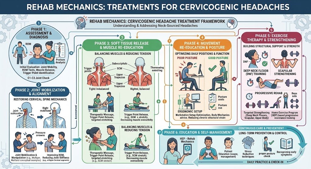
The Cervical Disc Bulge
When you adopt a "Tech Neck" posture (staring down at a screen), your head translates forward. This places massive, uneven pressure on the front of the cervical discs in your neck.
Over time, the gel-like center of the lower cervical discs (typically C5-C6 or C6-C7) bulges backward into the spinal canal.
This bulging disc physically presses against the exiting nerve root.
The Phenomenon of Referred Pain
When a nerve is crushed at its root (in the neck), the brain becomes confused. It doesn't necessarily feel the pain in the neck. Instead, the brain projects the pain down the length of the nerve to the area the nerve is supposed to supply.
The nerves from the lower neck supply the skin and deep tissue directly over the shoulder blade.
Therefore, a crushed disc in your neck creates a phantom, burning "knot" in your mid-back.
The Dorsal Scapular Nerve Entrapment
There is a second structural pathway for this pain: the Dorsal Scapular Nerve.
This specific nerve exits your mid-neck, travels through the scalene muscles on the side of your throat, and tunnels all the way down to plug directly into the rhomboid muscles between your shoulder blades.
If your neck muscles are in severe spasm from holding your heavy head forward all day, they act like a vice grip, crushing the dorsal scapular nerve in your throat. This "strangulation" causes an intense, electrical, aching pain in the rhomboids.
Identifying the Clinical Red Flags: Neck vs. Back
How do our physiotherapists prove that your mid-back pain is actually a neck problem? We use targeted structural provocation tests.
The Neck Extension Test: If you sit up straight and tilt your head backward to look at the ceiling, does the pain between your shoulder blades sharply increase? If yes, it is a cervical disc issue. Tilting backward physically closes the space in your neck, crushing the nerve harder and instantly amplifying the mid-back pain.
The Spurling’s Test: If we apply gentle downward pressure to the top of your head while your neck is tilted, it compresses the cervical nerve roots. If this instantly triggers the familiar burning between your shoulder blades, the diagnosis is definitively cervical.
The Relief Posture: Does placing your hand on top of your head or resting your chin on your chest relieve the mid-back ache? These postures open the nerve spaces in the neck, temporarily stopping the signal.
Primary Source Proof: Cervical Decompression
Orthopedic and neurological literature unequivocally demonstrates that resolving chronic interscapular (shoulder blade) pain requires directed manual therapy, traction, and stabilization of the cervical spine, completely outperforming localized treatment of the mid-back muscles.
Note: The link above directs to external, peer-reviewed medical literature demonstrating our commitment to evidence-based practice and international clinical guidelines for neurological rehabilitation.
The Rehab Mechanics Diagnostic and Corrective Protocol
We do not waste time massaging your rhomboids. To cure the pain, we must immediately decompress the cervical nerve roots and rebuild the posture that caused the compression.
Phase 1: Cervical Nerve Decompression (Weeks 1-3)
Our absolute first priority is getting the pressure off the nerve in your neck.
Cervical Retraction (The McKenzie Method): We teach you highly specific, repeated movements (like the "chin tuck") to alter the hydraulic pressure inside your cervical discs. This physically milks the bulging disc material forward, away from the crushed nerve root, centralizing and eliminating the shoulder blade pain.
Manual Cervical Traction: Our physiotherapists apply gentle, sustained upward pulling to the base of your skull. This separates the cervical vertebrae by millimeters, instantly opening the nerve canals (foramina) and giving the suffocating nerve room to breathe.
Scalene Release: Deep myofascial release on the side of the neck to un-trap the dorsal scapular nerve.
Phase 2: Thoracic Mobility and Unlocking (Weeks 3-6)
Your neck cannot sit straight if your mid-back is frozen in a hunch.
Thoracic Extension Mobilization: Utilizing high-grade manual manipulations and fulcrum stretching over a foam roller to forcefully restore the ability of your upper back to arch backward. If the mid-back moves correctly, the neck does not have to hinge and crush the discs to look straight ahead.
Pectoral Fascial Lengthening: Releasing the tight chest muscles that act as heavy anchors, dragging the shoulder blades and the neck forward.
Phase 3: Deep Cervical and Scapular Fortification (Weeks 6-10)
We must build the biological endurance to keep the nerve spaces permanently open.
Deep Cervical Flexor Activation: Re-awakening the tiny muscles in the front of the throat that act as the internal core of the neck, holding the head back over the shoulders automatically.
Lower Trapezius Loading: Instead of stretching the rhomboids, we must strengthen the entire mid-back complex using heavy Y-raises and rows, building the muscular scaffolding that prevents the spine from collapsing into "Tech Neck."
Silence the False Alarm
Do not spend another year chasing a "muscle knot" that does not exist. By accurately mapping your neurological pathways and aggressively treating the structural failure in your cervical spine, specialized physical therapy can permanently eradicate the stabbing pain in your mid-back.
Book a comprehensive neurological and spinal assessment with our clinical team today. We are conveniently located inside the Prime Medical Centre at 68 Abell Street, offering advanced orthopedic recovery in Queen West.
Contact us to schedule your appointment:
Email: info@rehabmechanics.com
Phone: (416) 533-3900
About the Author
Mr. Sanjay Attwala (B.Sc., M.Sc., RPT) is a Registered Physiotherapist, clinical director, and the founder of Rehab Mechanics in Toronto. With over 15 years of registered clinical practice and a deep specialization in complex musculoskeletal rehabilitation, Sanjay synthesizes rigorous international academic training with advanced evidence-based therapeutics to guide his clinical practice and patient education initiatives.
Academic Background & Credentials
Master of Science (M.Sc.) in Physiotherapy – University of Keele, United Kingdom (2010).
Bachelor of Science (B.Sc.) – University of Waterloo, Ontario, Canada.
Registered Physiotherapist (RPT) – Regulated health professional in excellent standing with the College of Physiotherapists of Ontario (CPO).
Corporate Entity – Operating officially under the S. Attwala Physiotherapy Professional Corporation with a DBA of Rehab Mechanics.
Clinical Expertise & Philosophy
Sanjay’s clinical approach rejects passive symptom management in favor of identifying underlying biomechanical root causes. His diverse expertise spans advanced manual therapies, personalized corrective exercise prescription, and modern physical modalities. At the Rehab Mechanics Toronto Queen West clinic, he routinely diagnoses and treats complex conditions including:
Spinal & Discogenic Pathology – Cervical, thoracic, and lumbar disc injuries, sciatica, and sacroiliac joint (SIJ) dysfunction.
Upper & Lower Extremity Injuries – Rotator cuff tears, frozen shoulder, tennis/golfer’s elbow, carpal tunnel syndrome, and complex ankle/foot pathologies.
Perinatal & Pelvic Health Rehabilitation – Specialized assessment and rehabilitation protocols tailored specifically for women during pregnancy and the post-partum period, addressing pelvic girdle pain, diastasis recti, and core stabilization.
Specialized Rehabilitation – Pelvic health therapy, TMJ dysfunction, post-surgical rehabilitation (including Total Hip and Total Knee Replacements), and custom orthotics dispensing.
Shockwave Therapy: with advanced cutting edge technological devices to suit your needs.
Interdisciplinary Practice & Patient Care
Sanjay practices an integrated model of healthcare, working closely alongside medical doctors inside the Prime Medical Centre on Abell Street to streamline patient recovery pathways. He maintains a human-centric, communication-first clinical framework, ensuring that care remains fully customized rather than automated.
His clinical caseload encompasses a broad operational spectrum under Ontario's regulatory frameworks, including:
Motor Vehicle Accident (MVA) Claims – Rehabilitation navigating Ontario’s statutory accident benefits schedule.
Workplace Safety and Insurance Board (WSIB) – Occupational injury management and return-to-work screening.
Extended Health Care (EHC) & Private Practice – Multi-tier insurance coordination and long-term athletic development plans.
Commitment to Research & Community
Outside of his clinical caseload at Rehab Mechanics and his additional practice affiliations in Etobicoke, Sanjay is an active health writer and community educator. He translates contemporary peer-reviewed medical research into accessible, actionable guidance on his professional blog. As a dedicated father and husband, he mirrors his professional advice in his personal life, focusing on structural mobility, cross-training, and longevity to help his family and his community thrive. Naturally he takes he a keen interest in rehabilitation for women who are pregnant and post-partum.
Disclaimer: The information provided on this blog is intended for educational and informational purposes only. It does not constitute medical advice, diagnosis, or a treatment plan. Always seek the direct advice of a Registered Physiotherapist, physician, or other qualified health provider regarding any medical condition or physical rehabilitation routine.
Be sure to check out Rehab Mechanics’ Google My Business Profile for daily posts, location and directions in addition to the directions on the website!
We love interacting with prospective and current patients, and love honest feedback and even constructive criticism and areas for improvement!
Do I Really Need to Do My Prescribed Home Physiotherapy Exercises?
Skipping prescribed home physiotherapy exercises is one of the leading causes of delayed recovery. While in-clinic treatments help reduce acute symptoms and improve joint mobility, consistent home exercises support the long-term structural remodeling of tissues. The biomechanical root cause of recurring injuries is the failure to build adequate load capacity through daily mechanotransduction, leaving the body vulnerable to the same stresses.
The Patient’s Story
A common scenario at Rehab Mechanics in Queen West involves active patients experiencing profound relief after a clinical session, only to return a week later complaining that their stiffness has crept back after running through High Park or walking the dog in Liberty Village. When asked if they completed their prescribed home exercise program, the answer is frequently a sheepish, "I didn't have time." Parkdale creatives and Financial District professionals often find themselves caught in the "167-Hour" trap—spending one hour healing in the clinic, and 167 hours falling back into the repetitive, poor postures that caused the injury in the first place.
Structural / Biomechanical Analysis
Mechanotransduction (The Tissue Response)
Tendons, ligaments, and muscle fibers adapt to stress through a biological process called mechanotransduction. When you perform a specific resistance exercise, your cells convert that mechanical stretch into biochemical signals. These signals instruct your body to produce new, healthy collagen fibers to thicken the tissue and support recovery.
The Tipping Point of Non-Compliance
The cellular signal that triggers collagen production only lasts about 36 to 48 hours after an exercise session. If you only exercise once a week in the clinic, your tissues spend five days in a dormant state, causing structural progress to halt entirely.
Neuromuscular Re-Education
Chronic pain physically changes motor pathways, causing you to move dysfunctionally to avoid discomfort. Manual therapy alone cannot rewire a brain. To overwrite old, painful movement habits with healthy motor engrams, the nervous system requires thousands of repetitions.
The Joint Compression Pattern
Without daily home exercises, the weak stabilizing muscles quickly fatigue. This allows joints to slip back into harmful compression patterns under the constant stress of gravity, irritating the tissue all over again.
Clinical Red Flags
Rapid return of joint stiffness within 48 hours of clinical treatment.
Inability to progress to heavier loads during in-clinic testing over several weeks.
Persistent pain at rest despite consistent manual therapy.
Loss of motor control or shaking during isolated muscle activation tests.
Diffuse, wandering aches indicating generalized muscular deconditioning rather than an isolated, healing injury.
Primary Source Proof
Review the Clinical Evidence on PubMed: The Impact of Home Exercise Compliance on Long-Term Functional Outcomes in Musculoskeletal Physiotherapy (National Institutes of Health)
The Rehab Mechanics Corrective Protocol
Phase 1 — Load Modification
Prescribing "micro-doses" of movement. These are highly specific isometric holds that take less than 60 seconds, performed multiple times a day.
Preventing tissue stiffness without causing fatigue or overwhelming a busy schedule.
Utilizing simple visual cues at your desk to remind you to check your posture.
Phase 2 — Pelvic Fortification
Re-establishing the foundation by waking up deep core and gluteal stabilizers.
Implementing daily, low-barrier activation drills performed at home to support pelvic neutrality.
Addressing underlying mobility restrictions that limit home exercise performance.
Phase 3 — Gait Retraining / Mechanics Correction
Progressively increasing the load, volume, and complexity of the home exercises.
Challenging the nervous system to overwrite faulty movement patterns during functional tasks.
Regular in-clinic audits of your home exercise form to ensure optimal mechanics.
Phase 4 — Return-to-Activity Strategy
Transitioning isolated home exercises into complex, real-world movements (like squats, deadlifts, and loaded carries).
Building the biological endurance necessary so that healthy mechanics become a permanent, protective habit.
Graduating patients to a self-managed, long-term strengthening routine.
Related Conditions We Treat
Post-Surgical Rehabilitation
Chronic Lower Back Pain
Tendinopathies (Achilles, Patellar, Gluteal)
Post-Concussion Syndrome
Chronic Ankle Instability
Scapular Dyskinesis
Related Blogs
"Does Sitting All Day Cause Gluteal Amnesia and Lower Back Pain?"
"Can Physiotherapy Stop the Progression of Degenerative Disc Disease?"
"Why Do My Deep Muscle Knots Keep Coming Back After a Massage?"
Services Used in Treatment
Neuromuscular Re-Education
Strengthening Programs
Gait Retraining
Manual Therapy
Soft Tissue Release
Myofascial Release
Custom Orthotics
Shockwave Therapy
FAQ Section
1. Can doing my home exercises assist in managing my pain faster? Yes. Consistent home exercises support recovery by maintaining the mobility and muscular activation achieved during your in-clinic sessions, preventing setbacks between visits.
2. What if my prescribed exercises cause discomfort? Muscle fatigue is normal, but sharp pain is not. We can help reduce irritation by adjusting the volume, load, or modifying the movement to better optimize your mechanics.
3. How often should I do my home physiotherapy program? It depends on the tissue being treated. Tendons often need a day of rest between heavy loading, while postural activation drills are often prescribed daily to support continuous neuromuscular re-education.
4. Why is manual therapy not enough on its own? Manual therapy is excellent for reducing tissue irritation and improving mobility, but it does not build strength. Your body needs active mechanical loading to strengthen stabilizers and address contributing factors long-term.
5. How long will I need to do these exercises? Once you have rebuilt your tissue capacity, we aim to transition these exercises into your normal fitness routine so you can optimize movement in your daily life without needing a separate "rehab" program.
6. Do I need special equipment for home exercises? No. We initially design programs using body weight, gravity, and simple resistance bands to ensure there are no barriers to completing your routine at home.
7. How do home exercises support post-surgical recovery? After surgery, muscles atrophy quickly. Daily home exercises are vital to re-establish neuromuscular pathways and rebuild the foundational strength needed to support the healing joint.
8. What if I don't have time to exercise every day? We assist in managing this by prescribing "micro-doses" of movement that take mere minutes, allowing you to seamlessly integrate rehab into a busy Toronto schedule.
How Physiotherapy Helps
Strengthening stabilizers to maintain joint alignment
Reducing mechanical overload through improved tissue capacity
Correcting compensatory movement patterns
Improving neuromuscular control and reflex timing
Reducing tissue irritation by building resilience
Optimizing movement for long-term health
Contact Us Today — All you have to lose is the pain
Book a comprehensive biomechanical assessment with our clinical team today. Located inside the Prime Medical Centre at 68 Abell Street, Queen West. Email: info@rehabmechanics.com Phone: (416) 533-3900
About the Author
Mr. Sanjay Attwala (B.Sc., M.Sc., RPT) is a Registered Physiotherapist, clinical director, and the founder of Rehab Mechanics in Toronto. With over 15 years of registered clinical practice and a deep specialization in complex musculoskeletal rehabilitation, Sanjay synthesizes rigorous international academic training with advanced evidence-based therapeutics to guide his clinical practice and patient education initiatives.
Academic Background & Credentials
Master of Science (M.Sc.) in Physiotherapy – University of Keele, United Kingdom (2010).
Bachelor of Science (B.Sc.) – University of Waterloo, Ontario, Canada.
Registered Physiotherapist (RPT) – Regulated health professional in excellent standing with the College of Physiotherapists of Ontario (CPO).
Corporate Entity – Operating officially under the S. Attwala Physiotherapy Professional Corporation with a DBA of Rehab Mechanics.
Clinical Expertise & Philosophy
Sanjay’s clinical approach rejects passive symptom management in favor of identifying underlying biomechanical root causes. His diverse expertise spans advanced manual therapies, personalized corrective exercise prescription, and modern physical modalities. At the Rehab Mechanics Toronto Queen West clinic, he routinely diagnoses and treats complex conditions including:
Spinal & Discogenic Pathology – Cervical, thoracic, and lumbar disc injuries, sciatica, and sacroiliac joint (SIJ) dysfunction.
Upper & Lower Extremity Injuries – Rotator cuff tears, frozen shoulder, tennis/golfer’s elbow, carpal tunnel syndrome, and complex ankle/foot pathologies.
Perinatal & Pelvic Health Rehabilitation – Specialized assessment and rehabilitation protocols tailored specifically for women during pregnancy and the post-partum period, addressing pelvic girdle pain, diastasis recti, and core stabilization.
Specialized Rehabilitation – Pelvic health therapy, TMJ dysfunction, post-surgical rehabilitation (including Total Hip and Total Knee Replacements), and custom orthotics dispensing.
Shockwave Therapy: with advanced cutting edge technological devices to suit your needs.
Interdisciplinary Practice & Patient Care
Sanjay practices an integrated model of healthcare, working closely alongside medical doctors inside the Prime Medical Centre on Abell Street to streamline patient recovery pathways. He maintains a human-centric, communication-first clinical framework, ensuring that care remains fully customized rather than automated.
His clinical caseload encompasses a broad operational spectrum under Ontario's regulatory frameworks, including:
Motor Vehicle Accident (MVA) Claims – Rehabilitation navigating Ontario’s statutory accident benefits schedule.
Workplace Safety and Insurance Board (WSIB) – Occupational injury management and return-to-work screening.
Extended Health Care (EHC) & Private Practice – Multi-tier insurance coordination and long-term athletic development plans.
Commitment to Research & Community
Outside of his clinical caseload at Rehab Mechanics and his additional practice affiliations in Etobicoke, Sanjay is an active health writer and community educator. He translates contemporary peer-reviewed medical research into accessible, actionable guidance on his professional blog. As a dedicated father and husband, he mirrors his professional advice in his personal life, focusing on structural mobility, cross-training, and longevity to help his family and his community thrive. Naturally he takes he a keen interest in rehabilitation for women who are pregnant and post-partum.
Disclaimer: The information provided on this blog is intended for educational and informational purposes only. It does not constitute medical advice, diagnosis, or a treatment plan. Always seek the direct advice of a Registered Physiotherapist, physician, or other qualified health provider regarding any medical condition or physical rehabilitation routine.
Be sure to check out Rehab Mechanics’ Google My Business Profile for daily posts, location and directions in addition to the directions on the website!
We love interacting with prospective and current patients, and love honest feedback and even constructive criticism and areas for improvement!
Does the Grinding Sound in My Neck Mean I Have Severe Arthritis?
No. The grinding or "sandpaper" sound in your neck (crepitus) is usually caused by stiff facet joints and gas bubbles shifting in the joint fluid, not bone-on-bone arthritis. Physiotherapy resolves this by releasing suboccipital tension, mobilizing locked cervical joints, and correcting "Tech Neck" to restore silent, smooth spinal motion.
The Terror of the "Sandpaper" Spine
In the digital workspaces of Liberty Village and Toronto's Financial District, professionals spend thousands of hours staring downward at monitors and smartphones. Aside from the inevitable muscle stiffness and tension headaches, this posture often produces a highly disturbing physical symptom.
When these patients attempt to roll their head in a circle or turn their neck to check a blind spot while driving, they hear a loud, gritty, grinding, or "sandpaper-like" noise originating deep inside their spine. Sometimes it sounds like Rice Krispies popping right at the base of the skull.
The medical term for this sound is crepitus. For a patient, hearing their own spine grind is incredibly frightening. The immediate, terrifying assumption is that their neck cartilage has completely disintegrated, that they are grinding raw bone-on-bone, and that they are suffering from severe, irreversible Cervical Osteoarthritis.
At Rehab Mechanics in Queen West, we frequently encounter patients who are terrified to move their necks out of fear that they are "destroying their spine." We want to deliver profound clinical reassurance: in the vast majority of cases, a noisy neck is an annoying mechanical issue, not a dangerous degenerative disease. By unlocking the stiffened spinal joints and reversing the postural collapse of "Tech Neck," expert physical therapy can silence the grinding and restore frictionless mobility.
Structural Analysis: The Mechanics of a Noisy Neck
To conquer the fear of movement, we must perform a detailed biomechanical and acoustic analysis of the cervical spine to understand exactly what is making the noise.
The Anatomy of the Cervical Facet Joints
Your neck consists of seven vertebrae (C1 to C7). At the back of each vertebra, there are tiny, overlapping hinges called the facet joints.
The Joint Capsule: These tiny joints dictate how your neck moves. They are encased in a watertight capsule filled with thick, lubricating synovial fluid.
The Normal Glide: In a healthy neck, these joints slide perfectly over one another, allowing silent, smooth rotation and bending.
The Three Causes of Neck Crepitus
When the mechanics of the neck break down due to poor posture, the environment inside and around these joints changes drastically, leading to three distinct sources of noise.
1. Cavitation (The Harmless "Pop")
This is the same phenomenon as "cracking" your knuckles.
When your neck is locked in a forward-head posture for hours, the pressure inside the synovial fluid of the facet joints builds up.
When you finally stretch or turn your head sharply, the joint capsule stretches, causing a rapid pressure drop. This forms tiny bubbles of nitrogen and carbon dioxide gas within the fluid, which instantly collapse (pop), creating a loud clicking or popping sound. This is completely harmless.
2. Tendon Snapping (The "Clunk")
Your neck is wrapped in thick, powerful muscles (like the upper trapezius and levator scapulae).
When you sit with rounded shoulders, these muscles lock into dense, rigid spasms.
As you roll your head, the tight, rigid tendons physically snap back and forth over the bony edges of the vertebrae, creating a loud, dull "clunking" sound. The noise is simply a tight guitar string snapping over a bone.
3. Facet Joint Stiffness (The "Sandpaper" Grind)
This is the noise that terrifies patients into thinking they have arthritis.
The "Rusty Hinge": When the facet joints are jammed in a slouched position for years, the synovial fluid dries up, and the joint capsules become stiff and fibrotic.
The Friction: When you finally try to move the stiff joint, the surfaces do not glide smoothly. The rough, stiffened tissue rubs together, creating a gritty, grinding, "sandpaper" sound. While it sounds like bone grinding on bone, it is usually just highly restricted, dry connective tissue dragging against itself.
Identifying the Clinical Red Flags: Crepitus vs. Danger
A noisy neck is almost always harmless unless it is accompanied by specific neurological symptoms.
Safe Crepitus: Loud popping, grinding, or snapping that is accompanied by general muscle stiffness but no sharp pain.
Dangerous Crepitus (Radiculopathy): If the grinding sound is immediately accompanied by a sharp, electrical shock of pain shooting down your shoulder blade, into your arm, or causing numbness in your fingers, the structural space for the nerve is compromised (stenosis or a herniated disc). This requires immediate clinical intervention.
Primary Source Proof: Cervical Kinematics
Orthopedic literature confirms that asymptomatic cervical crepitus is a normal physiological phenomenon, often exacerbated by joint stiffness, and that targeted manual joint mobilization effectively restores fluid dynamics and normalizes cervical kinematics.
Note: The link above directs to external, peer-reviewed medical literature demonstrating our commitment to evidence-based practice and international clinical guidelines for spinal rehabilitation.
The Rehab Mechanics "Silencing" Protocol
We cannot stop your neck from occasionally popping, but we can eliminate the severe stiffness and grinding by radically overhauling your cervical posture and joint mobility.
Phase 1: Joint Unlocking and Fluid Restoration
We must physically un-jam the "rusty hinges" to restore the lubricating synovial fluid.
Cervical Joint Mobilization: Our Registered Physiotherapists use precise, gentle Grade II and III manual glides to free up the stiffened facet joints of the neck. Restoring the micro-movement of these joints acts like WD-40, pulling fresh fluid into the joint capsule and silencing the sandpaper grind.
Suboccipital Release: We apply targeted, deep ischemic compression to the base of the skull to melt away the dense muscle spasms that are snapping over the bones.
Phase 2: Restoring the Thoracic Foundation
Your neck cannot move silently if your upper back is frozen in a slouch.
Thoracic Extension Mobilization: Utilizing high-grade joint manipulations and foam roller drills to forcefully restore the ability of your mid-back to arch backward. If the mid-back moves correctly, the neck does not have to strain and grind to look upward.
Pectoral Myofascial Release: Lengthening the tight chest muscles that are physically dragging the shoulder blades forward and jamming the lower neck.
Phase 3: Deep Cervical Fortification
We must build the biological endurance necessary to hold your head perfectly balanced over your spine.
Deep Cervical Flexor (DCF) Activation: The tiny muscles in the front of your throat act as the core of your neck. We use specific biofeedback drills to re-awaken these crucial stabilizers. When the DCF fires properly, it automatically pulls the head back over the shoulders, decompressing the facet joints.
Lower Trapezius Endurance: Implementing targeted rows and prone holds to build massive endurance in the lower shoulder blades, physically anchoring the shoulders down and away from the ears, preventing the tight tendons from snapping.
Move Without Fear
You do not have to live in fear of your own spine. By diagnosing the mechanical cause of your neck crepitus and reversing the forces of "Tech Neck," expert physical therapy can restore smooth, frictionless movement to your joints.
Book a comprehensive spinal biomechanical assessment with our clinical team today. We are conveniently located inside the Prime Medical Centre at 68 Abell Street, offering advanced orthopedic recovery in Queen West.
Contact us to schedule your appointment:
Email: info@rehabmechanics.com
Phone: (416) 533-3900
About the Author
Mr. Sanjay Attwala (B.Sc., M.Sc., RPT) is a Registered Physiotherapist, clinical director, and the founder of Rehab Mechanics in Toronto. With over 15 years of registered clinical practice and a deep specialization in complex musculoskeletal rehabilitation, Sanjay synthesizes rigorous international academic training with advanced evidence-based therapeutics to guide his clinical practice and patient education initiatives.
Academic Background & Credentials
Master of Science (M.Sc.) in Physiotherapy – University of Keele, United Kingdom (2010).
Bachelor of Science (B.Sc.) – University of Waterloo, Ontario, Canada.
Registered Physiotherapist (RPT) – Regulated health professional in excellent standing with the College of Physiotherapists of Ontario (CPO).
Corporate Entity – Operating officially under the S. Attwala Physiotherapy Professional Corporation with a DBA of Rehab Mechanics.
Clinical Expertise & Philosophy
Sanjay’s clinical approach rejects passive symptom management in favor of identifying underlying biomechanical root causes. His diverse expertise spans advanced manual therapies, personalized corrective exercise prescription, and modern physical modalities. At the Rehab Mechanics Toronto Queen West clinic, he routinely diagnoses and treats complex conditions including:
Spinal & Discogenic Pathology – Cervical, thoracic, and lumbar disc injuries, sciatica, and sacroiliac joint (SIJ) dysfunction.
Upper & Lower Extremity Injuries – Rotator cuff tears, frozen shoulder, tennis/golfer’s elbow, carpal tunnel syndrome, and complex ankle/foot pathologies.
Perinatal & Pelvic Health Rehabilitation – Specialized assessment and rehabilitation protocols tailored specifically for women during pregnancy and the post-partum period, addressing pelvic girdle pain, diastasis recti, and core stabilization.
Specialized Rehabilitation – Pelvic health therapy, TMJ dysfunction, post-surgical rehabilitation (including Total Hip and Total Knee Replacements), and custom orthotics dispensing.
Shockwave Therapy: with advanced cutting edge technological devices to suit your needs.
Interdisciplinary Practice & Patient Care
Sanjay practices an integrated model of healthcare, working closely alongside medical doctors inside the Prime Medical Centre on Abell Street to streamline patient recovery pathways. He maintains a human-centric, communication-first clinical framework, ensuring that care remains fully customized rather than automated.
His clinical caseload encompasses a broad operational spectrum under Ontario's regulatory frameworks, including:
Motor Vehicle Accident (MVA) Claims – Rehabilitation navigating Ontario’s statutory accident benefits schedule.
Workplace Safety and Insurance Board (WSIB) – Occupational injury management and return-to-work screening.
Extended Health Care (EHC) & Private Practice – Multi-tier insurance coordination and long-term athletic development plans.
Commitment to Research & Community
Outside of his clinical caseload at Rehab Mechanics and his additional practice affiliations in Etobicoke, Sanjay is an active health writer and community educator. He translates contemporary peer-reviewed medical research into accessible, actionable guidance on his professional blog. As a dedicated father and husband, he mirrors his professional advice in his personal life, focusing on structural mobility, cross-training, and longevity to help his family and his community thrive. Naturally he takes he a keen interest in rehabilitation for women who are pregnant and post-partum.
Disclaimer: The information provided on this blog is intended for educational and informational purposes only. It does not constitute medical advice, diagnosis, or a treatment plan. Always seek the direct advice of a Registered Physiotherapist, physician, or other qualified health provider regarding any medical condition or physical rehabilitation routine.
Be sure to check out Rehab Mechanics’ Google My Business Profile for daily posts, location and directions in addition to the directions on the website!
We love interacting with prospective and current patients, and love honest feedback and even constructive criticism and areas for improvement!
Is My Shin Pain Just Shin Splints, or a Stress Fracture?
Shin splints cause a diffuse, dull ache along the shin bone during activity, while a stress fracture causes sharp, highly localized pinpoint pain that aches even at rest. Physiotherapy differentiates the two and prevents stress fractures by correcting pelvic drop, altering running cadence, and progressively loading the tibial bone to rebuild its density.
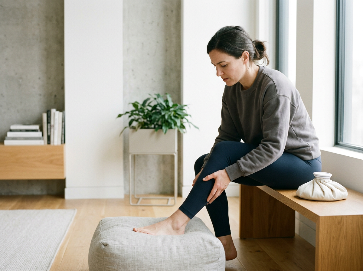
Shin pain can be devestating. At Rehab Mechanics, we are here to help!
The Runner's Breaking Point
Toronto has a massive, highly dedicated running community. When the weather clears, thousands of athletes hit the Martin Goodman Trail, the paths of High Park, and the pavement of Queen West. However, ramping up running mileage too quickly frequently leads to a deeply frustrating lower leg injury: pain radiating down the front or inside edge of the shin bone.
Initially, athletes brush it off as standard "shin splints." They stretch their calves, push through the dull ache, and assume their legs will eventually adapt. But when the pain suddenly changes—morphing from a broad, dull throb into a sharp, blinding, focal point of agony that hurts even when walking to the kitchen—panic sets in.
The terrifying question immediately arises: "Did I actually break a bone in my leg?"
At Rehab Mechanics, we specialize in high-level running biomechanics and differential diagnosis. The line between severe shin splints and a true Tibial Stress Fracture is incredibly thin. Both conditions exist on the exact same continuum of mechanical overload. By expertly identifying exactly where your shin bone is failing, we can implement targeted load management and biomechanical correction to save you from months in a walking boot.
Whether you are running in Trinity Bellwoods, down by our beautiful Lake Ontario, Liberty Village or jogging the historic streets of Parkdale, shin splints can make a great day miserable. Let’s lose the misery and regain the enjoyment of the runner’s high whether running solo or with a loved one!

The Cherry Blossoms in the Bellwoods
Structural Analysis: The Pathology of Tibial Overload
To understand how a muscle ache turns into a broken bone, we must perform a detailed structural analysis of how the lower leg absorbs the massive shock of running.
Medial Tibial Stress Syndrome (Shin Splints)
"Shin splints" is the layman’s term for Medial Tibial Stress Syndrome (MTSS). It is a traction (pulling) injury.
The Muscular Anchors: Your deep calf muscles (the soleus and tibialis posterior) attach directly to the periosteum—the thin, highly sensitive sheath of tissue that completely covers your shin bone (tibia).
The Whiplash Effect: If your foot severely overpronates (flattens) when it strikes the ground, these muscles are violently stretched. They yank aggressively on the periosteum with every single stride.
The Inflammation: This constant, violent tugging causes the periosteum to become severely inflamed, resulting in a dull, burning ache that spreads along the bottom third of the shin bone.
The Cortical Stress Fracture (The Bone Failure)
If you ignore MTSS and continue to run, the injury evolves from a muscle/fascia problem into a literal bone failure.
Bone Remodeling: Bone is living tissue. When you run, the impact creates microscopic damage (micro-cracks) in the bone. When you rest, specialized cells (osteoblasts) build new bone to patch the cracks, making the bone denser and stronger.
The Tipping Point: If you run too many miles without adequate rest days, the body cannot patch the micro-cracks fast enough. The microscopic damage accumulates in one specific spot on the tibia.
The Fracture: Eventually, these micro-cracks merge into a true stress fracture—a distinct crack in the cortical shell of the bone.
Identifying the Clinical Red Flags: MTSS vs. Fracture
How do our physiotherapists distinguish between an inflamed periosteum and a cracked bone without an MRI? We rely on highly specific mechanical loading tests.
The Palpation Test (The 5-Centimeter Rule): In standard shin splints, pressing your thumb along the inside of the shin bone causes a dull tenderness that is spread out over a large area (usually 5 to 10 centimeters long). If it is a stress fracture, the pain is exquisitely sharp, breathtaking, and localized to one exact, pinpoint spot (often the size of a dime).
The "Hop" Test: Patients with shin splints can usually perform 10 single-leg hops. It will ache, but it is manageable. A patient with a tibial stress fracture physically cannot hop on the leg due to sharp, shooting, structural bone pain.
Pain at Rest: Shin splints usually warm up during a run and throb afterward, but feel fine when resting on the couch. A stress fracture causes deep, unrelenting, throbbing bone pain even when you are lying in bed at night.

We may not be experiencing your pain but we feel it!
Primary Source Proof: Biomechanical Running Rehabilitation
Orthopedic sports medicine literature confirms that treating both MTSS and tibial stress fractures requires correcting the proximal kinematics (hip and pelvic control) and manipulating running cadence to reduce the vertical loading rate on the tibia.
Note: The link above directs to external, peer-reviewed medical literature demonstrating our commitment to evidence-based practice and international clinical guidelines for sports rehabilitation.
The Rehab Mechanics Corrective Protocol
If you have a true stress fracture, you must spend 4 to 8 weeks in a rigid CAM walking boot. However, the physical therapy to prevent it from happening again is identical to the treatment for severe shin splints. We must alter how your leg hits the ground.
Mechanical and scientific
Phase 1: Load Modification and Tissue Calming
We must stop the bone and periosteum from taking damage.
Relative Rest: Replacing pavement running with deep-water running, swimming, or stationary cycling to maintain elite cardiovascular fitness without vertical impact.
Advanced Modalities: Utilizing Extracorporeal Shockwave Therapy (ESWT) along the medial tibial border to stimulate massive local blood flow and accelerate cellular bone and fascial healing.
Phase 2: Pelvic Fortification (The Top-Down Fix)
If your hip is weak, your shin takes the punishment.
Gluteus Medius Activation: If your pelvis drops when you run, your knee caves inward, violently twisting the shin bone and ripping the calf muscles away from the tibia. We aggressively strengthen the lateral hip (banded lateral walks, single-leg deadlifts) to keep the leg perfectly straight during impact.
Heavy Calf Resistance: Rebuilding the soleus muscle using heavy, slow, bent-knee calf raises. A strong soleus acts as a massive shock absorber, taking the impact force before it reaches the shin bone.
Phase 3: Gait Retraining and Cadence Manipulation
Before you return to the Martin Goodman Trail, we fundamentally alter your running mechanics.
Cadence Optimization: We analyze your step rate. Over-striding (taking long, loping steps) causes a massive "braking force" that shatters the tibia. We use a metronome to increase your step cadence by 5% to 10%. Taking slightly shorter, faster steps drastically reduces the vertical impact force on the bone.
Custom Orthotics Assessment: If severe, rigid flat feet are the root cause, Sanjay is qualified to dispense custom medical orthotics to alter the mechanical pull on the tibial periosteum permanently.
Run Smarter, Not Harder
Do not attempt to "push through" pinpoint shin pain. Ignoring the warning signs of mechanical overload will result in a fractured bone and months of forced inactivity. By optimizing your running mechanics and building an indestructible pelvic foundation, expert physical therapy can eliminate shin splints and bulletproof your legs.
Avoiding the “dreaded” heel strike may be the cause
Related Conditions We Treat
Medial Tibial Stress Syndrome (MTSS)
Tibial Stress Fracture
Plantar Fasciitis
Achilles Tendinopathy
Patellofemoral Pain Syndrome
Hip Drop / Trendelenburg Gait
Overpronation / Flat Feet
Related Blogs
At the bottom, add:
Services Used in Treatments
Manual Therapy
Soft Tissue Release
Myofascial Release
Shockwave Therapy
Gait Retraining
Neuromuscular Re‑Education
Custom Orthotics
Strengthening Programs
FAQ’s

That is some frequency…
1. Is shin pain always shin splints?
No. Shin pain can come from several structures — the periosteum, the deep calf muscles, the tibialis posterior tendon, or the cortical bone itself. Shin splints (MTSS) create a diffuse, broad ache along the inside edge of the tibia. If the pain becomes sharp, pinpoint, and constant, it may indicate a tibial stress reaction or stress fracture.
2. How do I know if I have a stress fracture?
A stress fracture produces highly localized, focal pain that you can pinpoint with one finger. It often hurts during walking, standing, or even at rest. Patients with stress fractures typically cannot perform single‑leg hopping due to sharp, structural bone pain. Shin splints, by contrast, feel more spread out and usually improve slightly as you warm up.
3. Do I need an X‑ray or MRI to diagnose a stress fracture?
Not always. Physiotherapists use mechanical loading tests — palpation, hop testing, and rest‑pain assessment — to differentiate MTSS from a stress fracture with high accuracy. Imaging is recommended only when symptoms strongly suggest bone involvement or when pain persists despite proper load management.
4. Can physiotherapy fix shin splints?
Yes. Physiotherapy resolves shin splints by reducing periosteal irritation, correcting foot mechanics, strengthening the soleus and tibialis posterior, improving pelvic stability, and adjusting running cadence. These changes reduce the traction forces that cause MTSS and prevent the injury from progressing into a stress fracture.
5. How long does a stress fracture take to heal?
Most tibial stress fractures require 4–8 weeks of reduced impact loading, often supported by a CAM walking boot. During this period, physiotherapy focuses on maintaining cardiovascular fitness, restoring hip and calf strength, and correcting running mechanics to prevent recurrence once the bone has healed.
6. Should I stop running if I have shin pain?
If the pain is diffuse and improves as you warm up, you may be dealing with MTSS and can often continue training with modified mileage. If the pain becomes sharp, pinpoint, or present at rest, you must stop running immediately — continuing will worsen the bone stress and may lead to a full fracture.
7. Do flat feet or overpronation cause shin splints?
They can. Excessive pronation increases traction on the tibial periosteum, especially during long‑distance running. Custom orthotics, gait retraining, and strengthening the intrinsic foot muscles can significantly reduce this mechanical overload.
8. Why do shin splints happen when I increase mileage?
Your tibia experiences microscopic stress with every stride. If mileage increases too quickly, the bone does not have enough time to remodel and strengthen. This imbalance between impact load and bone recovery causes periosteal irritation (shin splints) and can progress to a stress fracture if ignored.
9. Can shockwave therapy help shin splints?
Yes. Shockwave therapy increases local blood flow, stimulates cellular repair, and accelerates healing along the medial tibial border. It is especially effective for chronic MTSS cases that have not responded to stretching or strengthening alone.
10. When should I see a physiotherapist?
You should seek assessment if shin pain:
lasts longer than 1–2 weeks
becomes sharp or pinpoint
hurts during walking or at rest
prevents you from running normally
worsens with increased mileage
returns every time you train
Early intervention prevents MTSS from progressing into a stress fracture.
How Physiotherapy Helps
Physiotherapy helps by:
reducing periosteal inflammation
correcting pelvic drop
improving cadence
strengthening soleus
reducing tibial loading
improving foot mechanics
Contact Us Today - All you have to lose is the pain
Book a comprehensive runner's biomechanical assessment with our clinical team today. We are conveniently located inside the Prime Medical Centre at 68 Abell Street, offering elite sports recovery in Queen West.
Contact us to schedule your appointment:
Email: info@rehabmechanics.com
Phone: (416) 533-3900
About the Author
Mr. Sanjay Attwala (B.Sc., M.Sc., RPT) is a Registered Physiotherapist, clinical director, and the founder of Rehab Mechanics in Toronto. With over 15 years of registered clinical practice and a deep specialization in complex musculoskeletal rehabilitation, Sanjay synthesizes rigorous international academic training with advanced evidence-based therapeutics to guide his clinical practice and patient education initiatives.
Academic Background & Credentials
Master of Science (M.Sc.) in Physiotherapy – University of Keele, United Kingdom (2010).
Bachelor of Science (B.Sc.) – University of Waterloo, Ontario, Canada.
Registered Physiotherapist (RPT) – Regulated health professional in excellent standing with the College of Physiotherapists of Ontario (CPO).
Corporate Entity – Operating officially under the S. Attwala Physiotherapy Professional Corporation with a DBA of Rehab Mechanics.
Clinical Expertise & Philosophy
Sanjay’s clinical approach rejects passive symptom management in favor of identifying underlying biomechanical root causes. His diverse expertise spans advanced manual therapies, personalized corrective exercise prescription, and modern physical modalities. At the Rehab Mechanics Toronto Queen West clinic, he routinely diagnoses and treats complex conditions including:
Spinal & Discogenic Pathology – Cervical, thoracic, and lumbar disc injuries, sciatica, and sacroiliac joint (SIJ) dysfunction.
Upper & Lower Extremity Injuries – Rotator cuff tears, frozen shoulder, tennis/golfer’s elbow, carpal tunnel syndrome, and complex ankle/foot pathologies.
Perinatal & Pelvic Health Rehabilitation – Specialized assessment and rehabilitation protocols tailored specifically for women during pregnancy and the post-partum period, addressing pelvic girdle pain, diastasis recti, and core stabilization.
Specialized Rehabilitation – Pelvic health therapy, TMJ dysfunction, post-surgical rehabilitation (including Total Hip and Total Knee Replacements), and custom orthotics dispensing.
Shockwave Therapy: with advanced cutting edge technological devices to suit your needs.
Interdisciplinary Practice & Patient Care
Sanjay practices an integrated model of healthcare, working closely alongside medical doctors inside the Prime Medical Centre on Abell Street to streamline patient recovery pathways. He maintains a human-centric, communication-first clinical framework, ensuring that care remains fully customized rather than automated.
His clinical caseload encompasses a broad operational spectrum under Ontario's regulatory frameworks, including:
Motor Vehicle Accident (MVA) Claims – Rehabilitation navigating Ontario’s statutory accident benefits schedule.
Workplace Safety and Insurance Board (WSIB) – Occupational injury management and return-to-work screening.
Extended Health Care (EHC) & Private Practice – Multi-tier insurance coordination and long-term athletic development plans.
Commitment to Research & Community
Outside of his clinical caseload at Rehab Mechanics and his additional practice affiliations in Etobicoke, Sanjay is an active health writer and community educator. He translates contemporary peer-reviewed medical research into accessible, actionable guidance on his professional blog. As a dedicated father and husband, he mirrors his professional advice in his personal life, focusing on structural mobility, cross-training, and longevity to help his family and his community thrive. Naturally he takes he a keen interest in rehabilitation for women who are pregnant and post-partum.
Disclaimer: The information provided on this blog is intended for educational and informational purposes only. It does not constitute medical advice, diagnosis, or a treatment plan. Always seek the direct advice of a Registered Physiotherapist, physician, or other qualified health provider regarding any medical condition or physical rehabilitation routine.
Be sure to check out Rehab Mechanics’ Google My Business Profile for daily posts, location and directions in addition to the directions on the website!
We love interacting with prospective and current patients, and love honest feedback and even constructive criticism and areas for improvement!
Is a Bony Heel Spur the Real Cause of My Foot Pain?
No. A heel spur is a biological response to tension, not the root cause of pain. The pain comes from the micro-tearing of the plantar fascia attached to it. Physiotherapy cures this by restoring ankle mobility, strengthening the foot arch, and using shockwave therapy to heal the fascia without surgical spur removal.
The X-Ray That Causes Panic
For active Torontonians—whether you are a nurse working a 12-hour shift at a downtown hospital or a commuter walking blocks to the King streetcar—waking up with sharp, stabbing pain in the bottom of your foot is an agonizing way to start the day.
When the pain persists for weeks, patients inevitably visit a walk-in clinic and request an X-ray. The doctor looks at the film, points to a sharp, jagged piece of bone growing out of the bottom of the heel, and says, "You have a heel spur."
The immediate, terrifying assumption is that this sharp spike of bone is literally stabbing into the flesh of the foot with every step, like a nail inside a shoe. Patients assume that the only way to stop the pain is to undergo invasive foot surgery to chisel the bone spur away.
At Rehab Mechanics in Queen West, we want to completely dismantle this myth. The heel spur is almost never the actual pain generator. It is merely a symptom of a much larger biomechanical failure happening in the arch of your foot. By utilizing advanced human mechanics physical therapy to rehabilitate the plantar fascia, we can permanently eliminate your foot pain—even while the bone spur remains perfectly intact on your X-ray.
Structural Analysis: The Mechanics of the Heel Spur Myth
To understand why shaving down the bone is unnecessary, we must perform a detailed biomechanical analysis of the bottom of your foot and why the body decides to grow extra bone in the first place.
The Windlass Mechanism and the Plantar Fascia
Your foot is stabilized by a massive, incredibly thick band of connective tissue called the plantar fascia. It anchors into the bottom of your heel bone (the calcaneus) and fans out to attach to the base of your toes.
The Shock Absorber: When you step down on the hard Toronto pavement, your arch flattens, stretching the plantar fascia to absorb the impact.
The Lever: When you push off your toes to walk, the fascia winds tight (the windlass mechanism), turning your foot into a rigid, powerful lever.
The Pathology of a Heel Spur (Wolff's Law)
A heel spur is not a random, malicious growth. It is a highly predictable biological adaptation governed by a principle called Wolff’s Law, which states that bone grows and thickens in response to the mechanical stress placed upon it.
The Chronic Overload
If your foot mechanics are faulty—such as extreme overpronation (flat feet) or incredibly stiff ankles—the plantar fascia is violently stretched beyond its capacity with every single step.
The Tugging Force: This creates a massive, chronic pulling force directly at the anchor point on the heel bone.
The Micro-Tearing: The fascia begins to suffer microscopic tears at the attachment site (Plantar Fasciopathy).
The Biological "Cement": Because the fascia is constantly ripping away from the bone, the body panics. It attempts to reinforce the attachment site by laying down extra calcium. Over years, this calcium hardens into a bony hook—the heel spur.
The True Pain Generator
The pain you feel is not the bone spur stabbing you. The spur actually grows horizontally, in the exact direction of the fascia, not vertically into the skin.
The excruciating, glass-like pain you feel in the morning is the inflamed, highly sensitive, micro-torn plantar fascia tissue stretching and ripping after stiffening up overnight. If a surgeon removes the spur but you do not fix the tight calf and weak arch, the fascia will simply tear again.
Identifying the Clinical Reality
Massive clinical studies have X-rayed thousands of people with absolutely zero foot pain. A staggering percentage of them have massive heel spurs and don't even know it. Conversely, many patients with excruciating plantar fasciitis have perfectly smooth heel bones. The spur is evidence of past tension, not the current cause of pain.
Primary Source Proof: Non-Operative Fascial Rehabilitation
Orthopedic and podiatric research confirms that heavy-load strength training and extracorporeal shockwave therapy target the cellular degradation of the fascia directly, resulting in profound pain resolution without the need for surgical spur excision.
Note: The link above directs to external, peer-reviewed medical literature demonstrating our commitment to evidence-based practice and international clinical guidelines for foot and ankle rehabilitation.
The Rehab Mechanics Fascial Reconstruction Protocol
We do not treat the bone; we treat the failing mechanical tissue attached to it. Our protocol aggressively targets the cellular health of the plantar fascia.
Phase 1: Cellular Regeneration and Decompression (Weeks 1-3)
We must force the degenerated, scarred fascial tissue to heal.
Extracorporeal Shockwave Therapy (ESWT): Our physiotherapists utilize high-energy acoustic sound waves. This mechanical energy blasts through the fibrotic scar tissue at the heel and stimulates profound neovascularization (the growth of brand-new blood vessels), finally allowing the starving fascia to heal.
Calf and Ankle Unlocking: If your calf muscles (gastrocnemius and soleus) are tight, your ankle cannot bend forward. This forces your foot to severely overpronate to compensate, ripping at the fascia. We use deep myofascial release and joint mobilizations to restore full ankle dorsiflexion, instantly taking the pulling force off the heel.
Phase 2: High-Load Fascial Remodeling (Weeks 4-8)
Passive stretching is useless for a degenerated tendon or fascia. It must be loaded heavily to rebuild its structural integrity.
The Rathleff Protocol: We implement a highly specific, evidence-based strengthening protocol. You will perform heavy, slow calf raises with a rolled-up towel placed specifically under your toes. This pre-stretches the fascia (engaging the windlass mechanism) while you lift your body weight, forcing the body to lay down incredibly strong, parallel collagen fibers.
Phase 3: Intrinsic Support and Mechanical Offloading
We must build biological arch support so the fascia is never overloaded again.
Intrinsic Foot Muscle Activation: We prescribe targeted micro-movements (like "foot doming") to resurrect the tiny muscles deep inside the foot. These muscles act as a suspension bridge, actively holding the arch up and taking the stress off the plantar fascia.
Custom Orthotic Dispensing: If your foot architecture is fundamentally collapsed, Sanjay is licensed to cast and dispense true custom medical orthotics. These devices mechanically block the arch from collapsing, acting as a crucial brace while the tissue heals.
Walk Pain-Free, Keep Your Heel Bone
You do not have to live with the morning hobble or subject yourself to risky foot surgery to remove a harmless bone spur. By fixing the biomechanics of your ankle and heavily rehabilitating the plantar fascia, expert physical therapy can permanently resolve your heel pain.
Book a comprehensive foot and ankle assessment with our clinical team today. We are conveniently located inside the Prime Medical Centre at 68 Abell Street, offering advanced orthopedic recovery in Queen West.
Contact us to schedule your appointment:
Email: info@rehabmechanics.com
Phone: (416) 533-3900
About the Author
Mr. Sanjay Attwala (B.Sc., M.Sc., RPT) is a Registered Physiotherapist, clinical director, and the founder of Rehab Mechanics in Toronto. With over 15 years of registered clinical practice and a deep specialization in complex musculoskeletal rehabilitation, Sanjay synthesizes rigorous international academic training with advanced evidence-based therapeutics to guide his clinical practice and patient education initiatives.
Academic Background & Credentials
Master of Science (M.Sc.) in Physiotherapy – University of Keele, United Kingdom (2010).
Bachelor of Science (B.Sc.) – University of Waterloo, Ontario, Canada.
Registered Physiotherapist (RPT) – Regulated health professional in excellent standing with the College of Physiotherapists of Ontario (CPO).
Corporate Entity – Operating officially under the S. Attwala Physiotherapy Professional Corporation with a DBA of Rehab Mechanics.
Clinical Expertise & Philosophy
Sanjay’s clinical approach rejects passive symptom management in favor of identifying underlying biomechanical root causes. His diverse expertise spans advanced manual therapies, personalized corrective exercise prescription, and modern physical modalities. At the Rehab Mechanics Toronto Queen West clinic, he routinely diagnoses and treats complex conditions including:
Spinal & Discogenic Pathology – Cervical, thoracic, and lumbar disc injuries, sciatica, and sacroiliac joint (SIJ) dysfunction.
Upper & Lower Extremity Injuries – Rotator cuff tears, frozen shoulder, tennis/golfer’s elbow, carpal tunnel syndrome, and complex ankle/foot pathologies.
Perinatal & Pelvic Health Rehabilitation – Specialized assessment and rehabilitation protocols tailored specifically for women during pregnancy and the post-partum period, addressing pelvic girdle pain, diastasis recti, and core stabilization.
Specialized Rehabilitation – Pelvic health therapy, TMJ dysfunction, post-surgical rehabilitation (including Total Hip and Total Knee Replacements), and custom orthotics dispensing.
Shockwave Therapy: with advanced cutting edge technological devices to suit your needs.
Interdisciplinary Practice & Patient Care
Sanjay practices an integrated model of healthcare, working closely alongside medical doctors inside the Prime Medical Centre on Abell Street to streamline patient recovery pathways. He maintains a human-centric, communication-first clinical framework, ensuring that care remains fully customized rather than automated.
His clinical caseload encompasses a broad operational spectrum under Ontario's regulatory frameworks, including:
Motor Vehicle Accident (MVA) Claims – Rehabilitation navigating Ontario’s statutory accident benefits schedule.
Workplace Safety and Insurance Board (WSIB) – Occupational injury management and return-to-work screening.
Extended Health Care (EHC) & Private Practice – Multi-tier insurance coordination and long-term athletic development plans.
Commitment to Research & Community
Outside of his clinical caseload at Rehab Mechanics and his additional practice affiliations in Etobicoke, Sanjay is an active health writer and community educator. He translates contemporary peer-reviewed medical research into accessible, actionable guidance on his professional blog. As a dedicated father and husband, he mirrors his professional advice in his personal life, focusing on structural mobility, cross-training, and longevity to help his family and his community thrive. Naturally he takes he a keen interest in rehabilitation for women who are pregnant and post-partum.
Disclaimer: The information provided on this blog is intended for educational and informational purposes only. It does not constitute medical advice, diagnosis, or a treatment plan. Always seek the direct advice of a Registered Physiotherapist, physician, or other qualified health provider regarding any medical condition or physical rehabilitation routine.
Be sure to check out Rehab Mechanics’ Google My Business Profile for daily posts, location and directions in addition to the directions on the website!
We love interacting with prospective and current patients, and love honest feedback and even constructive criticism and areas for improvement!
Can My Insurance Company Deny Long-Term Disability for Chronic Pain?
Yes, insurers routinely deny Long-Term Disability (LTD) claims for chronic pain due to a "lack of objective evidence." Overturning these denials requires expert medical-legal strategy, utilizing functional capacity evaluations to prove how central sensitization legally impairs your ability to work.
The Insurance Industry's "Broken Part" Fallacy
If you are a professional living in Toronto, your ability to earn a living relies entirely on your physical and cognitive stamina. When an injury or illness occurs, you expect the Long-Term Disability (LTD) insurance policy you have paid into for years to protect you.
However, if your disability is driven by conditions like Fibromyalgia, Complex Regional Pain Syndrome (CRPS), or chronic, non-specific lower back pain, you are about to face a brutal legal battle.
Insurance companies operate on a highly outdated, "broken part" model of medicine. They want to see a severed limb, a massive tumor, or a shattered spine on an X-ray. If you submit a claim for severe, debilitating chronic pain, but your MRI scans come back relatively "normal," the insurance adjuster will almost certainly send you a denial letter citing a "lack of objective medical evidence."
At LawCap, we specialize in litigating complex, "invisible" disability claims. We understand that chronic pain is not a psychological invention; it is a profound, documentable failure of the central nervous system. Defeating an insurance company requires shifting the legal argument away from the initial injury and focusing entirely on the biological reality of pain sensitization and functional impairment.
Structural Analysis: The Reality of Central Sensitization
To force an insurer to reinstate your benefits, your legal team must clearly explain the modern medical science of chronic pain to mediators and judges.
Acute Pain vs. Chronic Pain
Acute Pain: This is an alarm system. If you touch a hot stove, the nerves in your hand send a rapid signal to your brain to pull away. Once the burn heals, the alarm turns off.
Chronic Pain: This occurs when the alarm system breaks and gets permanently stuck in the "on" position, long after the original tissue injury (like a car accident or a surgical procedure) has structurally healed.
The Biology of Central Sensitization (Neurological Wind-Up)
When pain signals bombard the central nervous system for months on end, the spinal cord and the brain physically change.
The Lowered Threshold: The nervous system becomes hyper-excitable. The threshold for what the brain perceives as "dangerous" drops drastically.
Allodynia and Hyperalgesia: Eventually, completely normal, non-threatening stimuli—like the light brush of clothing against your skin, sitting in an office chair, or the mild stress of a workplace meeting—are interpreted by your brain as agonizing pain.
The Legal Implication: The insurance company is looking for a broken bone to explain the pain. We must prove to them that the nervous system itself is the broken part.
The Legal Battle: Why Insurers Deny Chronic Pain
Insurance companies utilize several highly aggressive tactics to undermine and deny chronic pain claims in Ontario.
The Independent Medical Examination (IME)
The insurer will force you to attend an assessment with their own "independent" doctor. These doctors are paid by the insurance company. They will conduct a brief, 15-minute physical exam, note that your joints move normally and your reflexes are intact, and author a report claiming you are perfectly fit to return to your desk job.
The Surveillance Trap
Insurers frequently hire private investigators to film you in public. If they catch you carrying a small bag of groceries in Queen West or briefly smiling while walking your dog in Trinity Bellwoods, they will use this footage to argue that your pain is exaggerated and that you are perfectly capable of working 40 hours a week.
Winning the Appeal: Medical-Legal Documentation
To overturn a denial, we cannot simply rely on notes from your family doctor saying "the patient is in pain." We must build an overwhelming, objective wall of functional evidence.
1. Functional Capacity Evaluations (FCE)
This is our most powerful legal tool. A specialized occupational therapist will test your physical and cognitive limits over a grueling 1 to 2-day period.
The FCE tests your ability to sit, stand, concentrate, and handle stress over time.
It documents your heart rate spikes, your muscle spasms, and your cognitive degradation as the pain flares up.
This provides the court with hard, objective data proving that while you might be able to lift a coffee mug once, you absolutely cannot sustain the focus or posture required for an 8-hour workday.
2. The Biopsychosocial Approach
Chronic pain destroys your sleep, triggers severe depression, and fuels anxiety. We bring in specialized psychologists and psychiatrists to document the mental and emotional fallout of your physical pain. By proving that the pain has fundamentally altered your psychological capacity to handle a workplace environment, we build an airtight case for total disability.
Do Not Let an Insurer Invalidate Your Pain
Living with chronic pain is exhausting enough without having to fight a multi-billion-dollar insurance corporation for the benefits you are rightfully owed. If your LTD claim has been denied due to a "lack of objective evidence," you need a legal strategy that bridges the gap between modern pain science and Canadian disability law.
About the Author
Mr. Sanjay Attwala (B.Sc., M.Sc., RPT) is a Registered Physiotherapist, clinical director, and the founder of Rehab Mechanics in Toronto. With over 15 years of registered clinical practice and a deep specialization in complex musculoskeletal rehabilitation, Sanjay synthesizes rigorous international academic training with advanced evidence-based therapeutics to guide his clinical practice and patient education initiatives.
Academic Background & Credentials
Master of Science (M.Sc.) in Physiotherapy – University of Keele, United Kingdom (2010).
Bachelor of Science (B.Sc.) – University of Waterloo, Ontario, Canada.
Registered Physiotherapist (RPT) – Regulated health professional in excellent standing with the College of Physiotherapists of Ontario (CPO).
Corporate Entity – Operating officially under the S. Attwala Physiotherapy Professional Corporation with a DBA of Rehab Mechanics.
Clinical Expertise & Philosophy
Sanjay’s clinical approach rejects passive symptom management in favor of identifying underlying biomechanical root causes. His diverse expertise spans advanced manual therapies, personalized corrective exercise prescription, and modern physical modalities. At the Rehab Mechanics Toronto Queen West clinic, he routinely diagnoses and treats complex conditions including:
Spinal & Discogenic Pathology – Cervical, thoracic, and lumbar disc injuries, sciatica, and sacroiliac joint (SIJ) dysfunction.
Upper & Lower Extremity Injuries – Rotator cuff tears, frozen shoulder, tennis/golfer’s elbow, carpal tunnel syndrome, and complex ankle/foot pathologies.
Perinatal & Pelvic Health Rehabilitation – Specialized assessment and rehabilitation protocols tailored specifically for women during pregnancy and the post-partum period, addressing pelvic girdle pain, diastasis recti, and core stabilization.
Specialized Rehabilitation – Pelvic health therapy, TMJ dysfunction, post-surgical rehabilitation (including Total Hip and Total Knee Replacements), and custom orthotics dispensing.
Shockwave Therapy: with advanced cutting edge technological devices to suit your needs.
Interdisciplinary Practice & Patient Care
Sanjay practices an integrated model of healthcare, working closely alongside medical doctors inside the Prime Medical Centre on Abell Street to streamline patient recovery pathways. He maintains a human-centric, communication-first clinical framework, ensuring that care remains fully customized rather than automated.
His clinical caseload encompasses a broad operational spectrum under Ontario's regulatory frameworks, including:
Motor Vehicle Accident (MVA) Claims – Rehabilitation navigating Ontario’s statutory accident benefits schedule.
Workplace Safety and Insurance Board (WSIB) – Occupational injury management and return-to-work screening.
Extended Health Care (EHC) & Private Practice – Multi-tier insurance coordination and long-term athletic development plans.
Commitment to Research & Community
Outside of his clinical caseload at Rehab Mechanics and his additional practice affiliations in Etobicoke, Sanjay is an active health writer and community educator. He translates contemporary peer-reviewed medical research into accessible, actionable guidance on his professional blog. As a dedicated father and husband, he mirrors his professional advice in his personal life, focusing on structural mobility, cross-training, and longevity to help his family and his community thrive. Naturally he takes he a keen interest in rehabilitation for women who are pregnant and post-partum.
Disclaimer: The information provided on this blog is intended for educational and informational purposes only. It does not constitute medical advice, diagnosis, or a treatment plan. Always seek the direct advice of a Registered Physiotherapist, physician, or other qualified health provider regarding any medical condition or physical rehabilitation routine.
Be sure to check out Rehab Mechanics’ Google My Business Profile for daily posts, location and directions in addition to the directions on the website!
We love interacting with prospective and current patients, and love honest feedback and even constructive criticism and areas for improvement!
Will a "Minor" Concussion From a Car Accident Affect My Injury Settlement?
Yes. Insurance companies frequently dismiss concussions as "minor," but Post-Concussion Syndrome causes severe, long-term neurological deficits. Securing a fair settlement requires aggressive medical-legal documentation linking your biomechanical brain trauma directly to your loss of income and diminished quality of life.
The "Invisible Injury" Bias in Ontario Auto Claims
If you have been involved in a motor vehicle accident (MVA) on the Gardiner Expressway or a sudden collision at a busy downtown Toronto intersection, the immediate focus is usually on visible injuries: broken bones, lacerations, or crushed joints. When the paramedics arrive, if you are conscious and speaking, you are often discharged with a diagnosis of a "mild concussion" or "minor whiplash."
Unfortunately, this early medical classification is frequently weaponized by auto insurance companies.
Under Ontario's Statutory Accident Benefits Schedule (SABS), insurers default to placing "mild" brain injuries into the Minor Injury Guideline (MIG), which severely caps your available rehabilitation funds to a fraction of what you actually need. They expect you to rest for a few weeks and return to work.
However, at LawCap, we know that a concussion is never "minor." Months after the accident, you may still be suffering from blinding light sensitivity, severe vertigo, cognitive fog, and an inability to stare at a computer screen for more than ten minutes. This is Post-Concussion Syndrome (PCS). To force the insurance company to properly value your tort claim and accident benefits, we must legally prove the profound, invisible biomechanical damage inside your skull.
Structural Analysis: The Neurometabolic Cascade of a Concussion
To legally defeat an insurance adjuster’s claim that you are "faking" or exaggerating your symptoms, your legal team must clearly articulate the objective, biological reality of a traumatic brain injury (TBI).
The Physics of the Impact
A concussion does not require your head to physically strike the steering wheel or the window. It is caused by acceleration and deceleration forces.
The Coup-Contrecoup: When your car is rear-ended, your skull is whipped violently forward and backward. Your brain, which floats in cerebrospinal fluid, crashes into the hard, bony inside of your skull, then rebounds and crashes into the opposite side.
Axonal Shearing: This violent shaking causes the brain tissue to twist. The delicate, microscopic nerve fibers (axons) that connect different regions of your brain are stretched, frayed, and sheared.
The Energy Crisis (Why MRI Scans Are Often "Normal")
Insurance companies love to point out that your CT scan or MRI came back "clear" as proof that you are not injured. This is a deliberate medical-legal fallacy.
Standard MRIs look for macro-structural damage, like massive bleeding or tumors. They cannot see microscopic axonal shearing or cellular chemical imbalances.
The Chemical Chaos: The physical shearing of the cells causes massive amounts of toxic chemicals (like glutamate) to flood the brain. The brain goes into an absolute panic, demanding massive amounts of energy (glucose) to repair the cells.
The Chokehold: However, the trauma of the accident temporarily restricts cerebral blood flow. The brain is starving for energy precisely when it needs it most. This metabolic mismatch is the exact physiological cause of the exhaustion, confusion, and chronic headaches that prevent you from working.
The Legal Framework: Breaking Out of the Minor Injury Guideline
In Ontario, dealing with an MVA claim involves navigating a two-part system: your Accident Benefits (no-fault) and your Tort Claim (suing the at-fault driver).
Accident Benefits (SABS)
If your insurer places you in the Minor Injury Guideline (MIG), your medical and rehabilitation benefits are drastically capped.
The Legal Strategy: To access higher funding tiers (Non-Catastrophic or Catastrophic Impairment), your legal team must assemble a network of specialized medical experts—such as neuropsychologists, vestibular physiotherapists, and occupational therapists.
We must generate hard, objective data proving that your concussion has resulted in a neurological deficit that falls outside the legal definition of a "minor" injury.
The Tort Claim (Loss of Competitive Advantage)
If your concussion prevents you from returning to your fast-paced corporate job in the Financial District, or if you can no longer handle the multitasking required to manage a busy household, you are entitled to compensation for lost future earning capacity.
Insurance defense lawyers will argue that your cognitive fog is just "stress" or a pre-existing psychological issue.
Our role is to utilize complex neurocognitive testing to prove a direct, undeniable causal link between the biomechanical trauma of the crash and your current inability to compete in the open labor market.
Proving the Invisible: How We Build the Case
To secure the maximum settlement value for a traumatic brain injury, we leave nothing to chance. We combat insurance skepticism with overwhelming, interdisciplinary medical evidence.
Functional Capacity Evaluations (FCE)
A subjective complaint of "I feel dizzy" is easily dismissed by an adjuster. We utilize objective Functional Capacity Evaluations to quantify exactly how your brain injury limits you. If your heart rate spikes and your balance fails after five minutes of reading, the FCE generates the hard data required to prove you cannot sustain an 8-hour workday.
Neuro-Optometry and Vestibular Mapping
A massive percentage of post-concussion syndrome is driven by damage to the vestibular system (inner ear) and the oculomotor nerve pathways. We partner with specialized clinicians who can map the exact tracking delays in your eye movements, providing undeniable, objective proof of the brain injury.
Protect Your Legal Rights and Your Future
A concussion is a complex neurological event that can derail your career and your quality of life. Do not let an insurance company rush you into an early, undervalued settlement by dismissing your symptoms as a "minor headache."
You need a legal team that understands the deep human mechanics of brain trauma and knows how to translate that biological reality into maximum legal compensation. We work in tandem with them all under the umbrella of litigation privilege.

About the Author
Mr. Sanjay Attwala (B.Sc., M.Sc., RPT) is a Registered Physiotherapist, clinical director, and the founder of Rehab Mechanics in Toronto. With over 15 years of registered clinical practice and a deep specialization in complex musculoskeletal rehabilitation, Sanjay synthesizes rigorous international academic training with advanced evidence-based therapeutics to guide his clinical practice and patient education initiatives.
Academic Background & Credentials
Master of Science (M.Sc.) in Physiotherapy – University of Keele, United Kingdom (2010).
Bachelor of Science (B.Sc.) – University of Waterloo, Ontario, Canada.
Registered Physiotherapist (RPT) – Regulated health professional in excellent standing with the College of Physiotherapists of Ontario (CPO).
Corporate Entity – Operating officially under the S. Attwala Physiotherapy Professional Corporation with a DBA of Rehab Mechanics.
Clinical Expertise & Philosophy
Sanjay’s clinical approach rejects passive symptom management in favor of identifying underlying biomechanical root causes. His diverse expertise spans advanced manual therapies, personalized corrective exercise prescription, and modern physical modalities. At the Rehab Mechanics Toronto Queen West clinic, he routinely diagnoses and treats complex conditions including:
Spinal & Discogenic Pathology – Cervical, thoracic, and lumbar disc injuries, sciatica, and sacroiliac joint (SIJ) dysfunction.
Upper & Lower Extremity Injuries – Rotator cuff tears, frozen shoulder, tennis/golfer’s elbow, carpal tunnel syndrome, and complex ankle/foot pathologies.
Perinatal & Pelvic Health Rehabilitation – Specialized assessment and rehabilitation protocols tailored specifically for women during pregnancy and the post-partum period, addressing pelvic girdle pain, diastasis recti, and core stabilization.
Specialized Rehabilitation – Pelvic health therapy, TMJ dysfunction, post-surgical rehabilitation (including Total Hip and Total Knee Replacements), and custom orthotics dispensing.
Shockwave Therapy: with advanced cutting edge technological devices to suit your needs.
Interdisciplinary Practice & Patient Care
Sanjay practices an integrated model of healthcare, working closely alongside medical doctors inside the Prime Medical Centre on Abell Street to streamline patient recovery pathways. He maintains a human-centric, communication-first clinical framework, ensuring that care remains fully customized rather than automated.
His clinical caseload encompasses a broad operational spectrum under Ontario's regulatory frameworks, including:
Motor Vehicle Accident (MVA) Claims – Rehabilitation navigating Ontario’s statutory accident benefits schedule.
Workplace Safety and Insurance Board (WSIB) – Occupational injury management and return-to-work screening.
Extended Health Care (EHC) & Private Practice – Multi-tier insurance coordination and long-term athletic development plans.
Commitment to Research & Community
Outside of his clinical caseload at Rehab Mechanics and his additional practice affiliations in Etobicoke, Sanjay is an active health writer and community educator. He translates contemporary peer-reviewed medical research into accessible, actionable guidance on his professional blog. As a dedicated father and husband, he mirrors his professional advice in his personal life, focusing on structural mobility, cross-training, and longevity to help his family and his community thrive. Naturally he takes he a keen interest in rehabilitation for women who are pregnant and post-partum.
Disclaimer: The information provided on this blog is intended for educational and informational purposes only. It does not constitute medical advice, diagnosis, or a treatment plan. Always seek the direct advice of a Registered Physiotherapist, physician, or other qualified health provider regarding any medical condition or physical rehabilitation routine.
Be sure to check out Rehab Mechanics’ Google My Business Profile for daily posts, location and directions in addition to the directions on the website!
We love interacting with prospective and current patients, and love honest feedback and even constructive criticism and areas for improvement!
Can a Herniated Disc in My Lower Back Heal on Its Own Without Surgery?
Yes. Through a biological process called spontaneous resorption, the body's immune system can literally "eat" the herniated disc material. Physiotherapy accelerates this by utilizing directional preference therapy and spinal decompression to centralize nerve pain while the disc structurally heals.
The Terror of the MRI Report
From this….
To this with Rehab Mechanics
There are few diagnoses in modern medicine that strike as much fear into patients as a "herniated disc." If you have been struggling with a sharp, burning pain shooting from your lower back, down your glute, and into your calf (sciatica), you likely pushed for an MRI.
When the radiologist's report returns with terrifying words like "disc extrusion," "protrusion," or "sequestration," the immediate, panic-inducing assumption is that your spine is permanently broken. Patients assume that the piece of disc material currently crushing their spinal nerve will sit there forever unless a neurosurgeon physically cuts it out.
At Rehab Mechanics in Queen West, we specialize in advanced spinal pathology, and we want to deliver a message of profound clinical hope: your body has the miraculous ability to heal itself.
While severe neurological deficits (like losing control of your bowel/bladder or the ability to lift your foot) represent absolute surgical emergencies, the vast majority of severe lumbar disc herniations can naturally regress and disappear. By utilizing highly specific biomechanical physiotherapy, we can manage the crushing nerve pain while your immune system actively "eats" the herniated disc material, keeping you far away from the operating room.
Structural Analysis: The Biology of Disc Resorption
To understand how a "slipped disc" can disappear, we must dive deep into the cellular biology of your immune system and the unique anatomy of the intervertebral disc.
The Anatomy of the Herniation
Your spinal discs act as shock absorbers between your vertebrae.
The Nucleus Pulposus: The soft, highly pressurized, jelly-like center of the disc.
The Annulus Fibrosus: The tough, fibrous outer ring that holds the jelly in place.
When you bend forward heavily or lift with poor posture, the massive pressure forces the inner jelly backward. Over time, it tears through the outer ring and squirts out into the spinal canal. This is a disc herniation. The extruded jelly physically crushes the adjacent sciatic nerve root.
The Miracle of Spontaneous Resorption
For decades, surgeons believed that once the jelly was out, it stayed out. Modern MRI studies have proven this completely false. In fact, massive disc extrusions are more likely to heal spontaneously than small bulges. Why? Because of an aggressive immune response.
The Immune System Attack
The jelly inside your spinal disc is a closed system; it has never been exposed to your bloodstream.
The Alien Invader: When the disc herniates and the jelly bursts into the highly vascularized spinal canal, your body's immune system does not recognize it. It views the disc material as a hostile, foreign invader, like a virus or a splinter.
The Macrophage Swarm: The immune system immediately launches a massive inflammatory attack. It sends thousands of specialized white blood cells called macrophages to the site.
Phagocytosis (The Cleanup): The word macrophage literally means "big eater." These cells swarm the extruded disc material, secrete enzymes to break it down, and physically digest (phagocytize) the herniated jelly.
The Shrinking Mass: Over a period of weeks to months, the immune system literally eats the herniation away. The mass shrinks, pulls away from the spinal nerve, and the excruciating sciatica vanishes.
Identifying the Clinical Timeline: Mechanical vs. Chemical Pain
While the immune system is eating the disc, you are still in severe pain. We must manage the symptoms during the biological healing window.
Chemical Pain (Weeks 1-4): The initial, blinding pain is heavily driven by the massive chemical inflammation of the immune attack itself. This pain is constant and severe.
Mechanical Pain (Weeks 4-12): As the inflammation settles, the pain becomes purely mechanical—meaning it only hurts when you move into positions that pinch the remaining disc material against the nerve.
Primary Source Proof: Spontaneous Disc Resorption
Extensive neurological and orthopedic research universally confirms that the phenomenon of spontaneous regression of herniated disc tissue is highly prevalent, making conservative physiotherapy the mandated first-line treatment over early surgical intervention.
Note: The link above directs to external, peer-reviewed medical literature demonstrating our commitment to evidence-based practice and international clinical guidelines for spinal rehabilitation.
Clinical evidence indicates that lumbar disc herniations may undergo spontaneous resorption with conservative management, with reported overall rates of approximately two-thirds to 70%. This process most commonly occurs within the first three to six months and is mediated by inflammatory activity, neovascularization, macrophage infiltration and phagocytosis, enzymatic matrix degradation, and related cellular mechanisms such as apoptosis and autophagy.
The likelihood of spontaneous regression varies substantially according to herniation morphology. Sequestered fragments demonstrate the highest probability of regression, reported at approximately 88% to 96%, followed by extrusions at approximately 66% to 70%, protrusions at approximately 38% to 41%, and disc bulges at approximately 13%.
The principal biological mechanism appears to involve immune recognition of exposed nucleus pulposus material as foreign tissue, prompting inflammatory recruitment and macrophage-mediated breakdown of the herniated fragment. Larger, extruded, and sequestered disc fragments tend to provoke a stronger inflammatory and vascular response, which may explain their greater tendency toward resorption. Although the timeline for radiological resolution varies among patients, clinical improvement often precedes complete imaging-confirmed regression.
For a more detailed discussion of the underlying physiological mechanisms, see the review on spontaneous resorption of lumbar disc herniation available through NIH PubMed Central. For population-level incidence data, refer to the published meta-analyses on the incidence of spontaneous resorption of lumbar disc herniation indexed in PubMed.
The Rehab Mechanics Decompression Protocol
We cannot make your immune system eat the disc faster, but we can mechanically alter the pressure in your spine to pull the disc away from the nerve and stop the agonizing leg pain while nature does its work.
Phase 1: Centralization and Directional Preference
Our absolute first priority is to "centralize" the pain—meaning we want to draw the burning pain out of your calf/foot and move it back up to your lower back.
The McKenzie Method: We utilize highly specific, repeated movements to alter the hydraulic pressure inside the disc. For a posterior herniation, we typically use repeated lumbar extensions (like a sloppy push-up or "cobra" pose). This movement physically milks the remaining disc jelly forward, away from the crushed nerve root.
Lateral Shift Correction: If the disc is pushing you completely crooked, we manually glide your hips back under your shoulders to safely clear the mechanical block before extending the spine.
Phase 2: Spinal Unloading and Core Bracing
We must protect the healing tear in the outer disc wall.
Spinal Decompression: Utilizing targeted manual traction to physically separate the vertebrae, relieving the ischemic (blood-starved) nerve roots.
Intra-Abdominal Pressure (IAP): We teach you how to fire your deep transversus abdominis to create a biological weight belt. When contracted, this muscle rigidifies the spine, preventing the micro-movements that aggravate the healing disc wall.
Phase 3: Hip Hinge Mechanics and Functional Return
To ensure you never herniate a disc again, we must fundamentally change how you move through the world.
The Hip Hinge: We meticulously teach you how to bend forward by pushing your hips back (like a deadlift), rather than rounding your lower spine. By relying entirely on massive glute and hamstring power, we ensure the lumbar spine remains a rigid, unmoving lever during athletic and daily lifting activities.
Fear-Avoidance Eradication: We systematically expose you to the movements you fear most, proving to your brain that your spine is healed, strong, and capable of handling load.
Trust Your Body's Capacity to Heal
A herniated disc is a severe injury, but it is not a permanent sentence. By managing the mechanical pressure on your nerves and allowing your immune system to aggressively resorb the herniated material, expert physical therapy can guide you to a complete recovery without spinal surgery.
Book an immediate, comprehensive spinal diagnostic assessment with our clinical team today. We are conveniently located inside the Prime Medical Centre at 68 Abell Street, offering elite, rapid-response orthopedic care in Toronto Queen West.
Contact us to schedule your appointment:
Email: info@rehabmechanics.com
Phone: (416) 533-3900
About the Author
Mr. Sanjay Attwala (B.Sc., M.Sc., RPT) is a Registered Physiotherapist, clinical director, and the founder of Rehab Mechanics in Toronto. With over 15 years of registered clinical practice and a deep specialization in complex musculoskeletal rehabilitation, Sanjay synthesizes rigorous international academic training with advanced evidence-based therapeutics to guide his clinical practice and patient education initiatives.
Academic Background & Credentials
Master of Science (M.Sc.) in Physiotherapy – University of Keele, United Kingdom (2010).
Bachelor of Science (B.Sc.) – University of Waterloo, Ontario, Canada.
Registered Physiotherapist (RPT) – Regulated health professional in excellent standing with the College of Physiotherapists of Ontario (CPO).
Corporate Entity – Operating officially under the S. Attwala Physiotherapy Professional Corporation with a DBA of Rehab Mechanics.
Clinical Expertise & Philosophy
Sanjay’s clinical approach rejects passive symptom management in favor of identifying underlying biomechanical root causes. His diverse expertise spans advanced manual therapies, personalized corrective exercise prescription, and modern physical modalities. At the Rehab Mechanics Toronto Queen West clinic, he routinely diagnoses and treats complex conditions including:
Spinal & Discogenic Pathology – Cervical, thoracic, and lumbar disc injuries, sciatica, and sacroiliac joint (SIJ) dysfunction.
Upper & Lower Extremity Injuries – Rotator cuff tears, frozen shoulder, tennis/golfer’s elbow, carpal tunnel syndrome, and complex ankle/foot pathologies.
Perinatal & Pelvic Health Rehabilitation – Specialized assessment and rehabilitation protocols tailored specifically for women during pregnancy and the post-partum period, addressing pelvic girdle pain, diastasis recti, and core stabilization.
Specialized Rehabilitation – Pelvic health therapy, TMJ dysfunction, post-surgical rehabilitation (including Total Hip and Total Knee Replacements), and custom orthotics dispensing.
Shockwave Therapy: with advanced cutting edge technological devices to suit your needs.
Interdisciplinary Practice & Patient Care
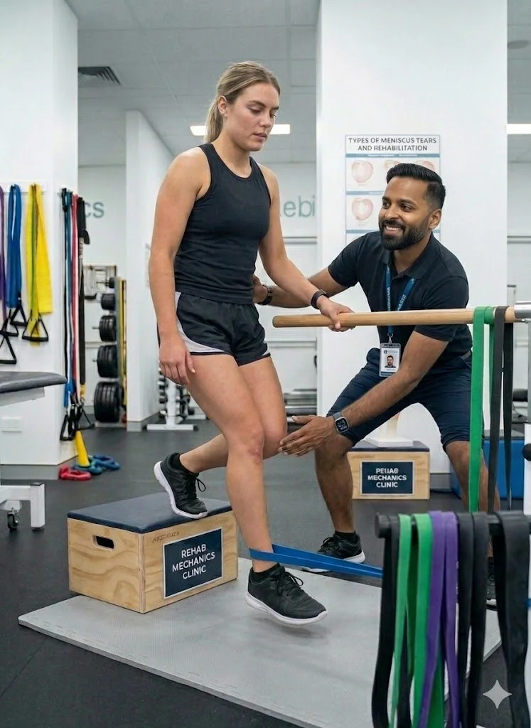
Sanjay practices an integrated model of healthcare, working closely alongside medical doctors inside the Prime Medical Centre on Abell Street to streamline patient recovery pathways. He maintains a human-centric, communication-first clinical framework, ensuring that care remains fully customized rather than automated.
His clinical caseload encompasses a broad operational spectrum under Ontario's regulatory frameworks, including:
Motor Vehicle Accident (MVA) Claims – Rehabilitation navigating Ontario’s statutory accident benefits schedule.
Workplace Safety and Insurance Board (WSIB) – Occupational injury management and return-to-work screening.
Extended Health Care (EHC) & Private Practice – Multi-tier insurance coordination and long-term athletic development plans.
Commitment to Research & Community
Outside of his clinical caseload at Rehab Mechanics and his additional practice affiliations in Etobicoke, Sanjay is an active health writer and community educator. He translates contemporary peer-reviewed medical research into accessible, actionable guidance on his professional blog. As a dedicated father and husband, he mirrors his professional advice in his personal life, focusing on structural mobility, cross-training, and longevity to help his family and his community thrive. Naturally he takes he a keen interest in rehabilitation for women who are pregnant and post-partum.
Disclaimer: The information provided on this blog is intended for educational and informational purposes only. It does not constitute medical advice, diagnosis, or a treatment plan. Always seek the direct advice of a Registered Physiotherapist, physician, or other qualified health provider regarding any medical condition or physical rehabilitation routine.